
Article Figures & Data
Figures
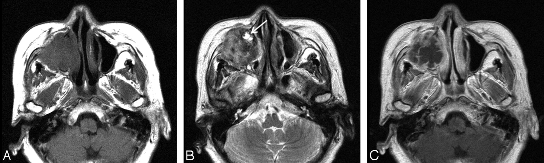
- Fig 1.
Case 1. Inflammatory type of MFH of the maxillary sinus in a 65-year-old woman. Axial MR images show a large ill-defined mass in the right maxillary sinus, which causes destruction of the sinus walls, extending into the anterior cheek and retromaxillary fat. A and B, Compared with the adjacent muscle, the signal intensity of the mass is mixed isointense and slightly hypointense on the T1-weighted image (A) and mixed hyperintense, isointense, and hypointense on the T2-weighted image (B). C, On the contrast-enhanced T1-weighted image, there is moderate heterogeneous enhancement at the periphery of the mass with the central areas remaining unenhanced. Also note that the mass invades the nasolacrimal duct, which is enlarged and filled with fluid (arrow in B).
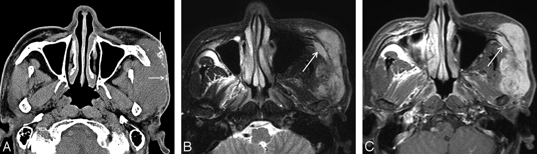
- Fig 2.
Case 7. MFH of the lateral cheek in a 57-year-old man. A, Precontrast axial CT scan shows a large ill-defined soft-tissue mass involving the left masticator space with extension to the skin. The mass is isoattenuated to the adjacent muscle and contains several coarse and irregular calcifications peripherally (arrows). Postcontrast CT scans demonstrated marked heterogeneous enhancement within the mass (data not shown). B, Compared with the cerebral cortex, the signal intensity of the mass is mixed isointense and hyperintense on the axial fat-suppressed T2-weighted MR image. T1-weighted images demonstrated homogeneous isointense signal intensity of the mass (data not shown). C, On the contrast-enhanced axial fat-suppressed T1-weighted MR image, there is marked heterogeneous enhancement within the mass. Also note the involvement of the zygoma by the mass (arrows).
- Fig 3.
Case 9. Myxoid type of MFH of the anterior cheek in a 39-year-old man. Axial MR images show a well-defined ovoid soft-tissue mass in the left anterior cheek. The mass causes erosion of the frontal process of the maxilla (arrows). A and B, Compared with the cerebral cortex, the signal intensity of the mass is slightly hypointense on the T1-weighted image (A) and markedly hyperintense on the T2-weighted image (B). C, Contrast-enhanced fat-suppressed T1-weighted image shows marked homogeneous enhancement throughout the mass.
- Fig 4.
Case 13. Storiform-pleomorphic type of MFH of the orbital roof secondary to fibrous dysplasia in a 45-year-old woman. A, Postcontrast axial CT scan with bone window setting shows a large ill-defined osteolytic lesion in the left orbital roof. The adjacent cranial bones demonstrate a diffuse ground-glass appearance, typical of fibrous dysplasia (asterisk). B and C, On the axial MR images, the lesion is seen as a large ill-defined mass extending to the anterior face. Compared with the adjacent muscle, the signal intensity of the mass is mixed isointense and hyperintense on the T2-weighted image (B). T1-weighted images demonstrated isointense signal intensity of the mass (data not shown). On the contrast-enhanced T1-weighted image (C), there is mild-to-moderate heterogeneous enhancement at the periphery of the mass with the central areas remaining unenhanced. The background bone with fibrous dysplasia is markedly hypointense on the T2-weighted image and enhances heterogeneously after administration of contrast material (asterisks).


Tables
Summary of CT and MR imaging features of 13 patients with MFH of the head and neck*
Patient No./Age(yr)/Sex CT/MR Image Center/Extent BD Size (cm) Margin Density on Precontrast CT T1WI T2WI Enhancement Pattern/Degree Histologic Subtype 1/65/F No/yes Maxillary sinus/nasal cavity, orbit, buccal space, anterior cheek Yes 5.5 Ill-defined – Mixed isointense, hypointense Mixed isointense, hyperintense, hypointense Heterogeneous/moderate Inflammatory 2/46/F Yes/yes Maxillary sinus/buccal space Yes 4.0 Ill-defined Isodense Mixed isointense, hyperintense Mixed isointense, hyperintense, hypointense Heterogeneous/moderate Inflammatory 3/42/M Yes/no Nasal cavity/maxillary sinus, ethmoid sinus, buccal space Yes 5.0 Ill-defined Mixed isodense, slightly hypodense – – Heterogeneous/mild to moderate Myxoid 4/58/M Yes/no Maxillary sinus/anterior cheek Yes 3.9 Ill-defined Mixed isodense, slightly hypodense – – Heterogeneous/moderate Storiform-pleomorphic 5/34/M Yes/yes Maxillary sinus/nasal cavity, ethmoid sinus, orbit, buccal space, anterior cheek Yes 4.0 Ill-defined Isodense Mixed isointense, hyperintense, hypointense Mixed hyperintense, isointense, hypointense Heterogeneous/moderate Storiform-pleomorphic 6/46/M Yes/yes Nasal cavity/nasopharynx, nasal septum Yes 4.1 Ill-defined – Isointense Mixed isointense, hypointense Heterogeneous/moderate Storiform-pleomorphic 7/57/M Yes/yes Masticator space/zygomatic arch Yes 7.8 Ill-defined Isodense Isointense Mixed isointense and hyperintense Heterogenous/marked – 8/30/M Yes/yes Masticator space/zygomatic arch, middle cranial base, middle cranial fossa Yes 5.8 Ill-defined Isodense Mixed isointense and hyperintense Mixed isointense and hyperintense Heterogeneous/moderate to marked Storiform-pleomorphic 9/39/M Yes/yes Anterior cheek/nasal cavity, frontal process of maxilla Yes 4.5 Well-defined Slightly hypodense Slightly hypointense Markedly hyperintense Homogeneous/marked Myxoid 10/24/M Yes/no Infraparotid lateral neck/sternocleidomastoid muscle, parotid gland No 6.8 Ill-defined – – – Heterogeneous/mild to moderate Mixed storiform-pleomorphic and myxoid 11/60/M Yes/no Poststyloid parapharyngeal space/prevertebral space/carotid artery No 5.2 Ill-defined – – – Heterogeneous/mild to moderate Storiform-pleomorphic 12/41/F No/yes Upper anterior gingiva/maxillary alveolar bone Yes 2.5 Well-defined – Isointense Hyperintense Homogeneous/moderate Storiform-pleomorphic 13/45/F Yes/yes Orbital roof/orbit, facial soft tissue Yes 5.0 Ill-defined Hypodense Isointense Mixed isointense and hyperintense Heterogenous/mild to moderate Storiform-pleomorphic Note:—BD indicates associated bone destruction; T1WI, T1-weighted imaging; T2WI, T2-weighted imaging; MFH, malignant fibrous histiocytoma.
* Size was denoted as greatest diameter; density of the lesion was compared with that of the adjacent muscle; signal intensity of the lesion was compared with that of the cerebral cortex.
In this issue


Jump to section
Related Articles
Cited By...
- No citing articles found.