Advertisement

Main menu

  • Home
  • Content
    • Current Issue
    • Accepted Manuscripts
    • Article Preview
    • Past Issue Archive
    • Video Articles
    • AJNR Case Collection
    • Case of the Week Archive
    • Case of the Month Archive
    • Classic Case Archive
  • Special Collections
    • AJNR Awards
    • Low-Field MRI
    • Alzheimer Disease
    • ASNR Foundation Special Collection
    • Photon-Counting CT
    • View All
  • Multimedia
    • AJNR Podcasts
    • AJNR SCANtastic
    • Trainee Corner
    • MRI Safety Corner
    • Imaging Protocols
  • For Authors
    • Submit a Manuscript
    • Submit a Video Article
    • Submit an eLetter to the Editor/Response
    • Manuscript Submission Guidelines
    • Statistical Tips
    • Fast Publishing of Accepted Manuscripts
    • Graphical Abstract Preparation
    • Imaging Protocol Submission
    • Author Policies
  • About Us
    • About AJNR
    • Editorial Board
    • Editorial Board Alumni
  • More
    • Become a Reviewer/Academy of Reviewers
    • Subscribers
    • Permissions
    • Alerts
    • Feedback
    • Advertisers
    • ASNR Home

User menu

  • Alerts
  • Log in

Search

  • Advanced search
American Journal of Neuroradiology

American Journal of Neuroradiology

ASHNR American Society of Functional Neuroradiology ASHNR American Society of Pediatric Neuroradiology ASSR
  • Alerts
  • Log in

Advanced Search

  • Home
  • Content
    • Current Issue
    • Accepted Manuscripts
    • Article Preview
    • Past Issue Archive
    • Video Articles
    • AJNR Case Collection
    • Case of the Week Archive
    • Case of the Month Archive
    • Classic Case Archive
  • Special Collections
    • AJNR Awards
    • Low-Field MRI
    • Alzheimer Disease
    • ASNR Foundation Special Collection
    • Photon-Counting CT
    • View All
  • Multimedia
    • AJNR Podcasts
    • AJNR SCANtastic
    • Trainee Corner
    • MRI Safety Corner
    • Imaging Protocols
  • For Authors
    • Submit a Manuscript
    • Submit a Video Article
    • Submit an eLetter to the Editor/Response
    • Manuscript Submission Guidelines
    • Statistical Tips
    • Fast Publishing of Accepted Manuscripts
    • Graphical Abstract Preparation
    • Imaging Protocol Submission
    • Author Policies
  • About Us
    • About AJNR
    • Editorial Board
    • Editorial Board Alumni
  • More
    • Become a Reviewer/Academy of Reviewers
    • Subscribers
    • Permissions
    • Alerts
    • Feedback
    • Advertisers
    • ASNR Home
  • Follow AJNR on Twitter
  • Visit AJNR on Facebook
  • Follow AJNR on Instagram
  • Join AJNR on LinkedIn
  • RSS Feeds

AJNR Awards, New Junior Editors, and more. Read the latest AJNR updates

Review ArticleReview Articles
Open Access

The Radiology of Referred Otalgia

R.C. Chen, A.S. Khorsandi, D.R. Shatzkes and R.A. Holliday
American Journal of Neuroradiology November 2009, 30 (10) 1817-1823; DOI: https://doi.org/10.3174/ajnr.A1605
R.C. Chen
  • Find this author on Google Scholar
  • Find this author on PubMed
  • Search for this author on this site
A.S. Khorsandi
  • Find this author on Google Scholar
  • Find this author on PubMed
  • Search for this author on this site
D.R. Shatzkes
  • Find this author on Google Scholar
  • Find this author on PubMed
  • Search for this author on this site
R.A. Holliday
  • Find this author on Google Scholar
  • Find this author on PubMed
  • Search for this author on this site
  • Article
  • Figures & Data
  • Info & Metrics
  • Responses
  • References
  • PDF
Loading

Article Figures & Data

Figures

  • Fig 1.
    • Download figure
    • Open in new tab
    • Download powerpoint
    Fig 1.

    Otalgia arising from head and neck sources. Essentially any pathology residing within the sensory net of cranial nerves V, VII, IX, and X and upper cervical nerves C2 and C3 can potentially cause pain referred to the ear. Reprinted with permission from the American Journal of Clinical Oncology (2003;26:e157–62).

  • Fig 2.
    • Download figure
    • Open in new tab
    • Download powerpoint
    Fig 2.

    Primary and referred otalgia pathways of the mandibular nerve (V3). Cranial nerve V is the most frequent pathway for referred otalgia via the auriculotemporal branch of the trigeminal nerve. Reprinted with permission from the American Journal of Clinical Oncology (2003;26:e157–62).

  • Fig 3.
    • Download figure
    • Open in new tab
    • Download powerpoint
    Fig 3.

    TMJ. Coronal reformatted CT scan demonstrates severe erosion and irregularity involving the head of the left condylar process. TMJ disease has been well documented to be associated with referred otalgia via the auriculotemporal and masseteric branches of V3.

  • Fig 4.
    • Download figure
    • Open in new tab
    • Download powerpoint
    Fig 4.

    Parotid malignancy. Positron-emission tomography image demonstrates abnormal hypermetabolic activity involving the left prestyloid parapharyngeal region. The corresponding anatomic images demonstrate a calcified mass within the deep lobe of the parotid gland. The parotid branches of the auriculotemporal nerve mediate referred otalgia.

  • Fig 5.
    • Download figure
    • Open in new tab
    • Download powerpoint
    Fig 5.

    Primary and referred otalgia pathways of the facial nerve (VII). Cranial nerve VII produces referred otalgia via the auricular branch of the facial nerve. Bell palsy can present as ear pain, antecedent to facial paralysis. Reprinted with permission from the American Journal of Clinical Oncology (2003;26:e157–62).

  • Fig 6.
    • Download figure
    • Open in new tab
    • Download powerpoint
    Fig 6.

    Sphenoid sinusitis. T2-weighted image demonstrates mucosal thickening in the left sphenoid sinus. The greater superficial petrosal nerve supplies afferents to this region.

  • Fig 7.
    • Download figure
    • Open in new tab
    • Download powerpoint
    Fig 7.

    Nasal spur. Coronal CT with bone algorithm demonstrates a nasal spur that touches the adjacent concha. Referred otalgia may be eliminated during the ENT examination via placement of a cocaine solution over the spur.
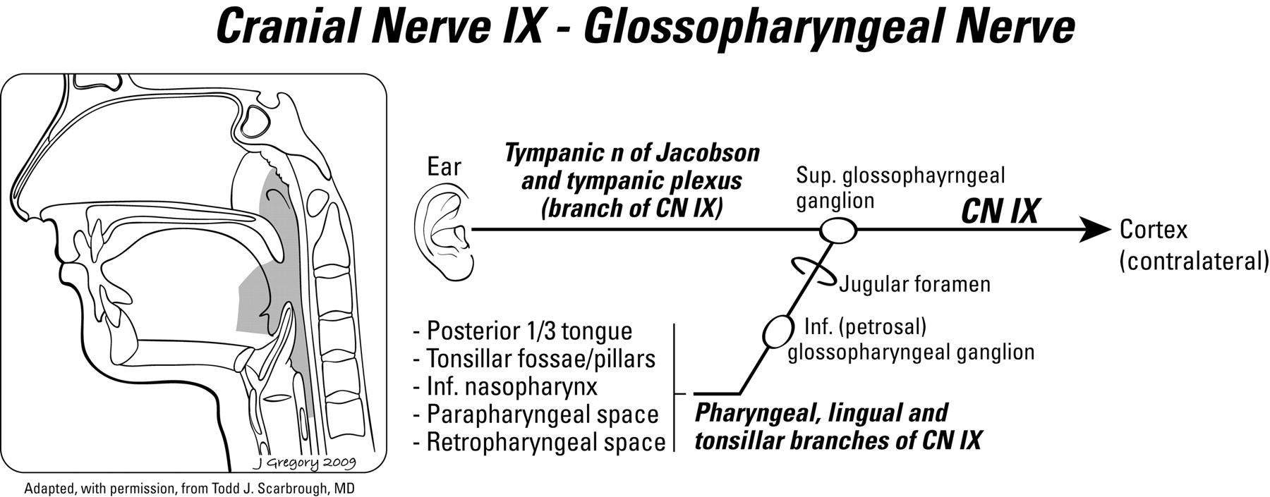
  • Fig 8.
    • Download figure
    • Open in new tab
    • Download powerpoint
    Fig 8.

    Primary and referred otalgia pathways of the glossopharyngeal nerve (IX). Cranial nerve IX mediates otalgia via the tympanic nerve of Jacobson. Reprinted with permission from the American Journal of Clinical Oncology (2003;26:e157–62).

  • Fig 9.
    • Download figure
    • Open in new tab
    • Download powerpoint
    Fig 9.

    Squamous cell carcinoma of the nasopharynx. Axial CT scan with contrast demonstrates an abnormal soft-tissue mass with relatively uniform contrast enhancement filling the left lateral fossa of Rosenmueller, affecting the pharyngeal plexus of cranial nerve IX.

  • Fig 10.
    • Download figure
    • Open in new tab
    • Download powerpoint
    Fig 10.

    Peritonsillar abscess. Axial CT scan with contrast demonstrates a right inferior peritonsillar abscess in association with edematous change in the adjacent right parapharyngeal space irritating the pharyngeal plexus (cranial nerve IX).

  • Fig 11.
    • Download figure
    • Open in new tab
    • Download powerpoint
    Fig 11.

    Retropharyngeal adenitis. Axial CT scan demonstrates suppurative adenitis involving the right retropharyngeal space, which can affect the pharyngeal plexus and produce referred otalgia.

  • Fig 12.
    • Download figure
    • Open in new tab
    • Download powerpoint
    Fig 12.

    Eagle syndrome. A 30-year-old man with otalgia. Plain film demonstrates a thickened and elongated styloid process, which can irritate the tonsillar bed via the tonsillar branch of cranial nerve IX. Patients classically have reproduction of pain on transoral palpation of the tonsils.

  • Fig 13.
    • Download figure
    • Open in new tab
    • Download powerpoint
    Fig 13.

    Primary and referred otalgia pathways of the vagus nerve (X). Cranial nerve X is involved with otalgia via the auricular nerve of Arnold. Reprinted with permission from the American Journal of Clinical Oncology (2003;26:e157–62)

  • Fig 14.
    • Download figure
    • Open in new tab
    • Download powerpoint
    Fig 14.

    Supraglottic squamous cell carcinoma. Positron-emission tomography/CT scan demonstrates an enhancing exophytic supraglottic mass arising from the posterior wall of the larynx. This area is innervated by the internal branch of the superior laryngeal nerve and can result in otalgia when irritated.

  • Fig 15.
    • Download figure
    • Open in new tab
    • Download powerpoint
    Fig 15.

    Papillary thyroid cancer. Axial CT scan demonstrates a hypoattenuated mass centered in the thyroid isthmus. Although most thyroid cancers are subclinical, some may present with pain or dysphagia. Because the sensory innervation to the thyroid gland is via the superior and recurrent laryngeal nerves, there have been documented cases of thyroid pathology manifesting as ear pain.

PreviousNext
Back to top

In this issue

American Journal of Neuroradiology: 30 (10)
American Journal of Neuroradiology
Vol. 30, Issue 10
1 Nov 2009
  • Table of Contents
  • Index by author
Advertisement
Print
Download PDF
Email Article

Thank you for your interest in spreading the word on American Journal of Neuroradiology.

NOTE: We only request your email address so that the person you are recommending the page to knows that you wanted them to see it, and that it is not junk mail. We do not capture any email address.

Enter multiple addresses on separate lines or separate them with commas.
The Radiology of Referred Otalgia
(Your Name) has sent you a message from American Journal of Neuroradiology
(Your Name) thought you would like to see the American Journal of Neuroradiology web site.
CAPTCHA
This question is for testing whether or not you are a human visitor and to prevent automated spam submissions.
Cite this article
R.C. Chen, A.S. Khorsandi, D.R. Shatzkes, R.A. Holliday
The Radiology of Referred Otalgia
American Journal of Neuroradiology Nov 2009, 30 (10) 1817-1823; DOI: 10.3174/ajnr.A1605

Citation Manager Formats

  • BibTeX
  • Bookends
  • EasyBib
  • EndNote (tagged)
  • EndNote 8 (xml)
  • Medlars
  • Mendeley
  • Papers
  • RefWorks Tagged
  • Ref Manager
  • RIS
  • Zotero
0 Responses
Respond to this article
Share
Bookmark this article
The Radiology of Referred Otalgia
R.C. Chen, A.S. Khorsandi, D.R. Shatzkes, R.A. Holliday
American Journal of Neuroradiology Nov 2009, 30 (10) 1817-1823; DOI: 10.3174/ajnr.A1605
del.icio.us logo Twitter logo Facebook logo Mendeley logo
  • Tweet Widget
  • Facebook Like
  • Google Plus One
Purchase

Jump to section

  • Article
    • Abstract
    • Significance
    • Diagnostic Work-Up for Referred Otalgia
    • Mechanism of Referred Otalgia
    • Sensory Innervation of the Ear
    • Sources of Referred Otalgia, Grouped According to Cranial and Cervical Nerves
    • Neuralgia as the Source of Otalgia
    • Conclusions
    • References
  • Figures & Data
  • Info & Metrics
  • Responses
  • References
  • PDF

Related Articles

  • No related articles found.
  • PubMed
  • Google Scholar

Cited By...

  • Otalgie referee: Causes habituelles et strategies devaluation et de prise en charge fondees sur des donnees probantes
  • Referred otalgia: Common causes and evidence-based strategies for assessment and management
  • Secondary Otalgia: Referred Pain Pathways and Pathologies
  • Diagnostic Yield and Therapeutic Impact of Face and Neck Imaging in Patients Referred with Otalgia without Clinically Overt Disease
  • Crossref
  • Google Scholar

This article has not yet been cited by articles in journals that are participating in Crossref Cited-by Linking.

More in this TOC Section

  • An Atlas of Neonatal Neurovascular Imaging Anatomy as Depicted with Microvascular Imaging: The Intracranial Arteries
  • An Atlas of Neonatal Neurovascular Imaging Anatomy as Depicted with Microvascular Imaging: The Intracranial Veins
  • Clinical Translation of Hyperpolarized 13C Metabolic Probes for Glioma Imaging
Show more Review Articles

Similar Articles

Advertisement

Indexed Content

  • Current Issue
  • Accepted Manuscripts
  • Article Preview
  • Past Issues
  • Editorials
  • Editor's Choice
  • Fellows' Journal Club
  • Letters to the Editor
  • Video Articles

Cases

  • Case Collection
  • Archive - Case of the Week
  • Archive - Case of the Month
  • Archive - Classic Case

More from AJNR

  • Trainee Corner
  • Imaging Protocols
  • MRI Safety Corner
  • Book Reviews

Multimedia

  • AJNR Podcasts
  • AJNR Scantastics

Resources

  • Turnaround Time
  • Submit a Manuscript
  • Submit a Video Article
  • Submit an eLetter to the Editor/Response
  • Manuscript Submission Guidelines
  • Statistical Tips
  • Fast Publishing of Accepted Manuscripts
  • Graphical Abstract Preparation
  • Imaging Protocol Submission
  • Evidence-Based Medicine Level Guide
  • Publishing Checklists
  • Author Policies
  • Become a Reviewer/Academy of Reviewers
  • News and Updates

About Us

  • About AJNR
  • Editorial Board
  • Editorial Board Alumni
  • Alerts
  • Permissions
  • Not an AJNR Subscriber? Join Now
  • Advertise with Us
  • Librarian Resources
  • Feedback
  • Terms and Conditions
  • AJNR Editorial Board Alumni

American Society of Neuroradiology

  • Not an ASNR Member? Join Now

© 2025 by the American Society of Neuroradiology All rights, including for text and data mining, AI training, and similar technologies, are reserved.
Print ISSN: 0195-6108 Online ISSN: 1936-959X

Powered by HighWire