Advertisement

Main menu

  • Home
  • Content
    • Current Issue
    • Accepted Manuscripts
    • Article Preview
    • Past Issue Archive
    • Video Articles
    • AJNR Case Collection
    • Case of the Week Archive
    • Case of the Month Archive
    • Classic Case Archive
  • Special Collections
    • AJNR Awards
    • Low-Field MRI
    • Alzheimer Disease
    • ASNR Foundation Special Collection
    • Photon-Counting CT
    • View All
  • Multimedia
    • AJNR Podcasts
    • AJNR SCANtastic
    • Trainee Corner
    • MRI Safety Corner
    • Imaging Protocols
  • For Authors
    • Submit a Manuscript
    • Submit a Video Article
    • Submit an eLetter to the Editor/Response
    • Manuscript Submission Guidelines
    • Statistical Tips
    • Fast Publishing of Accepted Manuscripts
    • Graphical Abstract Preparation
    • Imaging Protocol Submission
    • Author Policies
  • About Us
    • About AJNR
    • Editorial Board
    • Editorial Board Alumni
  • More
    • Become a Reviewer/Academy of Reviewers
    • Subscribers
    • Permissions
    • Alerts
    • Feedback
    • Advertisers
    • ASNR Home

User menu

  • Alerts
  • Log in

Search

  • Advanced search
American Journal of Neuroradiology

American Journal of Neuroradiology

ASHNR American Society of Functional Neuroradiology ASHNR American Society of Pediatric Neuroradiology ASSR
  • Alerts
  • Log in

Advanced Search

  • Home
  • Content
    • Current Issue
    • Accepted Manuscripts
    • Article Preview
    • Past Issue Archive
    • Video Articles
    • AJNR Case Collection
    • Case of the Week Archive
    • Case of the Month Archive
    • Classic Case Archive
  • Special Collections
    • AJNR Awards
    • Low-Field MRI
    • Alzheimer Disease
    • ASNR Foundation Special Collection
    • Photon-Counting CT
    • View All
  • Multimedia
    • AJNR Podcasts
    • AJNR SCANtastic
    • Trainee Corner
    • MRI Safety Corner
    • Imaging Protocols
  • For Authors
    • Submit a Manuscript
    • Submit a Video Article
    • Submit an eLetter to the Editor/Response
    • Manuscript Submission Guidelines
    • Statistical Tips
    • Fast Publishing of Accepted Manuscripts
    • Graphical Abstract Preparation
    • Imaging Protocol Submission
    • Author Policies
  • About Us
    • About AJNR
    • Editorial Board
    • Editorial Board Alumni
  • More
    • Become a Reviewer/Academy of Reviewers
    • Subscribers
    • Permissions
    • Alerts
    • Feedback
    • Advertisers
    • ASNR Home
  • Follow AJNR on Twitter
  • Visit AJNR on Facebook
  • Follow AJNR on Instagram
  • Join AJNR on LinkedIn
  • RSS Feeds

AJNR Awards, New Junior Editors, and more. Read the latest AJNR updates

Research ArticleSpine Imaging and Spine Image-Guided InterventionsT
Open Access

Subminute Fat-Water-Separated Dual-Echo Automated Spine Survey Iterative Scan Technique

K.L. Weiss, C.R. Richards, D. Sun and J.L. Weiss
American Journal of Neuroradiology November 2009, 30 (10) 1840-1846; DOI: https://doi.org/10.3174/ajnr.A1619
K.L. Weiss
  • Find this author on Google Scholar
  • Find this author on PubMed
  • Search for this author on this site
C.R. Richards
  • Find this author on Google Scholar
  • Find this author on PubMed
  • Search for this author on this site
D. Sun
  • Find this author on Google Scholar
  • Find this author on PubMed
  • Search for this author on this site
J.L. Weiss
  • Find this author on Google Scholar
  • Find this author on PubMed
  • Search for this author on this site
  • Article
  • Figures & Data
  • Info & Metrics
  • Responses
  • References
  • PDF
Loading

Article Figures & Data

Figures

  • Fig 1.
    • Download figure
    • Open in new tab
    • Download powerpoint
    Fig 1.

    A 61-year-old woman with metastatic breast cancer. Midline sagittal ASSIST DE precontrast images (A) and postcontrast images (B). From left to right: autolabeled water-decomposed, non-labeled water-decomposed, fat-decomposed, water plus fat (in-phase), color-encoded water (gray) plus fat (red), and colorized apparent-fat signal percentage map. Red arrows point to solitary L2 metastasis, visible on all sequences. The metastasis replaces fat and enhances with contrast. Incidental Modic type II degenerative changes (fatty marrow conversion) involving the anterior inferior endplate T8 are best appreciated on the precontrast fat-decomposed and fat percentage images (white arrows). Note that the 2D color bar for the water plus fat images has fat percentage on the y-axis (red hue) and signal intensity on the x-axis, whereas the spectral color bar for the fat ratio image goes from low (blue) to high (red) fat percentage.

  • Fig 2.
    • Download figure
    • Open in new tab
    • Download powerpoint
    Fig 2.

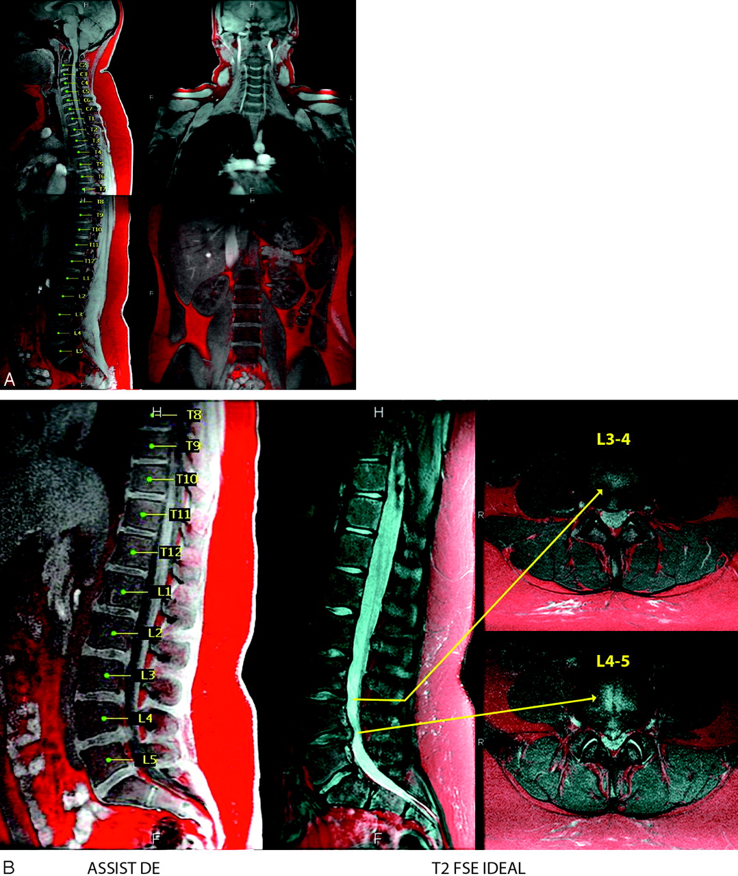
    A 13-year-old girl with back pain. A, Color-encoded autolabeled midline sagittal ASSIST DE and corresponding coronal image. B, Color-encoded lower station sagittal ASSIST DE and corresponding 2D T2 FSE IDEAL sagittal and axial oblique images, the latter obtained for confirmation and further elucidation. Sagittal ASSIST DE demonstrates a normal cervical and thoracic spine with premature lumbar degenerative disk disease at L2–3 through L4–5 with mild protrusions at L2–3, L3–4, and L5–S1 and a prominent central disk herniation at L4–5. T2-FSE IDEAL images were confirmatory and further elucidate the mild central protrusion L3-4 and moderate central extrusion L4-5.

  • Fig 3.
    • Download figure
    • Open in new tab
    • Download powerpoint
    Fig 3.

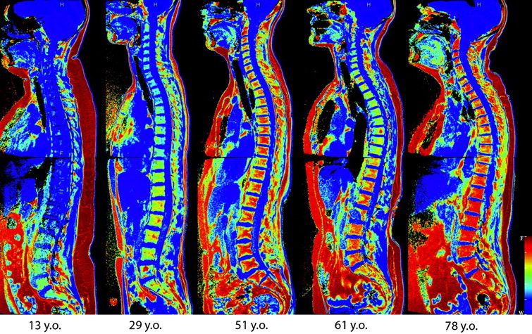
    Vertebral fat content and curvature as a function of age is demonstrated on midline sagittal ASSIST DE color-encoded apparent-fat signal-intensity percentage maps. The ages of the 5 female subjects (from left to right) are 13, 29, 51, 61, and 78 years. Note that as age increases, both kypholordosis and fatty marrow content tend to increase, the latter with a distal-to-proximal gradient. Note that the trend is not absolute because the 51-year-old woman appears to have a higher vertebral fat content than the 61-year-old woman. Additionally, percentage of fatty marrow appears relatively independent of subcutaneous fat because the youngest subject exhibits the highest subcutaneous fat volume but the lowest percentage of fatty marrow (mostly blue).

  • Fig 4.
    • Download figure
    • Open in new tab
    • Download powerpoint
    Fig 4.

    A 41-year-old woman with sarcoidosis. Midline lower station sagittal color-encoded ASSIST DE images from left to right: autolabeled water plus fat, postcontrast water plus fat, and precontrast apparent-fat signal-intensity percentage map. Note the diffuse fatty marrow replacement as evidenced by the paucity of reddish hue in the first 2 images and bluish color of the vertebrae on the third image. The middle image depicts heterogeneous abnormal enhancement of all visualized vertebrae related to extensive sarcoid involvement.

PreviousNext
Back to top

In this issue

American Journal of Neuroradiology: 30 (10)
American Journal of Neuroradiology
Vol. 30, Issue 10
1 Nov 2009
  • Table of Contents
  • Index by author
Advertisement
Print
Download PDF
Email Article

Thank you for your interest in spreading the word on American Journal of Neuroradiology.

NOTE: We only request your email address so that the person you are recommending the page to knows that you wanted them to see it, and that it is not junk mail. We do not capture any email address.

Enter multiple addresses on separate lines or separate them with commas.
Subminute Fat-Water-Separated Dual-Echo Automated Spine Survey Iterative Scan Technique
(Your Name) has sent you a message from American Journal of Neuroradiology
(Your Name) thought you would like to see the American Journal of Neuroradiology web site.
CAPTCHA
This question is for testing whether or not you are a human visitor and to prevent automated spam submissions.
Cite this article
K.L. Weiss, C.R. Richards, D. Sun, J.L. Weiss
Subminute Fat-Water-Separated Dual-Echo Automated Spine Survey Iterative Scan Technique
American Journal of Neuroradiology Nov 2009, 30 (10) 1840-1846; DOI: 10.3174/ajnr.A1619

Citation Manager Formats

  • BibTeX
  • Bookends
  • EasyBib
  • EndNote (tagged)
  • EndNote 8 (xml)
  • Medlars
  • Mendeley
  • Papers
  • RefWorks Tagged
  • Ref Manager
  • RIS
  • Zotero
0 Responses
Respond to this article
Share
Bookmark this article
Subminute Fat-Water-Separated Dual-Echo Automated Spine Survey Iterative Scan Technique
K.L. Weiss, C.R. Richards, D. Sun, J.L. Weiss
American Journal of Neuroradiology Nov 2009, 30 (10) 1840-1846; DOI: 10.3174/ajnr.A1619
del.icio.us logo Twitter logo Facebook logo Mendeley logo
  • Tweet Widget
  • Facebook Like
  • Google Plus One
Purchase

Jump to section

  • Article
    • Abstract
    • Materials and Methods
    • Results
    • Discussion
    • Acknowledgments
    • Footnotes
    • References
  • Figures & Data
  • Info & Metrics
  • Responses
  • References
  • PDF

Related Articles

  • No related articles found.
  • PubMed
  • Google Scholar

Cited By...

  • No citing articles found.
  • Crossref
  • Google Scholar

This article has not yet been cited by articles in journals that are participating in Crossref Cited-by Linking.

More in this TOC Section

  • Management Outcomes For VO Spine Biopsy
  • Characteristics of SIH Type I Culprit Lesions
  • Advanced Imaging of Type 2 Spinal CSF Leaks
Show more Spine Imaging and Spine Image-Guided Interventions

Similar Articles

Advertisement

Indexed Content

  • Current Issue
  • Accepted Manuscripts
  • Article Preview
  • Past Issues
  • Editorials
  • Editor's Choice
  • Fellows' Journal Club
  • Letters to the Editor
  • Video Articles

Cases

  • Case Collection
  • Archive - Case of the Week
  • Archive - Case of the Month
  • Archive - Classic Case

More from AJNR

  • Trainee Corner
  • Imaging Protocols
  • MRI Safety Corner
  • Book Reviews

Multimedia

  • AJNR Podcasts
  • AJNR Scantastics

Resources

  • Turnaround Time
  • Submit a Manuscript
  • Submit a Video Article
  • Submit an eLetter to the Editor/Response
  • Manuscript Submission Guidelines
  • Statistical Tips
  • Fast Publishing of Accepted Manuscripts
  • Graphical Abstract Preparation
  • Imaging Protocol Submission
  • Evidence-Based Medicine Level Guide
  • Publishing Checklists
  • Author Policies
  • Become a Reviewer/Academy of Reviewers
  • News and Updates

About Us

  • About AJNR
  • Editorial Board
  • Editorial Board Alumni
  • Alerts
  • Permissions
  • Not an AJNR Subscriber? Join Now
  • Advertise with Us
  • Librarian Resources
  • Feedback
  • Terms and Conditions
  • AJNR Editorial Board Alumni

American Society of Neuroradiology

  • Not an ASNR Member? Join Now

© 2025 by the American Society of Neuroradiology All rights, including for text and data mining, AI training, and similar technologies, are reserved.
Print ISSN: 0195-6108 Online ISSN: 1936-959X

Powered by HighWire