Advertisement

Main menu

  • Home
  • Content
    • Current Issue
    • Accepted Manuscripts
    • Article Preview
    • Past Issue Archive
    • AJNR Case Collection
    • Case of the Week Archive
    • Classic Case Archive
    • Case of the Month Archive
  • Special Collections
    • Spinal CSF Leak Articles (Jan 2020-June 2024)
    • 2024 AJNR Journal Awards
    • Most Impactful AJNR Articles
  • Multimedia
    • AJNR Podcast
    • AJNR Scantastics
    • Video Articles
  • For Authors
    • Submit a Manuscript
    • Author Policies
    • Fast publishing of Accepted Manuscripts
    • Graphical Abstract Preparation
    • Manuscript Submission Guidelines
    • Imaging Protocol Submission
    • Submit a Case for the Case Collection
  • About Us
    • About AJNR
    • Editorial Board
  • More
    • Become a Reviewer/Academy of Reviewers
    • Subscribers
    • Permissions
    • Alerts
    • Feedback
    • Advertisers
    • ASNR Home
  • Other Publications
    • ajnr

User menu

  • Alerts
  • Log in

Search

  • Advanced search
American Journal of Neuroradiology

American Journal of Neuroradiology

ASHNR American Society of Functional Neuroradiology ASHNR American Society of Pediatric Neuroradiology ASSR
  • Alerts
  • Log in

Advanced Search

  • Home
  • Content
    • Current Issue
    • Accepted Manuscripts
    • Article Preview
    • Past Issue Archive
    • AJNR Case Collection
    • Case of the Week Archive
    • Classic Case Archive
    • Case of the Month Archive
  • Special Collections
    • Spinal CSF Leak Articles (Jan 2020-June 2024)
    • 2024 AJNR Journal Awards
    • Most Impactful AJNR Articles
  • Multimedia
    • AJNR Podcast
    • AJNR Scantastics
    • Video Articles
  • For Authors
    • Submit a Manuscript
    • Author Policies
    • Fast publishing of Accepted Manuscripts
    • Graphical Abstract Preparation
    • Manuscript Submission Guidelines
    • Imaging Protocol Submission
    • Submit a Case for the Case Collection
  • About Us
    • About AJNR
    • Editorial Board
  • More
    • Become a Reviewer/Academy of Reviewers
    • Subscribers
    • Permissions
    • Alerts
    • Feedback
    • Advertisers
    • ASNR Home
  • Follow AJNR on Twitter
  • Visit AJNR on Facebook
  • Follow AJNR on Instagram
  • Join AJNR on LinkedIn
  • RSS Feeds

Welcome to the new AJNR, Updated Hall of Fame, and more. Read the full announcements.


AJNR is seeking candidates for the position of Associate Section Editor, AJNR Case Collection. Read the full announcement.


Research ArticleBrain

Is All “Communicating” Hydrocephalus Really Communicating? Prospective Study on the Value of 3D-Constructive Interference in Steady State Sequence at 3T

A. Dinçer, S. Kohan and M.M. Özek
American Journal of Neuroradiology November 2009, 30 (10) 1898-1906; DOI: https://doi.org/10.3174/ajnr.A1726
A. Dinçer
  • Find this author on Google Scholar
  • Find this author on PubMed
  • Search for this author on this site
S. Kohan
  • Find this author on Google Scholar
  • Find this author on PubMed
  • Search for this author on this site
M.M. Özek
  • Find this author on Google Scholar
  • Find this author on PubMed
  • Search for this author on this site
  • Article
  • Figures & Data
  • Info & Metrics
  • Responses
  • References
  • PDF
Loading

Article Figures & Data

Figures

  • Tables
  • Fig 1.
    • Download figure
    • Open in new tab
    • Download powerpoint
    Fig 1.

    Case 1. A, Sagittal TSE T2 image shows third ventricle enlargement with downward displacement of the floor of the third ventricle consistent with hydrocephalus. The cerebral aqueduct and foramen of Magendie are open widely, showing extensive flow void phenomenon. There is a mild enlargement in the fourth ventricle. There is no sign of obstructive membrane in the prepontine cistern. B, Left parasagittal TSE T2 images through the left lateral ventricular exit show no direct or indirect sign of membrane. C, Axial TSE T2 image through the fourth ventricular exits demonstrates prominent signal intensity void in the fourth ventricle, but there is no direct or indirect sign of obstructive membrane at the foramina of Luschka. D, Left parasagittal 3D-CISS image clearly points out the membrane itself in the foramen of Luschka. There is extensive intensity difference between the fourth ventricle and the neighboring cistern. E, Sagittal 3D-CISS image clearly demonstrates prepontine membranes extending from the clivus to the basilar artery. F, Left parasagittal follow-up 3D-CISS image indicates persistent membrane in the fourth ventricle exit foramen, though the intensity differences between cistern and ventricle have disappeared.

  • Fig 2.
    • Download figure
    • Open in new tab
    • Download powerpoint
    Fig 2.

    Case 2. A, Axial TSE T2 image through the posterior fossa shows left cerebellar hypoplasia and enlargement of the bilateral cerebellomedullary cistern without any evidence of membrane at the fourth ventricle exit foramina. B, Axial oblique reformatted image of sagittal 3D-CISS reveals obstructing membranes of foramina of Luschka, bulging into the cerebellomedullary cisterns.

  • Fig 3.
    • Download figure
    • Open in new tab
    • Download powerpoint
    Fig 3.

    Case 3. A, Sagittal TSE T2 image shows an enlarged third ventricle. The cerebral aqueduct seems to be open. B, Sagittal 3D-CISS demonstrates superior medullary velum synechia, causing triventricular hydrocephalus. There is a spontaneous third ventriculostomy at the floor of the ventricle, just behind the tip of the basilar artery. Sagittal CISS revealed the anatomic defect, whereas cine PC did not detect any flow through the defect.
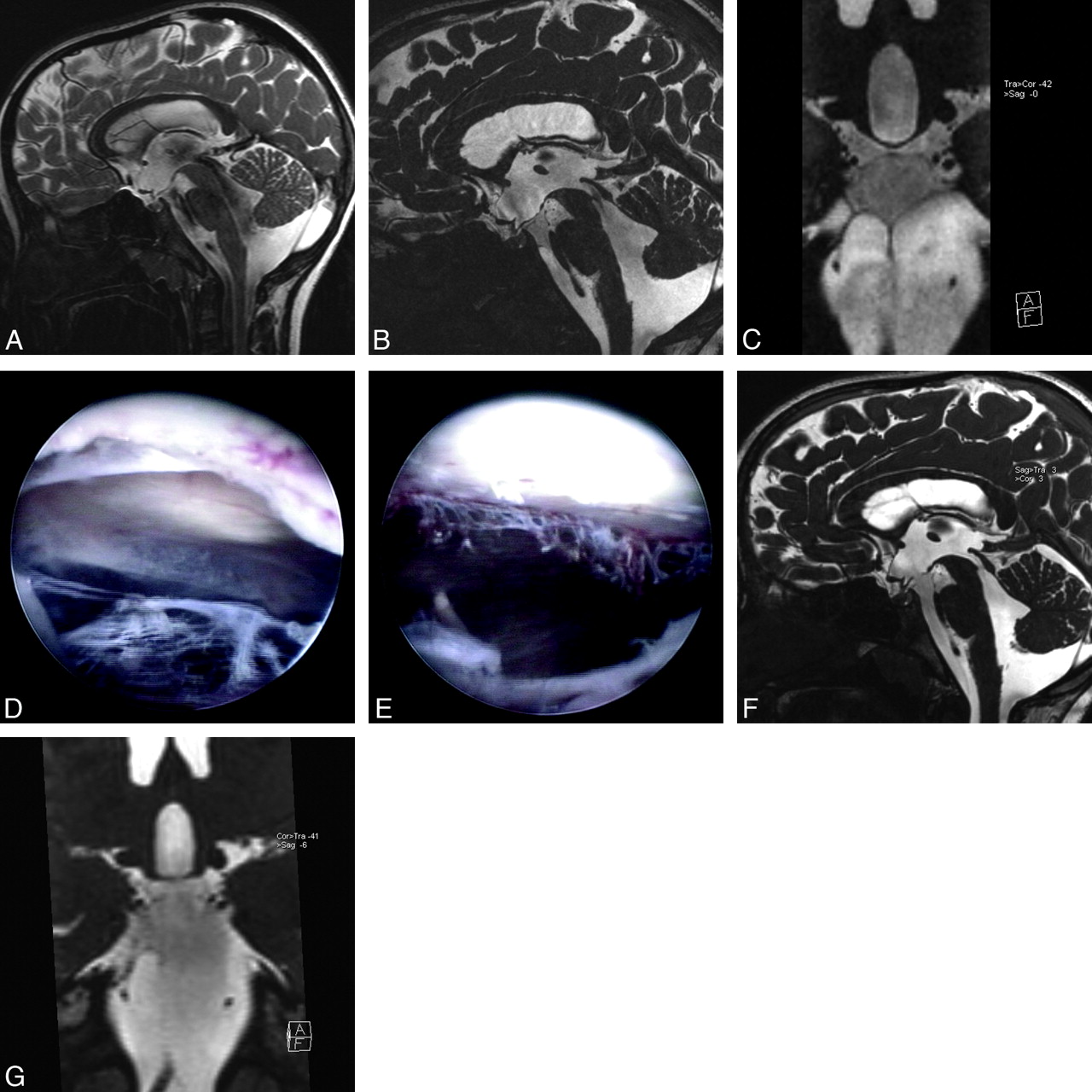
  • Fig 4.
    • Download figure
    • Open in new tab
    • Download powerpoint
    Fig 4.

    Case 4. A, Sagittal TSE T2 image demonstrates enlarged third ventricle, extensive flow void phenomenon in the cerebral aqueduct, the fourth ventricle, and prepontine/interpeduncular cisterns compatible with communicating hydrocephalus. B, Sagittal 3D-CISS image shows Liliequist membrane just below the downward bulging floor of the third ventricle. In addition to this, there is a thin membrane extending from the clivus to the basilar artery, dividing the prepontine cistern into upper and lower parts. The signal intensity difference between 2 parts with a sharp, linear zone of transition was seen. C, Coronal oblique reformatted image of 3D-CISS through the cisterns demonstrates lateral extension of the prepontine membrane, trapping CSF between the Liliequist membrane and the prepontine membrane. D, During ETV, after the floor of the third ventricle through the Liliequist membrane is opened, a complete membrane without any aperture is seen. E, The same view after the membrane is opened and removed, the cisternal part of the left abducens nerve is seen partly. F, On the follow-up sagittal 3D-CISS image, there is free communication between the third ventricle and the prepontine cistern through the interpeduncular cistern. G, Follow-up coronal oblique reformatted image of 3D-CISS after ETV through the cisterns (same view as in C) reveals nearly complete removal of the prepontine membrane. The difference in signal intensity between the 2 parts of the cisterns has almost completely disappeared.

  • Fig 5.
    • Download figure
    • Open in new tab
    • Download powerpoint
    Fig 5.

    Case 4. A, Axial TSE T2 image through the lateral ventricle demonstrates unilateral right lateral ventriculomegaly. B, Coronal TSE T2 image through the foramen of Monro falsely demonstrates a free communication between the right lateral ventricle and the third ventricle. C, Coronal reformatted sagittal 3D-CISS image points out a complete membranous obstruction in the right foramen of Monro. D, Follow-up axial-oblique reformatted image revealing complete removal of obstructing membrane and normal-appearing lateral ventricles.

Tables

  • Figures
    • View popup
    Table 1:

    Sequence parameters for cranial MR imaging

    SequencesImaging PlaneTR/TE/ETLTime of AcquisitionVoxel Size (mm3)Section/Partition Thickness/Gap (mm)FA/BW (Hz/Px)
    TSE T2Axial3590/101/131.531.755/0.590/100
    TSE T2Sagittal3000/131/131.411.22/0.290/100
    TSE T2Coronal4850/132/131.361.083/0.7590/100
    Cine PCOblique59/11/−3–51.08335/391
    3D Turbo flash T1Sagittal2000/3.9/−4.140.510.820/130
    TSE T1Axial500/13/222.055/0.570/155
    3D-CISSSagittal13.6/5.7/−4.440.2160.650/130
    • Note:—ETL indicates echo-train length; FA, flip angle; BW, bandwidth; TSE, turbo spin-echo; cine PC, cine phase-contrast; 3D turbo flash T1, 3D turbo fast low-angle shot T1; 3D-CISS, 3D constructive interference in the steady state.

    • View popup
    Table 2:

    Site of membranous obstruction on the basis of examination of different sequences

    SequencesLocalization of Obstructive MembranesTotal Number of Membranes
    Foramen of MonroCerebral AqueductsSuperior Velum Medullary SynechiaForamen of MagendieForamina of LuschkaForamen MagnumCisterns
    Conventional T1 and T27*411430157
    3D CISS74152846228157
    • * These cases were indirectly diagnosed on the basis of unilateral or bilateral lateral ventricular dilation. There are 2 bilateral membranes in 5 cases.

    • View popup
    Table 3:

    Classification of hydrocephalus on the basis of examination of different sequences

    SequencesNumber of Communicating Hydrocephalus CasesNumber of Noncommunicating Hydrocephalus Cases with Obstructive MembranesTotal Number of Cases with Obstructive Membranes
    Number of Cases with no Visible Obstructive MembraneNumber of Cases with Only an Obstructive Cisternal MembraneNumber of Cases with Only an Obstructive Intraventricular MembraneNumber of Cases with Obstructive Ventricular Membrane Plus Obstructive Cisternal Membrane
    Conventional T1 and T246047148
    3D-CISS137632292
PreviousNext
Back to top

In this issue

American Journal of Neuroradiology: 30 (10)
American Journal of Neuroradiology
Vol. 30, Issue 10
1 Nov 2009
  • Table of Contents
  • Index by author
Advertisement
Print
Download PDF
Email Article

Thank you for your interest in spreading the word on American Journal of Neuroradiology.

NOTE: We only request your email address so that the person you are recommending the page to knows that you wanted them to see it, and that it is not junk mail. We do not capture any email address.

Enter multiple addresses on separate lines or separate them with commas.
Is All “Communicating” Hydrocephalus Really Communicating? Prospective Study on the Value of 3D-Constructive Interference in Steady State Sequence at 3T
(Your Name) has sent you a message from American Journal of Neuroradiology
(Your Name) thought you would like to see the American Journal of Neuroradiology web site.
CAPTCHA
This question is for testing whether or not you are a human visitor and to prevent automated spam submissions.
Cite this article
A. Dinçer, S. Kohan, M.M. Özek
Is All “Communicating” Hydrocephalus Really Communicating? Prospective Study on the Value of 3D-Constructive Interference in Steady State Sequence at 3T
American Journal of Neuroradiology Nov 2009, 30 (10) 1898-1906; DOI: 10.3174/ajnr.A1726

Citation Manager Formats

  • BibTeX
  • Bookends
  • EasyBib
  • EndNote (tagged)
  • EndNote 8 (xml)
  • Medlars
  • Mendeley
  • Papers
  • RefWorks Tagged
  • Ref Manager
  • RIS
  • Zotero
0 Responses
Respond to this article
Share
Bookmark this article
Is All “Communicating” Hydrocephalus Really Communicating? Prospective Study on the Value of 3D-Constructive Interference in Steady State Sequence at 3T
A. Dinçer, S. Kohan, M.M. Özek
American Journal of Neuroradiology Nov 2009, 30 (10) 1898-1906; DOI: 10.3174/ajnr.A1726
del.icio.us logo Twitter logo Facebook logo Mendeley logo
  • Tweet Widget
  • Facebook Like
  • Google Plus One
Purchase

Jump to section

  • Article
    • Abstract
    • Materials and Methods
    • Results
    • Discussion
    • Conclusions
    • Footnotes
    • References
  • Figures & Data
  • Info & Metrics
  • Responses
  • References
  • PDF

Related Articles

  • No related articles found.
  • PubMed
  • Google Scholar

Cited By...

  • Arachnoid Membranes: Crawling Back into Radiologic Consciousness
  • Intrathecal Gadolinium-Enhanced MR Cisternography: A Comprehensive Review
  • Hydrocephalus in Patients with Neurofibromatosis Type 1: MR Imaging Findings and the Outcome of Endoscopic Third Ventriculostomy
  • Crossref (77)
  • Google Scholar

This article has been cited by the following articles in journals that are participating in Crossref Cited-by Linking.

  • Evaluation of hydrocephalus and other cerebrospinal fluid disorders with MRI: An update
    Merve Gulbiz Kartal, Oktay Algin
    Insights into Imaging 2014 5 4
  • Radiologic evaluation of pediatric hydrocephalus
    Alp Dinçer, M. Memet Özek
    Child's Nervous System 2011 27 10
  • Intrathecal Gadolinium-Enhanced MR Cisternography: A Comprehensive Review
    O. Algin, B. Turkbey
    American Journal of Neuroradiology 2013 34 1
  • Inferior vermian hypoplasia – preconception, misconception
    Ashley J. Robinson
    Ultrasound in Obstetrics & Gynecology 2014 43 2
  • Neuropathology
    Ville Leinonen, Ritva Vanninen, Tuomas Rauramaa
    2018 145
  • MR assessment of pediatric hydrocephalus: a road map
    Charles Raybaud
    Child's Nervous System 2016 32 1
  • Analysis of endoscopic third ventriculostomy patency by MRI: value of different pulse sequences, the sequence parameters, and the imaging planes for investigation of flow void
    Alp Dinçer, Erdem Yildiz, Saeed Kohan, M. Memet Özek
    Child's Nervous System 2011 27 1
  • Noncommunicating Hydrocephalus
    Vijetha V. Maller, Richard Ian Gray
    Seminars in Ultrasound, CT and MRI 2016 37 2
  • Microsurgical and endoscopic anatomy of Liliequist's membrane and the prepontine membranes: cadaveric study and clinical implications
    Ihsan Anik, Savas Ceylan, Kenan Koc, Mehtap Tugasaygi, Gozde Sirin, Nurperi Gazioglu, Bulent Sam
    Acta Neurochirurgica 2011 153 8
  • Phase-contrast MRI and 3D-CISS versus contrast-enhanced MR cisternography for the detection of spontaneous third ventriculostomy
    O. Algin, B. Hakyemez, M. Parlak
    Journal of Neuroradiology 2011 38 2

More in this TOC Section

  • Qualitative and Quantitative Analysis of MR Imaging Findings in Patients with Middle Cerebral Artery Stroke Implanted with Mesenchymal Stem Cells
  • Multimodal CT Provides Improved Performance for Lacunar Infarct Detection
  • Statin Therapy Does Not Affect the Radiographic and Clinical Profile of Patients with TIA and Minor Stroke
Show more Brain

Similar Articles

Advertisement

Indexed Content

  • Current Issue
  • Accepted Manuscripts
  • Article Preview
  • Past Issues
  • Editorials
  • Editors Choice
  • Fellow Journal Club
  • Letters to the Editor

Cases

  • Case Collection
  • Archive - Case of the Week
  • Archive - Case of the Month
  • Archive - Classic Case

Special Collections

  • Special Collections

Resources

  • News and Updates
  • Turn around Times
  • Submit a Manuscript
  • Author Policies
  • Manuscript Submission Guidelines
  • Evidence-Based Medicine Level Guide
  • Publishing Checklists
  • Graphical Abstract Preparation
  • Imaging Protocol Submission
  • Submit a Case
  • Become a Reviewer/Academy of Reviewers
  • Get Peer Review Credit from Publons

Multimedia

  • AJNR Podcast
  • AJNR SCANtastic
  • Video Articles

About Us

  • About AJNR
  • Editorial Board
  • Not an AJNR Subscriber? Join Now
  • Alerts
  • Feedback
  • Advertise with us
  • Librarian Resources
  • Permissions
  • Terms and Conditions

American Society of Neuroradiology

  • Not an ASNR Member? Join Now

© 2025 by the American Society of Neuroradiology All rights, including for text and data mining, AI training, and similar technologies, are reserved.
Print ISSN: 0195-6108 Online ISSN: 1936-959X

Powered by HighWire