Advertisement

Main menu

  • Home
  • Content
    • Current Issue
    • Accepted Manuscripts
    • Article Preview
    • Past Issue Archive
    • Video Articles
    • AJNR Case Collection
    • Case of the Week Archive
    • Case of the Month Archive
    • Classic Case Archive
  • Special Collections
    • AJNR Awards
    • Low-Field MRI
    • Alzheimer Disease
    • ASNR Foundation Special Collection
    • Photon-Counting CT
    • View All
  • Multimedia
    • AJNR Podcasts
    • AJNR SCANtastic
    • Trainee Corner
    • MRI Safety Corner
    • Imaging Protocols
  • For Authors
    • Submit a Manuscript
    • Submit a Video Article
    • Submit an eLetter to the Editor/Response
    • Manuscript Submission Guidelines
    • Statistical Tips
    • Fast Publishing of Accepted Manuscripts
    • Graphical Abstract Preparation
    • Imaging Protocol Submission
    • Author Policies
  • About Us
    • About AJNR
    • Editorial Board
    • Editorial Board Alumni
  • More
    • Become a Reviewer/Academy of Reviewers
    • Subscribers
    • Permissions
    • Alerts
    • Feedback
    • Advertisers
    • ASNR Home

User menu

  • Alerts
  • Log in

Search

  • Advanced search
American Journal of Neuroradiology

American Journal of Neuroradiology

ASHNR American Society of Functional Neuroradiology ASHNR American Society of Pediatric Neuroradiology ASSR
  • Alerts
  • Log in

Advanced Search

  • Home
  • Content
    • Current Issue
    • Accepted Manuscripts
    • Article Preview
    • Past Issue Archive
    • Video Articles
    • AJNR Case Collection
    • Case of the Week Archive
    • Case of the Month Archive
    • Classic Case Archive
  • Special Collections
    • AJNR Awards
    • Low-Field MRI
    • Alzheimer Disease
    • ASNR Foundation Special Collection
    • Photon-Counting CT
    • View All
  • Multimedia
    • AJNR Podcasts
    • AJNR SCANtastic
    • Trainee Corner
    • MRI Safety Corner
    • Imaging Protocols
  • For Authors
    • Submit a Manuscript
    • Submit a Video Article
    • Submit an eLetter to the Editor/Response
    • Manuscript Submission Guidelines
    • Statistical Tips
    • Fast Publishing of Accepted Manuscripts
    • Graphical Abstract Preparation
    • Imaging Protocol Submission
    • Author Policies
  • About Us
    • About AJNR
    • Editorial Board
    • Editorial Board Alumni
  • More
    • Become a Reviewer/Academy of Reviewers
    • Subscribers
    • Permissions
    • Alerts
    • Feedback
    • Advertisers
    • ASNR Home
  • Follow AJNR on Twitter
  • Visit AJNR on Facebook
  • Follow AJNR on Instagram
  • Join AJNR on LinkedIn
  • RSS Feeds

AJNR Awards, New Junior Editors, and more. Read the latest AJNR updates

Research ArticlePediatric Neuroimaging

Aicardi-Goutières Syndrome: Neuroradiologic Findings and Follow-Up

C. Uggetti, R. La Piana, S. Orcesi, M.G. Egitto, Y.J. Crow and E. Fazzi
American Journal of Neuroradiology November 2009, 30 (10) 1971-1976; DOI: https://doi.org/10.3174/ajnr.A1694
C. Uggetti
  • Find this author on Google Scholar
  • Find this author on PubMed
  • Search for this author on this site
R. La Piana
  • Find this author on Google Scholar
  • Find this author on PubMed
  • Search for this author on this site
S. Orcesi
  • Find this author on Google Scholar
  • Find this author on PubMed
  • Search for this author on this site
M.G. Egitto
  • Find this author on Google Scholar
  • Find this author on PubMed
  • Search for this author on this site
Y.J. Crow
  • Find this author on Google Scholar
  • Find this author on PubMed
  • Search for this author on this site
E. Fazzi
  • Find this author on Google Scholar
  • Find this author on PubMed
  • Search for this author on this site
  • Article
  • Figures & Data
  • Supplemental
  • Info & Metrics
  • Responses
  • References
  • PDF
Loading

Article Figures & Data

Figures

  • Fig 1.
    • Download figure
    • Open in new tab
    • Download powerpoint
    Fig 1.

    Leukoencephalopathy. A, Case 19. Axial spin-echo T2-weighted (1.5T, TR = 2554 ms, TE = 100 ms) MR image shows a faint T2 hyperintensity involving the cerebral white matter in a diffuse pattern. Calcifications are seen in the lobar white matter as focal hypointensities (arrows). B and C, Axial T2- (1.5T, turbo spin-echo, TR = 5022 ms, TE = 100 ms) (B) and T1-weighted images (1.5T, inversion recovery, TR = 3430 ms, TE = 15 ms, TI = 400 ms) (C), with the same parameters in case 10, demonstrate extensive cerebral white matter abnormalities, showing an anteroposterior gradient. The T1-weighted images better highlight areas of cavities in the frontal lobes, whereas the signal-intensity properties of the involved white matter are almost identical to those of the liquoral spaces (arrows).

  • Fig 2.
    • Download figure
    • Open in new tab
    • Download powerpoint
    Fig 2.

    Cerebral calcifications. A, Axial nonenhanced CT image of case 1 shows numerous punctuate calcifications within the basal ganglia and the cerebral white matter, a pattern typical in patients with AGS. B, Contrast-enhanced CT scan (case 19) shows large calcifications in the white matter. Although the CT examination was performed in the acute phase of the disease, no signs of contrast enhancement are seen. Atrophy, microcephaly, and areas of hypoattenuation in the periventricular white matter are also evident.

  • Fig 3.
    • Download figure
    • Open in new tab
    • Download powerpoint
    Fig 3.

    Coronal fast spin-echo T2-weighted MR image (1.5T, TR = 5022 ms, TE = 100 ms) of case 10 shows a diffuse signal-intensity abnormality of the cerebral lobar white matter. Note that the cerebellar white matter and the optic radiations (arrows) are spared, whereas the subcortical U-fibers are involved. The cortex shows an even thickness. Severe cerebral atrophy is evident.
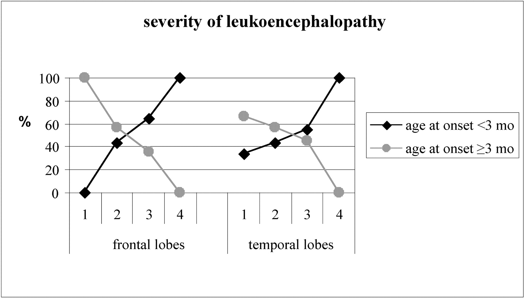
  • Fig 4.
    • Download figure
    • Open in new tab
    • Download powerpoint
    Fig 4.

    Correlation between age at onset and the severity of leukoencephalopathy involving the frontal and temporal lobes (leukoencephalopathy score: 1 = mild; 2 = moderate; 3 = severe; 4 = presence of cystic degeneration). Mths indicate months.

PreviousNext
Back to top

In this issue

American Journal of Neuroradiology: 30 (10)
American Journal of Neuroradiology
Vol. 30, Issue 10
1 Nov 2009
  • Table of Contents
  • Index by author
Advertisement
Print
Download PDF
Email Article

Thank you for your interest in spreading the word on American Journal of Neuroradiology.

NOTE: We only request your email address so that the person you are recommending the page to knows that you wanted them to see it, and that it is not junk mail. We do not capture any email address.

Enter multiple addresses on separate lines or separate them with commas.
Aicardi-Goutières Syndrome: Neuroradiologic Findings and Follow-Up
(Your Name) has sent you a message from American Journal of Neuroradiology
(Your Name) thought you would like to see the American Journal of Neuroradiology web site.
CAPTCHA
This question is for testing whether or not you are a human visitor and to prevent automated spam submissions.
Cite this article
C. Uggetti, R. La Piana, S. Orcesi, M.G. Egitto, Y.J. Crow, E. Fazzi
Aicardi-Goutières Syndrome: Neuroradiologic Findings and Follow-Up
American Journal of Neuroradiology Nov 2009, 30 (10) 1971-1976; DOI: 10.3174/ajnr.A1694

Citation Manager Formats

  • BibTeX
  • Bookends
  • EasyBib
  • EndNote (tagged)
  • EndNote 8 (xml)
  • Medlars
  • Mendeley
  • Papers
  • RefWorks Tagged
  • Ref Manager
  • RIS
  • Zotero
0 Responses
Respond to this article
Share
Bookmark this article
Aicardi-Goutières Syndrome: Neuroradiologic Findings and Follow-Up
C. Uggetti, R. La Piana, S. Orcesi, M.G. Egitto, Y.J. Crow, E. Fazzi
American Journal of Neuroradiology Nov 2009, 30 (10) 1971-1976; DOI: 10.3174/ajnr.A1694
del.icio.us logo Twitter logo Facebook logo Mendeley logo
  • Tweet Widget
  • Facebook Like
  • Google Plus One
Purchase

Jump to section

  • Article
    • Abstract
    • Materials and Methods
    • Results
    • Discussion
    • Conclusions
    • Acknowledgments
    • Footnotes
    • References
  • Figures & Data
  • Supplemental
  • Info & Metrics
  • Responses
  • References
  • PDF

Related Articles

  • PubMed
  • Google Scholar

Cited By...

  • Alternative cGAS signaling promotes herpes simplex encephalitis
  • Conserved chromatin regulators control the transcriptional immune response to intracellular pathogens in Caenorhabditis elegans
  • The 2021 European Alliance of Associations for Rheumatology/American College of Rheumatology points to consider for diagnosis and management of autoinflammatory type I interferonopathies: CANDLE/PRAAS, SAVI and AGS
  • Spectrum of Neuroradiologic Findings Associated with Monogenic Interferonopathies
  • Peripheral inflammation is associated with micro-structural and functional connectivity changes in depression-related brain networks
  • Combination of exome sequencing and immune testing confirms Aicardi-Goutieres syndrome type 5 in a challenging pediatric neurology case
  • Neuroradiologic patterns and novel imaging findings in Aicardi-Goutieres syndrome
  • Neuroimaging In Cockayne Syndrome
  • Reconciling Neuroimaging and Clinical Findings in Aicardi-Goutieres Syndrome: An Autoimmune-Mediated Encephalopathy
  • Reply:
  • Crossref
  • Google Scholar

This article has not yet been cited by articles in journals that are participating in Crossref Cited-by Linking.

More in this TOC Section

  • FRACTURE MR in Congenital Vertebral Anomalies
  • Comparing MRI Perfusion in Pediatric Brain Tumors
  • Sodium MRI in Pediatric Brain Tumors
Show more Pediatric Neuroimaging

Similar Articles

Advertisement

Indexed Content

  • Current Issue
  • Accepted Manuscripts
  • Article Preview
  • Past Issues
  • Editorials
  • Editor's Choice
  • Fellows' Journal Club
  • Letters to the Editor
  • Video Articles

Cases

  • Case Collection
  • Archive - Case of the Week
  • Archive - Case of the Month
  • Archive - Classic Case

More from AJNR

  • Trainee Corner
  • Imaging Protocols
  • MRI Safety Corner
  • Book Reviews

Multimedia

  • AJNR Podcasts
  • AJNR Scantastics

Resources

  • Turnaround Time
  • Submit a Manuscript
  • Submit a Video Article
  • Submit an eLetter to the Editor/Response
  • Manuscript Submission Guidelines
  • Statistical Tips
  • Fast Publishing of Accepted Manuscripts
  • Graphical Abstract Preparation
  • Imaging Protocol Submission
  • Evidence-Based Medicine Level Guide
  • Publishing Checklists
  • Author Policies
  • Become a Reviewer/Academy of Reviewers
  • News and Updates

About Us

  • About AJNR
  • Editorial Board
  • Editorial Board Alumni
  • Alerts
  • Permissions
  • Not an AJNR Subscriber? Join Now
  • Advertise with Us
  • Librarian Resources
  • Feedback
  • Terms and Conditions
  • AJNR Editorial Board Alumni

American Society of Neuroradiology

  • Not an ASNR Member? Join Now

© 2025 by the American Society of Neuroradiology All rights, including for text and data mining, AI training, and similar technologies, are reserved.
Print ISSN: 0195-6108 Online ISSN: 1936-959X

Powered by HighWire