Advertisement

Main menu

  • Home
  • Content
    • Current Issue
    • Accepted Manuscripts
    • Article Preview
    • Past Issue Archive
    • Video Articles
    • AJNR Case Collection
    • Case of the Week Archive
    • Case of the Month Archive
    • Classic Case Archive
  • Special Collections
    • AJNR Awards
    • Low-Field MRI
    • Alzheimer Disease
    • ASNR Foundation Special Collection
    • Photon-Counting CT
    • View All
  • Multimedia
    • AJNR Podcasts
    • AJNR SCANtastic
    • Trainee Corner
    • MRI Safety Corner
    • Imaging Protocols
  • For Authors
    • Submit a Manuscript
    • Submit a Video Article
    • Submit an eLetter to the Editor/Response
    • Manuscript Submission Guidelines
    • Statistical Tips
    • Fast Publishing of Accepted Manuscripts
    • Graphical Abstract Preparation
    • Imaging Protocol Submission
    • Author Policies
  • About Us
    • About AJNR
    • Editorial Board
    • Editorial Board Alumni
  • More
    • Become a Reviewer/Academy of Reviewers
    • Subscribers
    • Permissions
    • Alerts
    • Feedback
    • Advertisers
    • ASNR Home

User menu

  • Alerts
  • Log in

Search

  • Advanced search
American Journal of Neuroradiology

American Journal of Neuroradiology

ASHNR American Society of Functional Neuroradiology ASHNR American Society of Pediatric Neuroradiology ASSR
  • Alerts
  • Log in

Advanced Search

  • Home
  • Content
    • Current Issue
    • Accepted Manuscripts
    • Article Preview
    • Past Issue Archive
    • Video Articles
    • AJNR Case Collection
    • Case of the Week Archive
    • Case of the Month Archive
    • Classic Case Archive
  • Special Collections
    • AJNR Awards
    • Low-Field MRI
    • Alzheimer Disease
    • ASNR Foundation Special Collection
    • Photon-Counting CT
    • View All
  • Multimedia
    • AJNR Podcasts
    • AJNR SCANtastic
    • Trainee Corner
    • MRI Safety Corner
    • Imaging Protocols
  • For Authors
    • Submit a Manuscript
    • Submit a Video Article
    • Submit an eLetter to the Editor/Response
    • Manuscript Submission Guidelines
    • Statistical Tips
    • Fast Publishing of Accepted Manuscripts
    • Graphical Abstract Preparation
    • Imaging Protocol Submission
    • Author Policies
  • About Us
    • About AJNR
    • Editorial Board
    • Editorial Board Alumni
  • More
    • Become a Reviewer/Academy of Reviewers
    • Subscribers
    • Permissions
    • Alerts
    • Feedback
    • Advertisers
    • ASNR Home
  • Follow AJNR on Twitter
  • Visit AJNR on Facebook
  • Follow AJNR on Instagram
  • Join AJNR on LinkedIn
  • RSS Feeds

AJNR Awards, New Junior Editors, and more. Read the latest AJNR updates

Research ArticleHead and Neck Imaging

Can Quantitative Diffusion-Weighted MR Imaging Differentiate Benign and Malignant Cold Thyroid Nodules? Initial Results in 25 Patients

C. Schueller-Weidekamm, K. Kaserer, G. Schueller, C. Scheuba, H. Ringl, M. Weber, C. Czerny and A.M. Herneth
American Journal of Neuroradiology February 2009, 30 (2) 417-422; DOI: https://doi.org/10.3174/ajnr.A1338
C. Schueller-Weidekamm
  • Find this author on Google Scholar
  • Find this author on PubMed
  • Search for this author on this site
K. Kaserer
  • Find this author on Google Scholar
  • Find this author on PubMed
  • Search for this author on this site
G. Schueller
  • Find this author on Google Scholar
  • Find this author on PubMed
  • Search for this author on this site
C. Scheuba
  • Find this author on Google Scholar
  • Find this author on PubMed
  • Search for this author on this site
H. Ringl
  • Find this author on Google Scholar
  • Find this author on PubMed
  • Search for this author on this site
M. Weber
  • Find this author on Google Scholar
  • Find this author on PubMed
  • Search for this author on this site
C. Czerny
  • Find this author on Google Scholar
  • Find this author on PubMed
  • Search for this author on this site
A.M. Herneth
  • Find this author on Google Scholar
  • Find this author on PubMed
  • Search for this author on this site
  • Article
  • Figures & Data
  • Info & Metrics
  • Responses
  • References
  • PDF
Loading

Article Figures & Data

Figures

  • Tables
  • Fig 1.
    • Download figure
    • Open in new tab
    • Download powerpoint
    Fig 1.

    A patient with follicular thyroid carcinoma in the right thyroid lobe (T2, N0, M0; tumor size, 842 mm2). A, On sagittal DWI EPI MR image (b-value = 0 s/mm2), the tumor appears homogeneously hyperintense (arrow). B, The corresponding ADC map shows high values of the carcinoma (arrow) as a result of unhindered diffusion capability. The diagnosis was confirmed at histology.

  • Fig 2.
    • Download figure
    • Open in new tab
    • Download powerpoint
    Fig 2.

    A patient with histologically confirmed adenoma in the lower pole and follicular thyroid carcinoma in the upper pole of the left thyroid lobe (size, 130 mm2; 125 mm2). A, On sagittal DWI EPI MR image (b-value = 0 s/mm2), the adenoma (arrow) and the thyroid carcinoma (open arrow) appear slightly hyperintense. B, The corresponding ADC map shows low ADC values for adenoma (arrow) as a result of restricted diffusion capability. The ADC values for carcinoma (open arrow) are high as a result of unhindered diffusion capability.
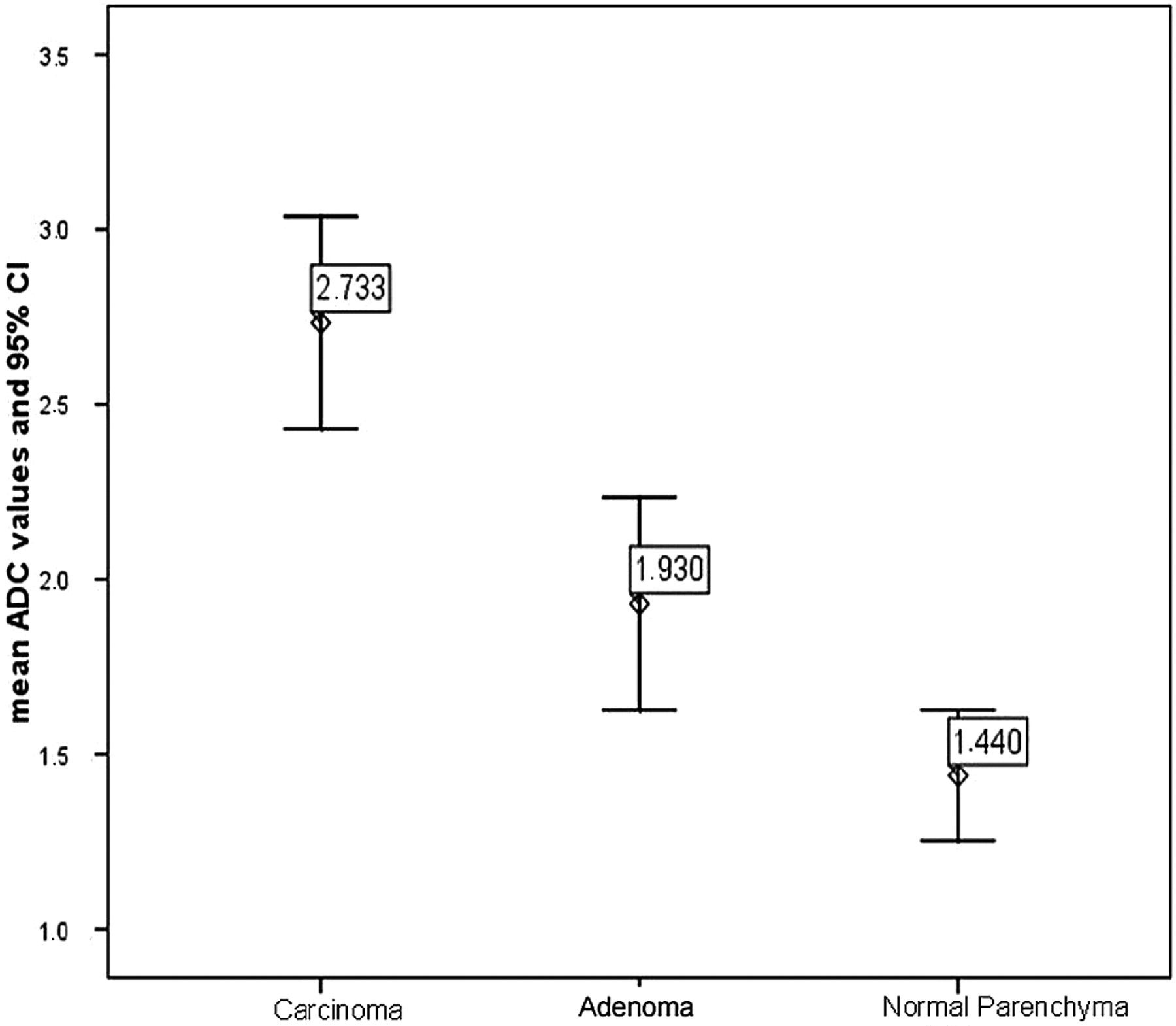
  • Fig 3.
    • Download figure
    • Open in new tab
    • Download powerpoint
    Fig 3.

    Boxplot of ADC values in thyroid cancer, adenoma, and normal thyroid parenchyma. The horizontal lines are median values, the boxes show minimum and maximum values, and the whiskers indicate 2 SDs. Median ADC values for cancer and adenoma are significantly different, and there is no overlap of absolute values.

Tables

  • Figures
  • Mean ADC values and 95% CI of carcinoma, adenoma, and normal parenchyma*

    DiagnosisADC of Lesions†ADC of Normal Parenchyma*
    Carcinoma (n = 20)2.73 ± 0.65 (2.43–3.04)1.439 ± 0.648 (1.25–1.60)
    Adenoma (n = 5)1.93 ± 0.25 (1.63–2.23)1.341 ± 0.245 (1.18–1.57)
    • Note:—ADC indicates apparent diffusion coefficient.

    • * Data are mean ± SD. Data in parentheses are the ranges of the 95% CI.

    • † ADC values are expressed as the (mean ± SD) × 10−3 mm2/s (P < .05).

PreviousNext
Back to top

In this issue

American Journal of Neuroradiology: 30 (2)
American Journal of Neuroradiology
Vol. 30, Issue 2
February 2009
  • Table of Contents
  • Index by author
Advertisement
Print
Download PDF
Email Article

Thank you for your interest in spreading the word on American Journal of Neuroradiology.

NOTE: We only request your email address so that the person you are recommending the page to knows that you wanted them to see it, and that it is not junk mail. We do not capture any email address.

Enter multiple addresses on separate lines or separate them with commas.
Can Quantitative Diffusion-Weighted MR Imaging Differentiate Benign and Malignant Cold Thyroid Nodules? Initial Results in 25 Patients
(Your Name) has sent you a message from American Journal of Neuroradiology
(Your Name) thought you would like to see the American Journal of Neuroradiology web site.
CAPTCHA
This question is for testing whether or not you are a human visitor and to prevent automated spam submissions.
Cite this article
C. Schueller-Weidekamm, K. Kaserer, G. Schueller, C. Scheuba, H. Ringl, M. Weber, C. Czerny, A.M. Herneth
Can Quantitative Diffusion-Weighted MR Imaging Differentiate Benign and Malignant Cold Thyroid Nodules? Initial Results in 25 Patients
American Journal of Neuroradiology Feb 2009, 30 (2) 417-422; DOI: 10.3174/ajnr.A1338

Citation Manager Formats

  • BibTeX
  • Bookends
  • EasyBib
  • EndNote (tagged)
  • EndNote 8 (xml)
  • Medlars
  • Mendeley
  • Papers
  • RefWorks Tagged
  • Ref Manager
  • RIS
  • Zotero
0 Responses
Respond to this article
Share
Bookmark this article
Can Quantitative Diffusion-Weighted MR Imaging Differentiate Benign and Malignant Cold Thyroid Nodules? Initial Results in 25 Patients
C. Schueller-Weidekamm, K. Kaserer, G. Schueller, C. Scheuba, H. Ringl, M. Weber, C. Czerny, A.M. Herneth
American Journal of Neuroradiology Feb 2009, 30 (2) 417-422; DOI: 10.3174/ajnr.A1338
del.icio.us logo Twitter logo Facebook logo Mendeley logo
  • Tweet Widget
  • Facebook Like
  • Google Plus One
Purchase

Jump to section

  • Article
    • Abstract
    • Materials and Methods
    • Results
    • Discussion
    • Conclusions
    • Acknowledgments
    • References
  • Figures & Data
  • Info & Metrics
  • Responses
  • References
  • PDF

Related Articles

  • No related articles found.
  • PubMed
  • Google Scholar

Cited By...

  • Diffusion-weighted MRI in differentiating malignant from benign thyroid nodules: a meta-analysis
  • 3T diffusion-weighted MRI of the thyroid gland with reduced distortion: preliminary results
  • Crossref
  • Google Scholar

This article has not yet been cited by articles in journals that are participating in Crossref Cited-by Linking.

More in this TOC Section

  • Hydrops Herniation into the Semicircular Canals
  • Post SRS Peritumoral Hyperintense Signal of VSs
  • DIVr of NPC Predicted Outcome
Show more HEAD AND NECK IMAGING

Similar Articles

Advertisement

Indexed Content

  • Current Issue
  • Accepted Manuscripts
  • Article Preview
  • Past Issues
  • Editorials
  • Editor's Choice
  • Fellows' Journal Club
  • Letters to the Editor
  • Video Articles

Cases

  • Case Collection
  • Archive - Case of the Week
  • Archive - Case of the Month
  • Archive - Classic Case

More from AJNR

  • Trainee Corner
  • Imaging Protocols
  • MRI Safety Corner
  • Book Reviews

Multimedia

  • AJNR Podcasts
  • AJNR Scantastics

Resources

  • Turnaround Time
  • Submit a Manuscript
  • Submit a Video Article
  • Submit an eLetter to the Editor/Response
  • Manuscript Submission Guidelines
  • Statistical Tips
  • Fast Publishing of Accepted Manuscripts
  • Graphical Abstract Preparation
  • Imaging Protocol Submission
  • Evidence-Based Medicine Level Guide
  • Publishing Checklists
  • Author Policies
  • Become a Reviewer/Academy of Reviewers
  • News and Updates

About Us

  • About AJNR
  • Editorial Board
  • Editorial Board Alumni
  • Alerts
  • Permissions
  • Not an AJNR Subscriber? Join Now
  • Advertise with Us
  • Librarian Resources
  • Feedback
  • Terms and Conditions
  • AJNR Editorial Board Alumni

American Society of Neuroradiology

  • Not an ASNR Member? Join Now

© 2025 by the American Society of Neuroradiology All rights, including for text and data mining, AI training, and similar technologies, are reserved.
Print ISSN: 0195-6108 Online ISSN: 1936-959X

Powered by HighWire