
Article Figures & Data
Figures
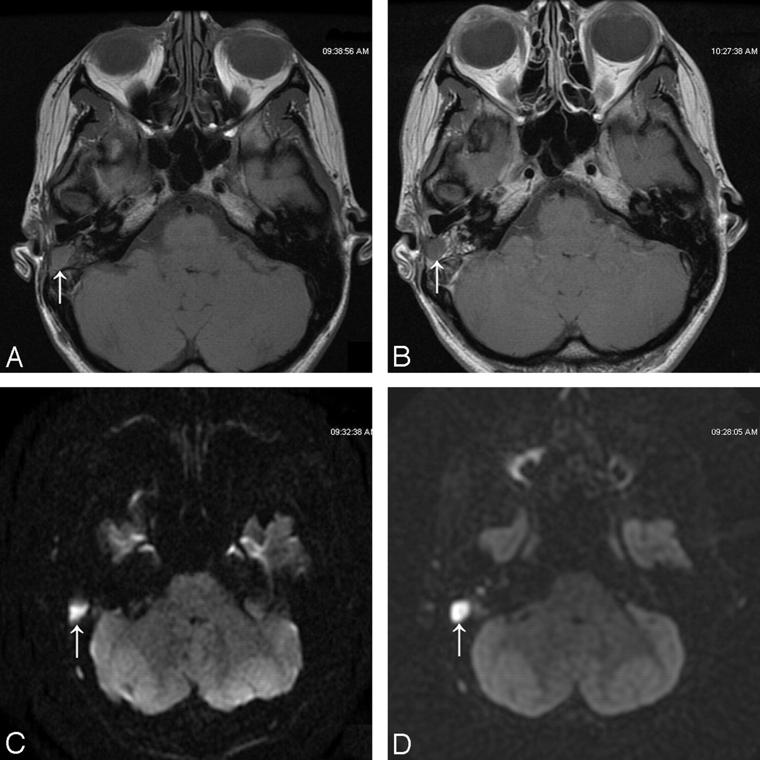
- Fig 1.
Recurrent cholesteatoma (white arrow). A, Unenhanced T1-weighted image with low signal intensity. B, Delayed contrast-enhanced T1-weighted image with no change in signal intensity; C and D, Echo-planar DWI (C) and PROPELLER DWI (D) show high signal intensity with more artifacts on the echo-planar DWI (C).
- Fig 2.
Patient 5 with a wrong diagnosis on delayed contrast-enhanced T1-weighted imaging. No recurrence of cholesteatoma was histologically proved (white arrow). A, Unenhanced T1-weighted image shows low signal intensity. B, Delayed contrast-enhanced T1-weighted image shows no change in signal intensity. C and D, Echo-planar DWI (C) and PROPELLER DWI (D) confirm no recurrence of cholesteatoma. Note the low signal intensity on DWI with more artifacts (C).
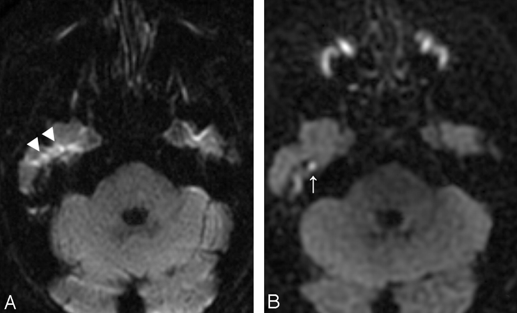
- Fig 3.
Recurrent cholesteatoma. A, Echo-planar DWI shows artifacts (double arrowhead) masking the recurrence. B, PROPELLER DWI shows small attical recurrent cholesteatoma (white arrow).


Tables
Sensitivity, specificity, PPV, NPV, and P value for the 3 observers
Sensitivity (%) Specificity (%) PPV (%) NPV (%) P Value S1 S2 F S1 S2 F S1 S2 F S1 S2 F S1 S2 F EPI 33.3 38.9 44.4 100.0 100.0 87.5 100.0 100.0 80.0 57.1 59.3 58.3 ≤0.02 ≤.01 ≤.05 PROP 100.0 100.0 89.5 100.0 100.0 100.0 100.0 100.0 100.0 100.0 100.0 88.9 ≤0.001 ≤.001 ≤.001 T1 94.1 88.2 88.2 93.7 68.7 81.2 94.1 75.0 83.3 93.7 84.6 86.7 ≤0.001 ≤.001 ≤.001 Note:—EPI indicates ASSET echo-planar DWI; PROP, PROPELLER DWI; T1, T1-weighted imaging; S1, senior1; S2, senior2; F, fellow; PPV, positive predictive value; NPV, negative predictive value.
In this issue


Jump to section
Related Articles
Cited By...
- Use of Non-Echo-Planar Diffusion-Weighted MR Imaging for the Detection of Cholesteatomas in High-Risk Tympanic Retraction Pockets
- Detection of Middle Ear Cholesteatoma by Diffusion-Weighted MR Imaging: Multishot Echo-Planar Imaging Compared with Single-Shot Echo-Planar Imaging
- The Utility of Diffusion-Weighted Imaging for Cholesteatoma Evaluation
- Neuroradiology of Cholesteatomas
- BLADE in Sagittal T2-Weighted MR Imaging of the Cervical Spine