Advertisement

Main menu

  • Home
  • Content
    • Current Issue
    • Accepted Manuscripts
    • Article Preview
    • Past Issue Archive
    • AJNR Case Collection
    • Case of the Week Archive
    • Classic Case Archive
    • Case of the Month Archive
  • Special Collections
    • Spinal CSF Leak Articles (Jan 2020-June 2024)
    • 2024 AJNR Journal Awards
    • Most Impactful AJNR Articles
  • Multimedia
    • AJNR Podcast
    • AJNR Scantastics
    • Video Articles
  • For Authors
    • Submit a Manuscript
    • Author Policies
    • Fast publishing of Accepted Manuscripts
    • Graphical Abstract Preparation
    • Manuscript Submission Guidelines
    • Imaging Protocol Submission
    • Submit a Case for the Case Collection
  • About Us
    • About AJNR
    • Editorial Board
  • More
    • Become a Reviewer/Academy of Reviewers
    • Subscribers
    • Permissions
    • Alerts
    • Feedback
    • Advertisers
    • ASNR Home
  • Other Publications
    • ajnr

User menu

  • Alerts
  • Log in

Search

  • Advanced search
American Journal of Neuroradiology

American Journal of Neuroradiology

ASHNR American Society of Functional Neuroradiology ASHNR American Society of Pediatric Neuroradiology ASSR
  • Alerts
  • Log in

Advanced Search

  • Home
  • Content
    • Current Issue
    • Accepted Manuscripts
    • Article Preview
    • Past Issue Archive
    • AJNR Case Collection
    • Case of the Week Archive
    • Classic Case Archive
    • Case of the Month Archive
  • Special Collections
    • Spinal CSF Leak Articles (Jan 2020-June 2024)
    • 2024 AJNR Journal Awards
    • Most Impactful AJNR Articles
  • Multimedia
    • AJNR Podcast
    • AJNR Scantastics
    • Video Articles
  • For Authors
    • Submit a Manuscript
    • Author Policies
    • Fast publishing of Accepted Manuscripts
    • Graphical Abstract Preparation
    • Manuscript Submission Guidelines
    • Imaging Protocol Submission
    • Submit a Case for the Case Collection
  • About Us
    • About AJNR
    • Editorial Board
  • More
    • Become a Reviewer/Academy of Reviewers
    • Subscribers
    • Permissions
    • Alerts
    • Feedback
    • Advertisers
    • ASNR Home
  • Follow AJNR on Twitter
  • Visit AJNR on Facebook
  • Follow AJNR on Instagram
  • Join AJNR on LinkedIn
  • RSS Feeds

Welcome to the new AJNR, Updated Hall of Fame, and more. Read the full announcements.


AJNR is seeking candidates for the position of Associate Section Editor, AJNR Case Collection. Read the full announcement.


OtherREVIEW ARTICLES
Open Access

Imaging Lesions of the Cavernous Sinus

A.A.K. Abdel Razek and M. Castillo
American Journal of Neuroradiology March 2009, 30 (3) 444-452; DOI: https://doi.org/10.3174/ajnr.A1398
A.A.K. Abdel Razek
  • Find this author on Google Scholar
  • Find this author on PubMed
  • Search for this author on this site
M. Castillo
  • Find this author on Google Scholar
  • Find this author on PubMed
  • Search for this author on this site
  • Article
  • Figures & Data
  • Info & Metrics
  • Responses
  • References
  • PDF
Loading

Article Figures & Data

Figures

  • Fig 1.
    • Download figure
    • Open in new tab
    • Download powerpoint
    Fig 1.

    Normal fat deposits. Axial noncontrast CT scan shows normal and incidentally found deposits of fat (arrowheads) in the posterior CSs. These deposits may be seen in obese individuals, those taking corticosteroids, or those with Cushing syndrome. In the absence of these conditions, they have no significance.
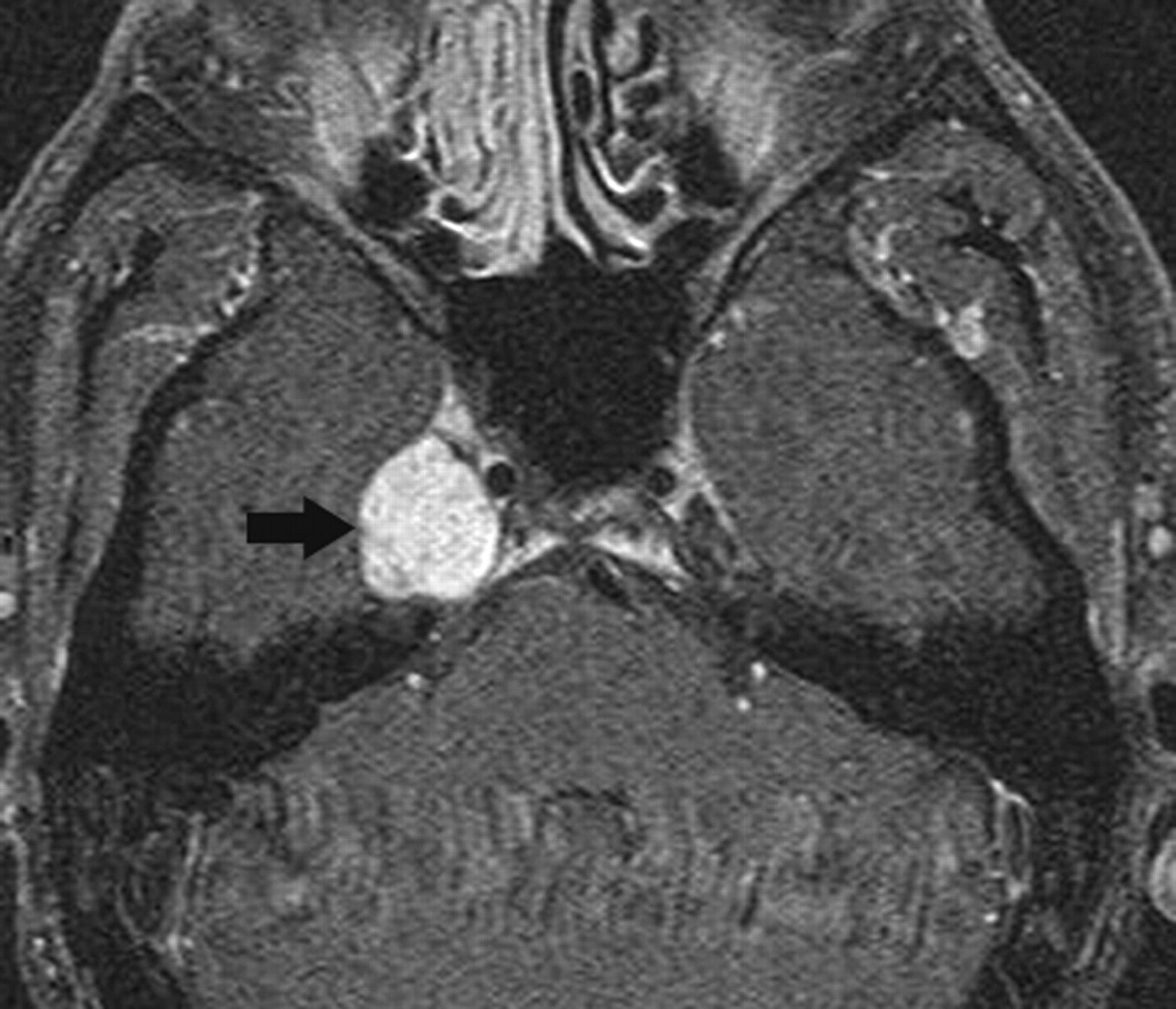
  • Fig 2.
    • Download figure
    • Open in new tab
    • Download powerpoint
    Fig 2.

    Schwannoma. Axial postcontrast T1-weighted image shows a well-defined enhancing mass (arrow) involving the Meckel cave on the right. Although the findings are nonspecific, the most common mass in this location is a schwannoma.

  • Fig 3.
    • Download figure
    • Open in new tab
    • Download powerpoint
    Fig 3.

    Malignant peripheral nerve sheath tumor. Coronal postcontrast T1-weighted image shows a large aggressive-appearing mass that involves the left CS, surrounds the ICA (arrow), erodes the middle cranial fossa floor, and extends into the infratemporal region.
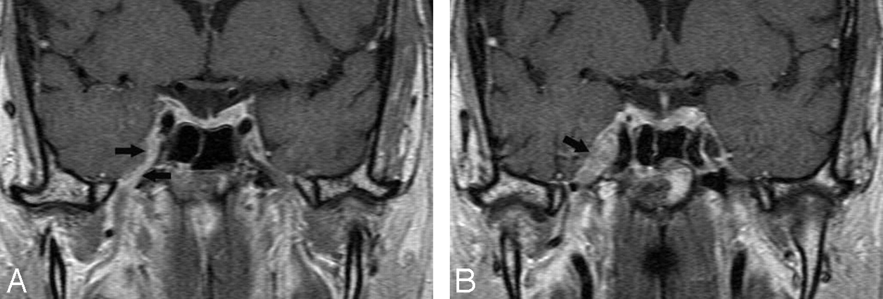
  • Fig 4.
    • Download figure
    • Open in new tab
    • Download powerpoint
    Fig 4.

    Cavernous hemangiomas. A, Axial postcontrast T1-weighted image shows a large and homogeneously enhancing mass arising from the lateral wall of the left CS. B, Axial postcontrast T1-weighted image in a different cavernoma, which shows inhomogeneous contrast enhancement but also arises from the lateral wall of the CS, pushing the ICA (arrow) medially. When a mass arises in the lateral wall of a CS, the most important differential diagnosis is that of meningioma versus cavernoma.

  • Fig 5.
    • Download figure
    • Open in new tab
    • Download powerpoint
    Fig 5.

    Meningiomas. A, Axial T2-weighted image shows a meningioma (arrow) that is isoattenuated to white matter involving the right CS and extending from the Meckel cave into the superior orbital fissure. B, Axial postcontrast T1-weighted image in a different patient shows the bulk of a homogeneously enhancing mass in the Meckel cave but extending through the porous trigeminus into the prepontine cistern. A schwannoma needs to be considered in the differential diagnosis.
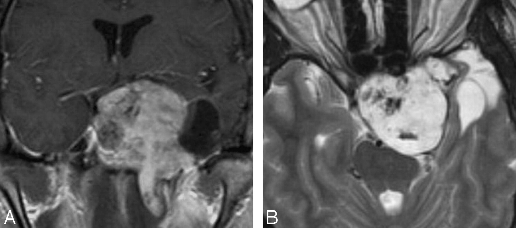
  • Fig 6.
    • Download figure
    • Open in new tab
    • Download powerpoint
    Fig 6.

    Chondrosarcoma. A, Coronal postcontrast T1-weighted image shows a large inhomogeneous-appearing mass involving the left CS, sella, suprasellar region, ipsilateral middle cranial fossa, and intratemporal region. The mass has a cystic lateral component. B, Axial T2-weighted image shows that the solid portion of the mass is hyperintense, a finding that is typical of chondrosarcoma.

  • Fig 7.
    • Download figure
    • Open in new tab
    • Download powerpoint
    Fig 7.

    Nasopharyngeal squamous cell carcinoma. Axial T2-weighted image shows a relatively hypointense mass involving the left CS and sella, extending into the posterior ethmoid air cells. Invasive T2 hypointense masses are generally either neoplasias or fungal infections.
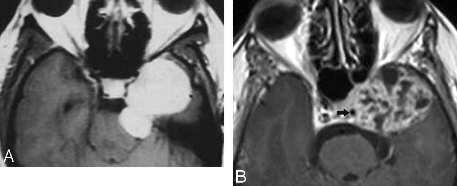
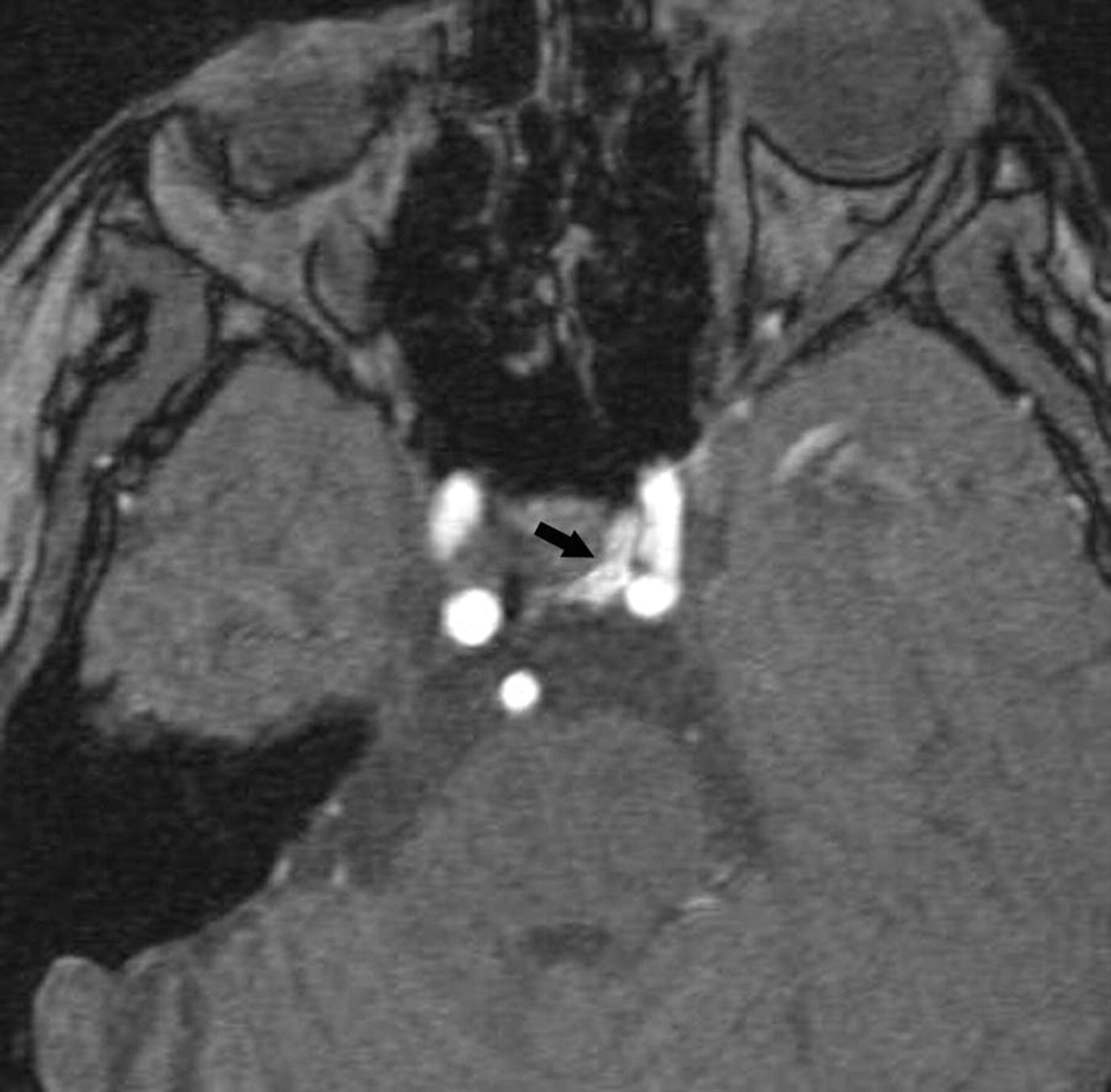
  • Fig 8.
    • Download figure
    • Open in new tab
    • Download powerpoint
    Fig 8.

    Juvenile angiofibroma. Axial postcontrast T1-weighted image shows a very large tumor involving both CSs and surrounding the ICAs (arrows). The mass extends into the sella, paranasal sinuses, right middle cranial fossa, and both orbits. Note flow voids (arrowheads) due to enlarged blood vessels.
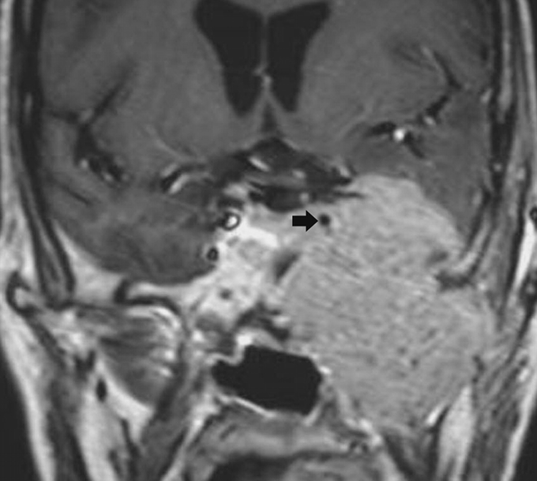
  • Fig 9.
    • Download figure
    • Open in new tab
    • Download powerpoint
    Fig 9.

    Metastasis. Axial postcontrast T1-weighted image shows an enhancing mass (from primary breast carcinoma) in the right the Meckel cave (arrowhead). In the absence of primary tumor elsewhere, schwannoma needs to be considered in the differential diagnosis.

  • Fig 10.
    • Download figure
    • Open in new tab
    • Download powerpoint
    Fig 10.

    Perineural tumor spread. A, Coronal postcontrast T1-weighted image shows a thick enhancing third division (arrows) of the right trigeminal nerve in a patient with a small adenoid cystic carcinoma in the nasopharynx. B, Slightly posterior to A, the tumor has invaded the Gasserian ganglion and fills the Meckel cave (arrow).
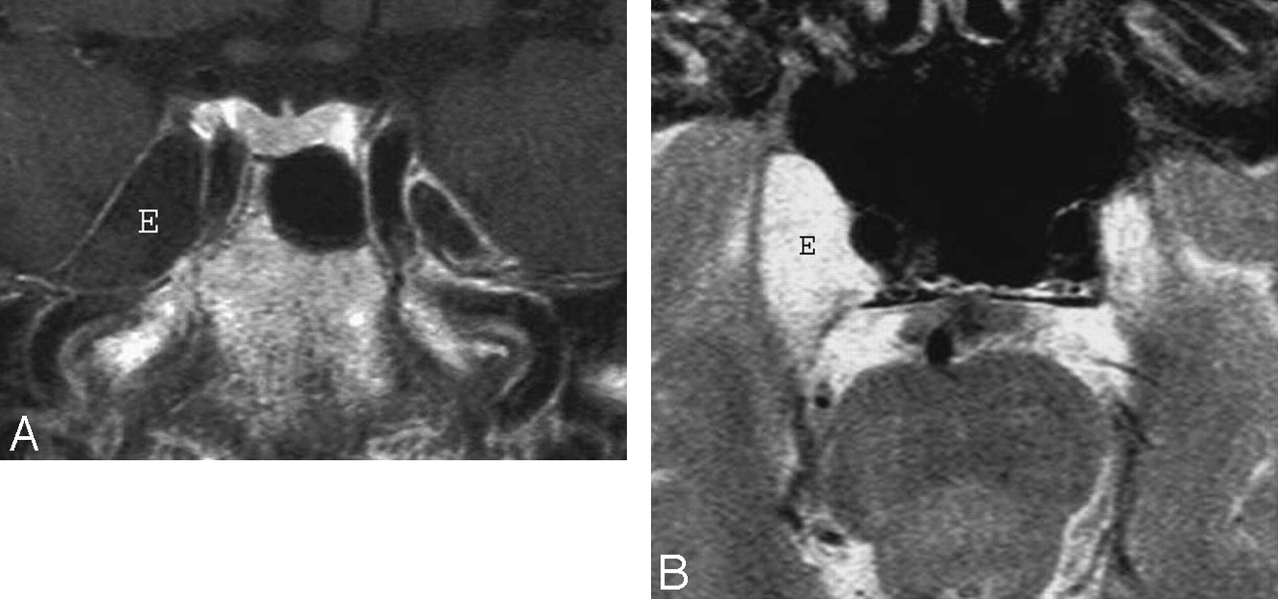
  • Fig 11.
    • Download figure
    • Open in new tab
    • Download powerpoint
    Fig 11.

    Epidermoid. A, Axial postcontrast T1-weighted image shows a large mass (E) inside the right the Meckel cave. The mass does not enhance and is nearly isointense to CSF. B, Axial T2-weighted image in the same patient shows that the mass (E) is nearly as bright as CSF. Despite the large size of the lesion, clinical symptoms were mild. The differential diagnosis includes an intratemporal meningocele.

  • Fig 12.
    • Download figure
    • Open in new tab
    • Download powerpoint
    Fig 12.

    Tolosa-Hunt syndrome. A, Axial T2-weighted image shows hypointense soft tissue throughout the right CS and extending into the superior orbital fissure. B, Coronal postcontrast T1-weighted image shows that the abnormal soft tissue enhances prominently and diffusely and involves the Meckel cave.
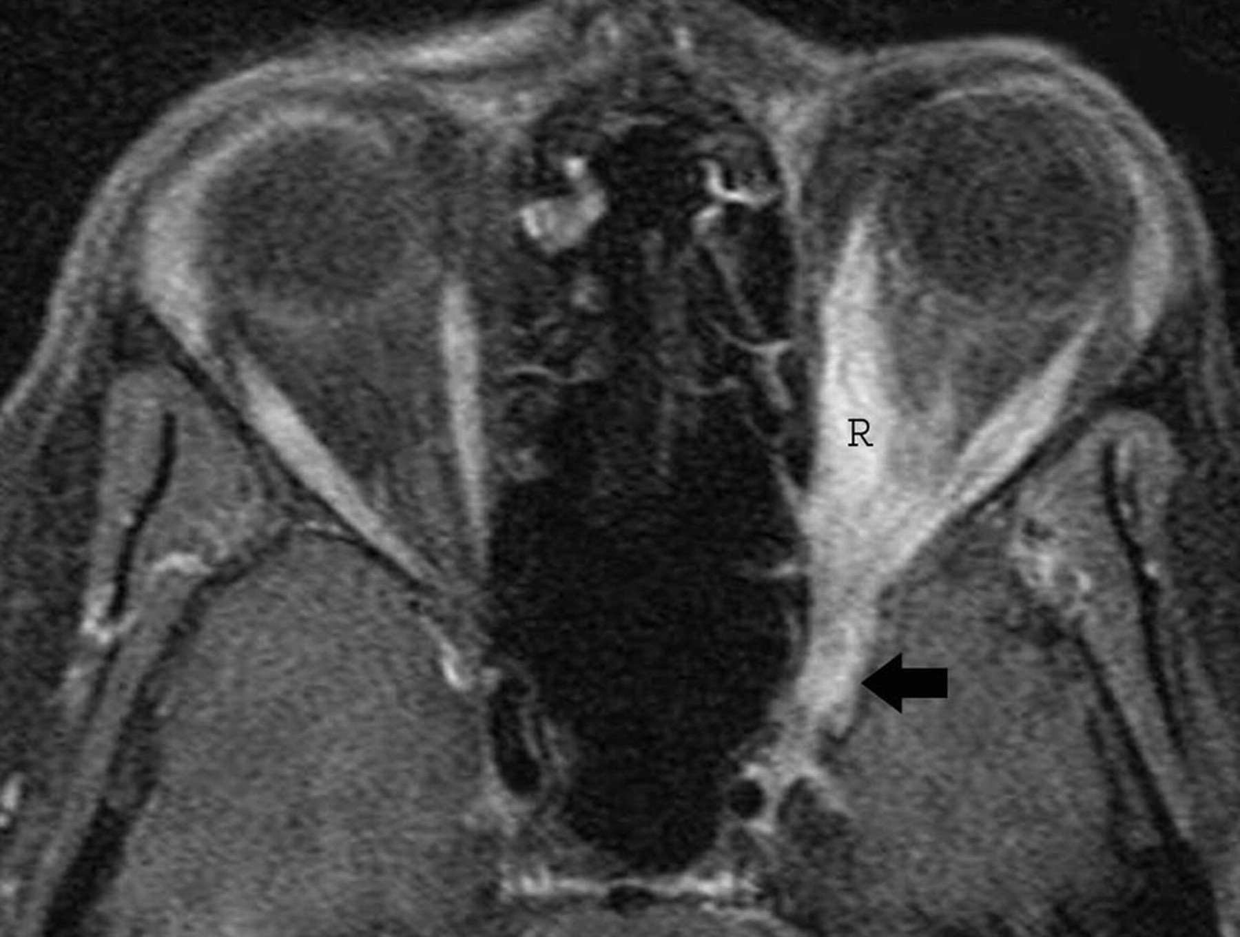
  • Fig 13.
    • Download figure
    • Open in new tab
    • Download powerpoint
    Fig 13.

    Inflammatory myofibroblastic tumor. A, Axial postcontrast T1-weighted image shows thickening and stranding of the left extraocular muscles more on medial rectus (R) and extension of the process into the ipsilateral CS (arrow). There is prominent enhancement of these muscles and of the retro-orbital fat.
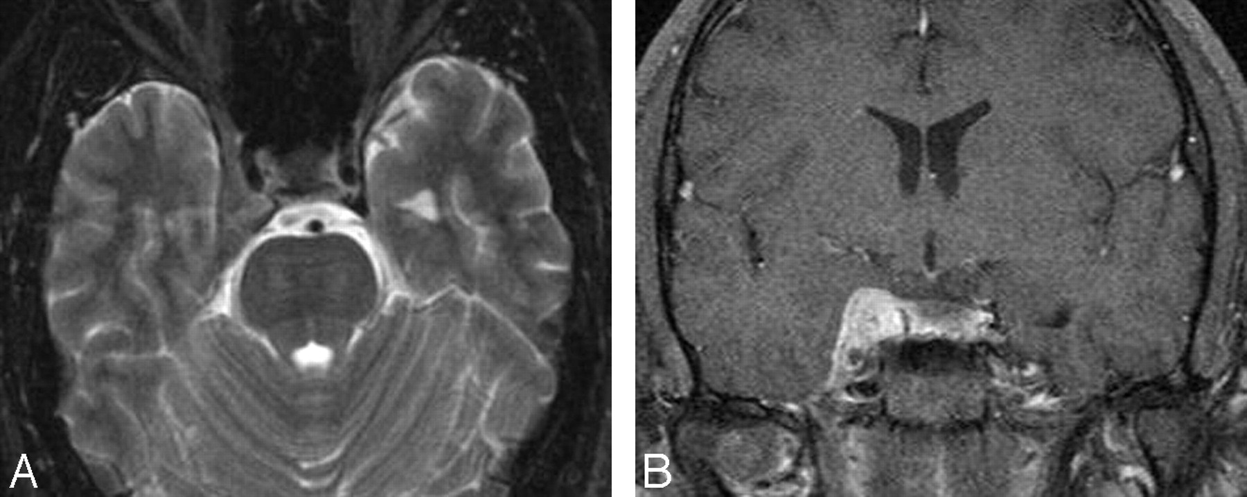
  • Fig 14.
    • Download figure
    • Open in new tab
    • Download powerpoint
    Fig 14.

    Idiopathic pachymeningitis. Coronal postcontrast T1-weighted image shows nonspecific thickening of the right CS at the level of the foramen ovale. There is perhaps slight thickening of the lateral dural wall of the contralateral CS.

  • Fig 15.
    • Download figure
    • Open in new tab
    • Download powerpoint
    Fig 15.

    Fungal infection. A, Coronal postcontrast T1-weighted image shows involvement by aspergillosis of the mucosa in the left sphenoid sinus (white arrow), which extends laterally and has resulted in thrombosis of the adjacent CS (black arrow). The intracavernous ICA (arrowhead) is narrowed, and its walls are significantly thickened. B, In a different patient, an axial postcontrast T1-weighted image shows enhancing mucosa in the right sphenoid sinus with enhancing soft tissues in the ipsilateral CS (including the Meckel cave), with narrowing of the ICA and significant thickening of its walls.

  • Fig 16.
    • Download figure
    • Open in new tab
    • Download powerpoint
    Fig 16.

    ICA to a CS fistula. Axial source image from an MR angiogram shows flow-related enhancement in the medial (arrow) left CS from a direct-type fistula.
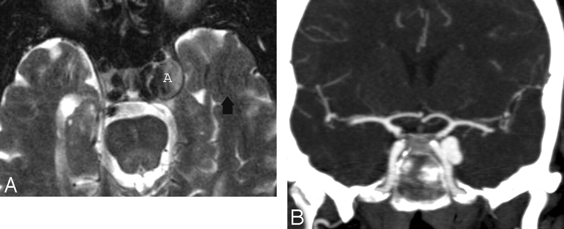
  • Fig 17.
    • Download figure
    • Open in new tab
    • Download powerpoint
    Fig 17.

    Intracavernous aneurysms. A, Axial T2-weighted image shows a left intracavernous ICA (A) aneurysm. Note flow artifacts (arrow) confirming the pulsatile nature of the lesions. B, Coronal postcontrast maximum-intensity image from a CT angiogram in the same patient shows the left intracavernous aneurysm.

  • Fig 18.
    • Download figure
    • Open in new tab
    • Download powerpoint
    Fig 18.

    CS thrombosis. A, Coronal postcontrast T1-weighted image shows an enlarged and inhomogeneous-appearing right CS that contains areas of low signal intensity (arrow) compatible with clot. B, Coronal postcontrast T1-weighted image in a different patient shows a large nonenhancing clot expanding the left CS. The ipsilateral ICA is slightly narrowed.

PreviousNext
Back to top

In this issue

American Journal of Neuroradiology: 30 (3)
American Journal of Neuroradiology
Vol. 30, Issue 3
March 2009
  • Table of Contents
  • Index by author
Advertisement
Print
Download PDF
Email Article

Thank you for your interest in spreading the word on American Journal of Neuroradiology.

NOTE: We only request your email address so that the person you are recommending the page to knows that you wanted them to see it, and that it is not junk mail. We do not capture any email address.

Enter multiple addresses on separate lines or separate them with commas.
Imaging Lesions of the Cavernous Sinus
(Your Name) has sent you a message from American Journal of Neuroradiology
(Your Name) thought you would like to see the American Journal of Neuroradiology web site.
CAPTCHA
This question is for testing whether or not you are a human visitor and to prevent automated spam submissions.
Cite this article
A.A.K. Abdel Razek, M. Castillo
Imaging Lesions of the Cavernous Sinus
American Journal of Neuroradiology Mar 2009, 30 (3) 444-452; DOI: 10.3174/ajnr.A1398

Citation Manager Formats

  • BibTeX
  • Bookends
  • EasyBib
  • EndNote (tagged)
  • EndNote 8 (xml)
  • Medlars
  • Mendeley
  • Papers
  • RefWorks Tagged
  • Ref Manager
  • RIS
  • Zotero
0 Responses
Respond to this article
Share
Bookmark this article
Imaging Lesions of the Cavernous Sinus
A.A.K. Abdel Razek, M. Castillo
American Journal of Neuroradiology Mar 2009, 30 (3) 444-452; DOI: 10.3174/ajnr.A1398
del.icio.us logo Twitter logo Facebook logo Mendeley logo
  • Tweet Widget
  • Facebook Like
  • Google Plus One
Purchase

Jump to section

  • Article
    • Abstract
    • Imaging Protocol and Anatomy
    • Neoplastic Lesions
    • Inflammatory, Infectious, and Granulomatous Lesions
    • Vascular Lesions
    • References
  • Figures & Data
  • Info & Metrics
  • Responses
  • References
  • PDF

Related Articles

  • No related articles found.
  • PubMed
  • Google Scholar

Cited By...

  • Cavernous Sinus Vascular Venous Malformation
  • Bilateral cavernous sinus thrombosis complicating acute unilateral pansinusitis in a 15-year-old boy
  • Diplopia: a unique presentation of recurrence of a salivary gland carcinoma
  • Actinomyces cavernous sinus infection: a case and systematic literature review
  • The parasellar region and central skull base
  • MRI findings in Tolosa-Hunt syndrome (THS)
  • Giant cavernous carotid artery aneurysm mimicking a fungal granuloma and presenting with massive epistaxis
  • Crossref (109)
  • Google Scholar

This article has been cited by the following articles in journals that are participating in Crossref Cited-by Linking.

  • Imaging Spectrum of CNS Vasculitis
    Ahmed Abdel Khalek Abdel Razek, Hortensia Alvarez, Stephen Bagg, Sherif Refaat, Mauricio Castillo
    RadioGraphics 2014 34 4
  • The fifth cranial nerve in headaches
    J. C. A. Edvinsson, A. Viganò, A. Alekseeva, E. Alieva, R. Arruda, C. De Luca, N. D’Ettore, I. Frattale, M. Kurnukhina, N. Macerola, E. Malenkova, M. Maiorova, A. Novikova, P. Řehulka, V. Rapaccini, O. Roshchina, G. Vanderschueren, L. Zvaune, A. P. Andreou, K. A. Haanes
    The Journal of Headache and Pain 2020 21 1
  • Imaging features of rhinocerebral mucormycosis: A study of 43 patients
    Jacob Therakathu, Shailesh Prabhu, Aparna Irodi, Sniya Valsa Sudhakar, Vikas K. Yadav, V. Rupa
    The Egyptian Journal of Radiology and Nuclear Medicine 2018 49 2
  • Imaging of cranial nerves: a pictorial overview
    Nicola Romano, Margherita Federici, Antonio Castaldi
    Insights into Imaging 2019 10 1
  • Radiologic Clues to Cerebral Venous Thrombosis
    María Canedo-Antelo, Sandra Baleato-González, Antonio J. Mosqueira, Jéssica Casas-Martínez, Laura Oleaga, Joan C. Vilanova, Antonio Luna-Alcalá, Roberto García-Figueiras
    RadioGraphics 2019 39 6
  • Neuroimaging of Meckel’s cave in normal and disease conditions
    Ajay Malhotra, Long Tu, Vivek B. Kalra, Xiao Wu, Ali Mian, Rajiv Mangla, Elias Michaelides, Pina Sanelli, Dheeraj Gandhi
    Insights into Imaging 2018 9 4
  • Sellar and Parasellar Imaging
    Carlos Zamora, Mauricio Castillo
    Neurosurgery 2017 80 1
  • Cavernous sinus lesions
    Kamran Munawar, Gopi Nayak, Girish M. Fatterpekar, Chandra Sen, David Zagzag, Elcin Zan, Mari Hagiwara
    Clinical Imaging 2020 68
  • CNS aspergilloma mimicking tumors: Review of CNS aspergillus infection imaging characteristics in the immunocompetent population
    Devendra Kumar, Pankaj Nepal, Sumit Singh, Subramaniyan Ramanathan, Maneesh Khanna, Rakesh Sheoran, Sanjay Kumar Bansal, Santosh Patil
    Journal of Neuroradiology 2018 45 3
  • A comparison of benign and inflammatory manifestations of Tolosa-Hunt syndrome
    Chih-Hsien Hung, Kuo-Hsuan Chang, Yi-Ming Wu, Yao-Liang Chen, Rong-Kuo Lyu, Hong-Shiu Chang, Yih-Ru Wu, Chiung-Mei Chen, Chin-Chang Huang, Chun-Che Chu, Ming-Feng Liao, Yau-Yau Wai, Shih-Pin Hsu, Long-Sun Ro
    Cephalalgia 2013 33 10

More in this TOC Section

  • Ultra-High-Field MR Neuroimaging
  • Mechanisms of Healing in Coiled Intracranial Aneurysms: A Review of the Literature
  • 4D-CTA in Neurovascular Disease: A Review
Show more Review Articles

Similar Articles

Advertisement

Indexed Content

  • Current Issue
  • Accepted Manuscripts
  • Article Preview
  • Past Issues
  • Editorials
  • Editors Choice
  • Fellow Journal Club
  • Letters to the Editor

Cases

  • Case Collection
  • Archive - Case of the Week
  • Archive - Case of the Month
  • Archive - Classic Case

Special Collections

  • Special Collections

Resources

  • News and Updates
  • Turn around Times
  • Submit a Manuscript
  • Author Policies
  • Manuscript Submission Guidelines
  • Evidence-Based Medicine Level Guide
  • Publishing Checklists
  • Graphical Abstract Preparation
  • Imaging Protocol Submission
  • Submit a Case
  • Become a Reviewer/Academy of Reviewers
  • Get Peer Review Credit from Publons

Multimedia

  • AJNR Podcast
  • AJNR SCANtastic
  • Video Articles

About Us

  • About AJNR
  • Editorial Board
  • Not an AJNR Subscriber? Join Now
  • Alerts
  • Feedback
  • Advertise with us
  • Librarian Resources
  • Permissions
  • Terms and Conditions

American Society of Neuroradiology

  • Not an ASNR Member? Join Now

© 2025 by the American Society of Neuroradiology All rights, including for text and data mining, AI training, and similar technologies, are reserved.
Print ISSN: 0195-6108 Online ISSN: 1936-959X

Powered by HighWire