Advertisement

Main menu

  • Home
  • Content
    • Current Issue
    • Accepted Manuscripts
    • Article Preview
    • Past Issue Archive
    • AJNR Case Collection
    • Case of the Week Archive
    • Classic Case Archive
    • Case of the Month Archive
  • Special Collections
    • Spinal CSF Leak Articles (Jan 2020-June 2024)
    • 2024 AJNR Journal Awards
    • Most Impactful AJNR Articles
  • Multimedia
    • AJNR Podcast
    • AJNR Scantastics
    • Video Articles
  • For Authors
    • Submit a Manuscript
    • Author Policies
    • Fast publishing of Accepted Manuscripts
    • Graphical Abstract Preparation
    • Manuscript Submission Guidelines
    • Imaging Protocol Submission
    • Submit a Case for the Case Collection
  • About Us
    • About AJNR
    • Editorial Board
  • More
    • Become a Reviewer/Academy of Reviewers
    • Subscribers
    • Permissions
    • Alerts
    • Feedback
    • Advertisers
    • ASNR Home
  • Other Publications
    • ajnr

User menu

  • Alerts
  • Log in

Search

  • Advanced search
American Journal of Neuroradiology

American Journal of Neuroradiology

ASHNR American Society of Functional Neuroradiology ASHNR American Society of Pediatric Neuroradiology ASSR
  • Alerts
  • Log in

Advanced Search

  • Home
  • Content
    • Current Issue
    • Accepted Manuscripts
    • Article Preview
    • Past Issue Archive
    • AJNR Case Collection
    • Case of the Week Archive
    • Classic Case Archive
    • Case of the Month Archive
  • Special Collections
    • Spinal CSF Leak Articles (Jan 2020-June 2024)
    • 2024 AJNR Journal Awards
    • Most Impactful AJNR Articles
  • Multimedia
    • AJNR Podcast
    • AJNR Scantastics
    • Video Articles
  • For Authors
    • Submit a Manuscript
    • Author Policies
    • Fast publishing of Accepted Manuscripts
    • Graphical Abstract Preparation
    • Manuscript Submission Guidelines
    • Imaging Protocol Submission
    • Submit a Case for the Case Collection
  • About Us
    • About AJNR
    • Editorial Board
  • More
    • Become a Reviewer/Academy of Reviewers
    • Subscribers
    • Permissions
    • Alerts
    • Feedback
    • Advertisers
    • ASNR Home
  • Follow AJNR on Twitter
  • Visit AJNR on Facebook
  • Follow AJNR on Instagram
  • Join AJNR on LinkedIn
  • RSS Feeds

Welcome to the new AJNR, Updated Hall of Fame, and more. Read the full announcements.


AJNR is seeking candidates for the position of Associate Section Editor, AJNR Case Collection. Read the full announcement.


OtherBRAIN

Posterior Reversible Encephalopathy Syndrome after Intravenous Caffeine for Post–Lumbar Puncture Headaches

G.A. Ortiz, N.A. Bianchi, M.P. Tiede and R.G. Bhatia
American Journal of Neuroradiology March 2009, 30 (3) 586-587; DOI: https://doi.org/10.3174/ajnr.A1321
G.A. Ortiz
  • Find this author on Google Scholar
  • Find this author on PubMed
  • Search for this author on this site
N.A. Bianchi
  • Find this author on Google Scholar
  • Find this author on PubMed
  • Search for this author on this site
M.P. Tiede
  • Find this author on Google Scholar
  • Find this author on PubMed
  • Search for this author on this site
R.G. Bhatia
  • Find this author on Google Scholar
  • Find this author on PubMed
  • Search for this author on this site
  • Article
  • Figures & Data
  • Info & Metrics
  • Responses
  • References
  • PDF
Loading

Abstract

SUMMARY: A 33-year-old woman developed severe post–lumbar puncture headaches in the course of work-up for multiple sclerosis. Immediately after receiving treatment with intravenous caffeine, she became blind and experienced a generalized tonic-clonic seizure. Brain MR imaging then showed vasogenic parieto-occipital edema. She recovered clinically and radiologically within 72 hours. After 1 year of follow-up, there was no recurrence of symptoms or radiologic changes.

The clinical presentation of posterior reversible encephalopathy syndrome (PRES) is characterized by encephalopathy, seizures, headache, and visual abnormalities. From a radiologic standpoint, it is characterized by reversible vasogenic subcortical edema without infarction.1 It usually occurs in patients with underlying predisposing medical conditions (hypertension, malignant tumor, renal disease, postpartum state, and organ transplantation, among many others2,3) in whom a triggering factor (hypertensive emergency, eclampsia, chemotherapy [calcineurin inhibitors], or use of midodrine or ephedrine) can be identified.3 We here report a patient with post-dural puncture headaches (PDPH) in whom PRES was triggered by intravenous (IV) caffeine.

Case Report

A 33-year-old woman with no previous medical history presented with left-sided numbness and weakness over her left arm, leg, and face. Brain MR imaging revealed scattered white matter lesions on fluid-attenuated inversion recovery (FLAIR) sequences with no significant enhancement on T1 sequences after contrast injection, and she was admitted with a presumptive diagnosis of multiple sclerosis. Blood pressure was 110/70 mm Hg, pulse was 84 beats per minute, and she was alert and oriented. Initial neurologic examination showed only mild left hemiparesis with hypoesthesia and ipsilateral Babinski sign. Lumbar puncture was performed, and 12 cc of clear CSF were obtained, with an opening pressure of 140 mm/H2O. She was treated with intravenous (IV) methylprednisolone, 250 mg every 6 hours, and 2 days later her neurologic deficits were minimal. On the third day, she experienced severe postural headaches (not associated with neck rigidity, photophobia, nausea, or emesis), and a PDPH was diagnosed. Initially, the PDPH responded partially to a horizontal position in bed, IV hydration, nonsteroidal anti-inflammatory agents, and opioids. However, the next day her headache became constant and refractory. Treatment with IV caffeine was recommended at that time (500 mg of caffeine sodium benzoate, diluted in 1000-mL NS, administered for 4 hours), and blood patch was considered as the following therapeutic alternative. Two hours after finishing caffeine administration, the patient became confused and complained of increased headaches and blindness. On examination, there was no light perception, pupils were reactive to light, and there was no papilledema. Her blood pressure was 134/83 mm Hg and pulse was 92 beats per minute. Brain MR imaging was done immediately: FLAIR sequences showed edema in the occipital lobes (Fig 1A), and new enhancement of the parieto-occipital lobes as well as previously nonenhancing lesions was seen on T1 images with contrast. Her vision improved spontaneously after the MR imaging examination, but 1 hour later she experienced a generalized tonic-clonic seizure, which resolved after IV administration of lorazepam and phosphenytoin.

  Fig 1.
  • Download figure
  • Open in new tab
  • Download powerpoint
Fig 1.

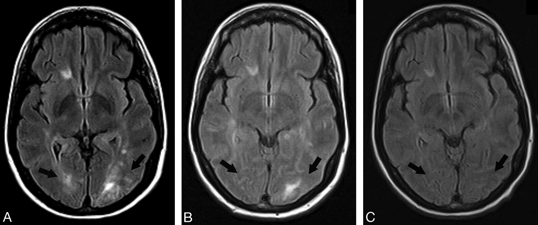
Brain MR imaging, comparison of axial FLAIR images. A, Brain MR imaging immediately after IV administration of caffeine, showing bilateral hyperintense signal intensity in the white matter of the occipital lobe, indicative of vasogenic edema (arrows). B, Follow-up brain MR imaging 72 hours later. There is resolution of the hyperintense signal intensity over the right occipital lobe and significantly reduced hyperintense signal intensity over the left occipital lobe, indicative of resolving vasogenic edema in both occipital lobes (arrows). C, Brain MR imaging 1 year later showing no hyperintense signal intensity in the posterior lobes (arrows).

The following morning, her headaches had resolved and her vision was normal. EEG was normal.

All treatments, except antiepileptic drugs, were stopped. She remained in bed at rest for 24 hours, and she was able to get up and ambulate without any headaches.

Follow-up brain MR imaging examination 72 hours later showed no enhancing lesions and near-resolution of the edema in the posterior lobes (Fig 1B). She was discharged after 10 days of hospitalization with no recurrence of headaches or seizures.

One year later, follow-up MR imaging examination did not show any signs of edema or abnormal enhancing in the posterior lobes (Fig 1C). She has remained asymptomatic since then and has been receiving treatment with interferon-beta for multiple sclerosis.

Discussion

The common denominator in PRES seems to be the presence of conditions that tend to provoke endothelial dysfunction leading to increased blood-brain barrier permeability or change in cerebrovascular autonomic control, or both. In this setting, a trigger factor, such as an increase in systemic blood pressure, generates segmental vasogenic edema.2,4 Despite the good overall prognosis, this condition is not completely benign because of the potential for bleeding into the lesion of reversible posterior leukoencephalopathy syndrome, as in cases of hypertensive encephalopathy. Also, there are atypical cases that may take longer than usual to reverse or may not be completely reversible.3,4

PDPH is believed to be caused by a CSF leak leading to intrathecal hypotension and reflex dilation and distention of the intracranial blood vessels, which are sensitive structures. This vasodilation has been shown to be asymmetric comparing posterior versus anterior circulation and also right versus left side.3,5

IV caffeine, probably by induction of vasoconstriction of the cerebral vasculature, has been documented to be an efficacious treatment of PDPH in many studies, without significant adverse effects.6,7 It is commonly used as first-line treatment because it is a less invasive technique than the epidural blood patch.

Multiple sclerosis may have been a predisposing condition to increased permeability in our patient, which, in combination with possible increased perfusion secondary to PDPH, could have been a favorable setting for IV caffeine to trigger a further asymmetric increase of local perfusion. The use of steroids may have also cooperated through fluid retention.

Why this phenomenon does not occur in many patients treated with IV caffeine for PDPH is probably because of the absence of an underlying predisposing condition, or perhaps PRES is underdiagnosed, as in the so-called postpartum seizures after treatment with IV caffeine reported in the medical literature before the more recent description of PRES.8–11 With this case report, we suggest adding multiple sclerosis and PDPH to the list of possible predisposing factors and IV caffeine to the list of trigger factors for PRES.

References

  1. ↵
    Hinchey J, Chaves C, Appignani B, et al. A reversible posterior leukoencephalopathy syndrome. N Engl J Med 1996;334:494–500
    CrossRefPubMed
  2. ↵
    Kim JS, Lee KS, Lim SC, et al. Reversible posterior leukoencephalopathy syndrome in a patient with multiple system atrophy: a possible association with oral midodrine treatment. Mov Disord 2007;22:1043–46
    CrossRefPubMed
  3. ↵
    Lee VH, Wijdicks EF, Manno EM, et al. Clinical spectrum of reversible posterior leukoencephalopathy syndrome. Arch Neurol 2008;65:205–10
    CrossRefPubMed
  4. ↵
    Schwartz RB. Hyperperfusion encephalopathies: hypertensive encephalopathy and related conditions. Neurologist 2002;8:22–34
    CrossRefPubMed
  5. ↵
    Gobel H, Klostermann H, Lindner V, et al. [Hemodynamic reactions in the area supplied by the middle cerebral artery in post-puncture headache]. Neurochirurgia (Stuttg) 1990;33 Suppl 1:4–7
    PubMed
  6. ↵
    Choi A, Laurito CE, Cunningham FE. Pharmacologic management of postdural puncture headache. Ann Pharmacother 1996;30:831–39
    PubMed
  7. ↵
    Yücel A, Ozyalçcin S, Talu GK, et al. Intravenous administration of caffeine sodium benzoate for postdural puncture headache. Reg Anesth Pain Med 1999;24:51–54
    PubMed
  8. ↵
    Bolton VE, Leicht CH, Scanlon TS. Postpartum seizure after epidural blood patch and intravenous caffeine sodium benzoate. Anesthesiology 1989;70:146–49
    CrossRefPubMed
  9. Cohen SM, Laurito CE, Curran MJ. Grand mal seizure in a postpartum patient following intravenous infusion of caffeine sodium benzoate to treat persistent headache. J Clin Anesth 1992;4:48–51
    CrossRefPubMed
  10. Paech M. Unexpected postpartum seizures associated with post-dural puncture headache treated with caffeine. Int J Obstet Anesth 1996;5:43–46
    CrossRefPubMed
  11. ↵
    Shearer VE, Jhaveri HS, Cunningham FG. Puerperal seizures after post-dural puncture headache. Obstet Gynecol 1995;85:255–60
    CrossRefPubMed
  • Received July 22, 2008.
  • Accepted after revision August 11, 2008.
  • Copyright © American Society of Neuroradiology
View Abstract
PreviousNext
Back to top

In this issue

American Journal of Neuroradiology: 30 (3)
American Journal of Neuroradiology
Vol. 30, Issue 3
March 2009
  • Table of Contents
  • Index by author
Advertisement
Print
Download PDF
Email Article

Thank you for your interest in spreading the word on American Journal of Neuroradiology.

NOTE: We only request your email address so that the person you are recommending the page to knows that you wanted them to see it, and that it is not junk mail. We do not capture any email address.

Enter multiple addresses on separate lines or separate them with commas.
Posterior Reversible Encephalopathy Syndrome after Intravenous Caffeine for Post–Lumbar Puncture Headaches
(Your Name) has sent you a message from American Journal of Neuroradiology
(Your Name) thought you would like to see the American Journal of Neuroradiology web site.
CAPTCHA
This question is for testing whether or not you are a human visitor and to prevent automated spam submissions.
Cite this article
G.A. Ortiz, N.A. Bianchi, M.P. Tiede, R.G. Bhatia
Posterior Reversible Encephalopathy Syndrome after Intravenous Caffeine for Post–Lumbar Puncture Headaches
American Journal of Neuroradiology Mar 2009, 30 (3) 586-587; DOI: 10.3174/ajnr.A1321

Citation Manager Formats

  • BibTeX
  • Bookends
  • EasyBib
  • EndNote (tagged)
  • EndNote 8 (xml)
  • Medlars
  • Mendeley
  • Papers
  • RefWorks Tagged
  • Ref Manager
  • RIS
  • Zotero
0 Responses
Respond to this article
Share
Bookmark this article
Posterior Reversible Encephalopathy Syndrome after Intravenous Caffeine for Post–Lumbar Puncture Headaches
G.A. Ortiz, N.A. Bianchi, M.P. Tiede, R.G. Bhatia
American Journal of Neuroradiology Mar 2009, 30 (3) 586-587; DOI: 10.3174/ajnr.A1321
del.icio.us logo Twitter logo Facebook logo Mendeley logo
  • Tweet Widget
  • Facebook Like
  • Google Plus One
Purchase

Jump to section

  • Article
    • Abstract
    • Case Report
    • Discussion
    • References
  • Figures & Data
  • Info & Metrics
  • Responses
  • References
  • PDF

Related Articles

  • No related articles found.
  • PubMed
  • Google Scholar

Cited By...

  • Pearls & Oy-sters: Spontaneous intracranial hypotension and posterior reversible encephalopathy syndrome
  • Crossref (13)
  • Google Scholar

This article has been cited by the following articles in journals that are participating in Crossref Cited-by Linking.

  • Annual Update in Intensive Care and Emergency Medicine 2011
    S. Legriel, F. Pico, E. Azoulay
    2011 1
  • Post-dural puncture headache and posterior reversible encephalopathy syndrome: a misdiagnosis or co-presentation?
    H. Doherty, S. Hameed, I. Ahmed, I.F. Russell
    International Journal of Obstetric Anesthesia 2014 23 3
  • Influenza A‐induced rhabdomyolysis and acute kidney injury complicated by posterior reversible encephalopathy syndrome
    R. A. Fearnley, S. W. Lines, A. J. P. Lewington, A. R. Bodenham
    Anaesthesia 2011 66 8
  • Posterior reversible encephalopathy syndrome following a thoracic discectomy–induced dural leak: case report
    Lisa B. E. Shields, John R. Johnson, Christopher B. Shields
    Journal of Neurosurgery: Spine 2016 25 5
  • Posterior reversible encephalopathy syndrome after depletive lumbar puncture: a case report
    Michael Grelat, Jean-Baptiste Debaux, Jean-Louis Sautreaux
    Journal of Medical Case Reports 2014 8 1
  • Pearls & Oy-sters: Spontaneous intracranial hypotension and posterior reversible encephalopathy syndrome
    Alejandro Santillan, Whitley Aamodt, Ratna Bhavaraju-Sanka
    Neurology 2016 86 6
  • Post-partum posterior reversible encephalopathy syndrome requiring decompressive craniectomy: case report and review of the literature
    Gennadiy A. Katsevman, Ryan C. Turner, Cletus Cheyuo, Charles L. Rosen, Matthew S. Smith
    Acta Neurochirurgica 2019 161 2
  • Cerebrospinal Fluid Hypovolemia and Posterior Reversible Encephalopathy Syndrome
    Yuan-yuan Zheng, Xiong-peng Weng, Fang-wang Fu, Yun-gang Cao, Yan Li, Guo-qing Zheng, Wei Chen
    Frontiers in Neurology 2020 11
  • Posterior Reversible Encephalopathy Syndrome Caused by Fioricet (Butalbital-Acetaminophen-Caffeine)
    Naomi Cret, Madalina Halalau, Shahab Rezvani, Alexandra Halalau
    Case Reports in Medicine 2019 2019
  • Des pathologies encéphaliques à connaître — Syndrome d’encéphalopathie postérieure réversible
    S. Legriel, F. Pico, F. Bruneel, G. Troché, J. -P. Bedos
    Réanimation 2011 20 S2

More in this TOC Section

  • Evaluating the Effects of White Matter Multiple Sclerosis Lesions on the Volume Estimation of 6 Brain Tissue Segmentation Methods
  • White Matter Alterations in the Brains of Patients with Active, Remitted, and Cured Cushing Syndrome: A DTI Study
  • Enhanced Axonal Metabolism during Early Natalizumab Treatment in Relapsing-Remitting Multiple Sclerosis
Show more Brain

Similar Articles

Advertisement

Indexed Content

  • Current Issue
  • Accepted Manuscripts
  • Article Preview
  • Past Issues
  • Editorials
  • Editors Choice
  • Fellow Journal Club
  • Letters to the Editor

Cases

  • Case Collection
  • Archive - Case of the Week
  • Archive - Case of the Month
  • Archive - Classic Case

Special Collections

  • Special Collections

Resources

  • News and Updates
  • Turn around Times
  • Submit a Manuscript
  • Author Policies
  • Manuscript Submission Guidelines
  • Evidence-Based Medicine Level Guide
  • Publishing Checklists
  • Graphical Abstract Preparation
  • Imaging Protocol Submission
  • Submit a Case
  • Become a Reviewer/Academy of Reviewers
  • Get Peer Review Credit from Publons

Multimedia

  • AJNR Podcast
  • AJNR SCANtastic
  • Video Articles

About Us

  • About AJNR
  • Editorial Board
  • Not an AJNR Subscriber? Join Now
  • Alerts
  • Feedback
  • Advertise with us
  • Librarian Resources
  • Permissions
  • Terms and Conditions

American Society of Neuroradiology

  • Not an ASNR Member? Join Now

© 2025 by the American Society of Neuroradiology All rights, including for text and data mining, AI training, and similar technologies, are reserved.
Print ISSN: 0195-6108 Online ISSN: 1936-959X

Powered by HighWire