Advertisement

Main menu

  • Home
  • Content
    • Current Issue
    • Accepted Manuscripts
    • Article Preview
    • Past Issue Archive
    • AJNR Case Collection
    • Case of the Week Archive
    • Classic Case Archive
    • Case of the Month Archive
  • Special Collections
    • Spinal CSF Leak Articles (Jan 2020-June 2024)
    • 2024 AJNR Journal Awards
    • Most Impactful AJNR Articles
  • Multimedia
    • AJNR Podcast
    • AJNR Scantastics
    • Video Articles
  • For Authors
    • Submit a Manuscript
    • Author Policies
    • Fast publishing of Accepted Manuscripts
    • Graphical Abstract Preparation
    • Manuscript Submission Guidelines
    • Imaging Protocol Submission
    • Submit a Case for the Case Collection
  • About Us
    • About AJNR
    • Editorial Board
  • More
    • Become a Reviewer/Academy of Reviewers
    • Subscribers
    • Permissions
    • Alerts
    • Feedback
    • Advertisers
    • ASNR Home
  • Other Publications
    • ajnr

User menu

  • Alerts
  • Log in

Search

  • Advanced search
American Journal of Neuroradiology

American Journal of Neuroradiology

ASHNR American Society of Functional Neuroradiology ASHNR American Society of Pediatric Neuroradiology ASSR
  • Alerts
  • Log in

Advanced Search

  • Home
  • Content
    • Current Issue
    • Accepted Manuscripts
    • Article Preview
    • Past Issue Archive
    • AJNR Case Collection
    • Case of the Week Archive
    • Classic Case Archive
    • Case of the Month Archive
  • Special Collections
    • Spinal CSF Leak Articles (Jan 2020-June 2024)
    • 2024 AJNR Journal Awards
    • Most Impactful AJNR Articles
  • Multimedia
    • AJNR Podcast
    • AJNR Scantastics
    • Video Articles
  • For Authors
    • Submit a Manuscript
    • Author Policies
    • Fast publishing of Accepted Manuscripts
    • Graphical Abstract Preparation
    • Manuscript Submission Guidelines
    • Imaging Protocol Submission
    • Submit a Case for the Case Collection
  • About Us
    • About AJNR
    • Editorial Board
  • More
    • Become a Reviewer/Academy of Reviewers
    • Subscribers
    • Permissions
    • Alerts
    • Feedback
    • Advertisers
    • ASNR Home
  • Follow AJNR on Twitter
  • Visit AJNR on Facebook
  • Follow AJNR on Instagram
  • Join AJNR on LinkedIn
  • RSS Feeds

Welcome to the new AJNR, Updated Hall of Fame, and more. Read the full announcements.


AJNR is seeking candidates for the position of Associate Section Editor, AJNR Case Collection. Read the full announcement.


Research ArticleBRAIN

Cerebral Blood Volume Measurements by Perfusion-Weighted MR Imaging in Gliomas: Ready for Prime Time in Predicting Short-Term Outcome and Recurrent Disease?

S. Bisdas, M. Kirkpatrick, P. Giglio, C. Welsh, M.V. Spampinato and Z. Rumboldt
American Journal of Neuroradiology April 2009, 30 (4) 681-688; DOI: https://doi.org/10.3174/ajnr.A1465
S. Bisdas
  • Find this author on Google Scholar
  • Find this author on PubMed
  • Search for this author on this site
M. Kirkpatrick
  • Find this author on Google Scholar
  • Find this author on PubMed
  • Search for this author on this site
P. Giglio
  • Find this author on Google Scholar
  • Find this author on PubMed
  • Search for this author on this site
C. Welsh
  • Find this author on Google Scholar
  • Find this author on PubMed
  • Search for this author on this site
M.V. Spampinato
  • Find this author on Google Scholar
  • Find this author on PubMed
  • Search for this author on this site
Z. Rumboldt
  • Find this author on Google Scholar
  • Find this author on PubMed
  • Search for this author on this site
  • Article
  • Figures & Data
  • Info & Metrics
  • Responses
  • References
  • PDF
Loading

Article Figures & Data

Figures

  • Tables
  • Fig 1.
    • Download figure
    • Open in new tab
    • Download powerpoint
    Fig 1.

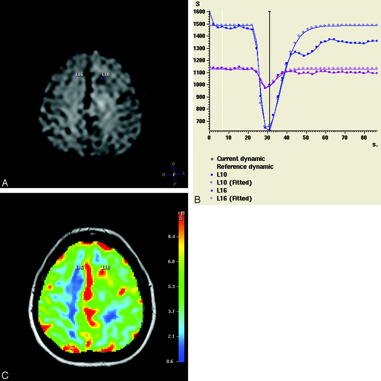
    Source dynamic MR image (A), signal intensity time curves (S) (B), and color-coded perfusion map (overlaid on the corresponding FLAIR image) (C) of a male patient with a supratentorial astrocytoma (WHO grade III) in the left hemisphere (patient 21). The tumor pixel with the highest CBV value is represented as a small blue cross (L10). The contralateral corresponding white matter pixel is a purple cross (L16). The 2 pixels have distinct signal intensity curves with markedly deeper signal intensity drop in the tumor pixel (which begins 24 seconds after perfusion-weighted MR imaging initiation).

  • Fig 2.
    • Download figure
    • Open in new tab
    • Download powerpoint
    Fig 2.

    Scatterplot of the PFS and rCBVmax of the astrocytoma population. The plot presents the 2 population subgroups: the recurrent (solid triangles) and the nonrecurrent (open circles) astrocytomas. Note the high perfusion values of the recurrent tumors combined with shorter survival.

  • Fig 3.
    • Download figure
    • Open in new tab
    • Download powerpoint
    Fig 3.

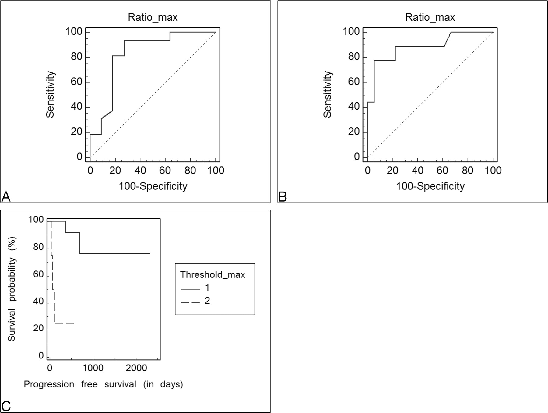
    The ROC curves analyzing the sensitivity and specificity of rCBVmax values in astrocytomas for 1-year survival (A) and recurrence (B) show the optimal threshold value as ≤3.8 (sensitivity, 93.7% [95% confidence interval (CI), 69.7–99%]; specificity, 72.7% [95% CI, 39.1–93.7%], P = .0002) for 1-year survival and as >4.2 (sensitivity, 77.8% [95% CI, 40–96.5%]; specificity, 94.4% [72.6–99.1%], P = .0001) for recurrence. C, Kaplan-Meier curve of PFS probability (hazard ratio [HR] = 0.09, P = .0007). The thresholdmax represents the rCBVmax cutoff value, in which 1 corresponds to rCBVmax ≤4.2.

  • Fig 4.
    • Download figure
    • Open in new tab
    • Download powerpoint
    Fig 4.

    A and B, The ROC curves analyzing the sensitivity and specificity of the WHO grading system (I-IV) for the 1-year survival (A) and recurrence (B) show the optimal threshold value as <2 (WHO grade II) (sensitivity, 81.2% [95% confidence interval (CI), 54.3–95.7%]; specificity, 90.9% [95% CI, 58.7–98.5%], P = .0002) for 1-year survival and as >2 (sensitivity, 88.9% [95% CI, 51.7–98.2%]; specificity, 72.2% [95% CI, 46.5–90.2%], P = .0001) for recurrence. C, Kaplan-Meier curve of PFS probability is based on stratification according to the WHO grade (1 = low grade [WHO I-II], 2 = high grade [WHO III and IV)]; HR = 0.15, P = .05).

Tables

  • Figures
    • View popup
    Table 1:

    Patient population demographics, histologic diagnosis with WHO grade, KPS, PFS, and maximum rCBV values

    PatientAge (yr)SexPathologyWHOKPSPFS (days)rCBVmax
    126MAstro21002981.1
    264FAstro470999.1
    361MAstro4801914.7
    443FAstro1906682.1
    545MAstro21004852.1
    669FAstro4607493.5
    779MAstro2602103.0
    837MAstro2501174.8
    956FAstro410015211.9
    1019FAstro1909751.8
    1146MOligoastro28010411.2
    1256FOligoastro29014026.2
    1343MAstro21004286.1
    1449MAstro4601344.2
    1553FAstro29016082.9
    1641MOligoastro31007911.2
    1719MAstro39013232.7
    1820MAstro1803923.8
    1931MAstro17018311.2
    2037MAstro3906936.2
    2130FAstro3905671.2
    2216FOligodendro37013022.2
    2340MOligodendro2805461.1
    2437FOligodendro210015405.6
    2543FOligodendro21003804.1
    2668FAstro380577.8
    2761MAstro4508511
    2881MAstro470288.0
    2938FAstro2805461.0
    3053MAstro4100764.4
    3151MAstro2903861.1
    3241MAstro31003341.5
    3341FAstro2809111.3
    3446MAstro2906992.4
    • Note:—Astro indicates astrocytoma; oligoastro, oligoastrocytoma; oligodendro, oligodendroglioma; KPS, Kornofsky performance score; PFS, progression-free survival; rCBV, relative cerebral blood volume.

    • View popup
    Table 2:

    Summary statistics of mean and maximum rCBV values, KPS, and PFS of the 3 subgroups of the patient population

    rCBVmeanrCBVmaxKPS, Median (range)PFS (average in days)
    Astrocytomas (n = 27)3.5 (±2.7)3.7 (±2.7)90 (50–100)576 (±505)
    Oligodendrogliomas (n = 4)3.1 (±2.2)3.3 (±2.0)90 (70–100)942 (±566)
    Oligoastrocytomas (n= 3)2.4 (±1.7)2.9 (±2.3)90 (70–90)1030 (±401)
PreviousNext
Back to top

In this issue

American Journal of Neuroradiology: 30 (4)
American Journal of Neuroradiology
Vol. 30, Issue 4
April 2009
  • Table of Contents
  • Index by author
Advertisement
Print
Download PDF
Email Article

Thank you for your interest in spreading the word on American Journal of Neuroradiology.

NOTE: We only request your email address so that the person you are recommending the page to knows that you wanted them to see it, and that it is not junk mail. We do not capture any email address.

Enter multiple addresses on separate lines or separate them with commas.
Cerebral Blood Volume Measurements by Perfusion-Weighted MR Imaging in Gliomas: Ready for Prime Time in Predicting Short-Term Outcome and Recurrent Disease?
(Your Name) has sent you a message from American Journal of Neuroradiology
(Your Name) thought you would like to see the American Journal of Neuroradiology web site.
CAPTCHA
This question is for testing whether or not you are a human visitor and to prevent automated spam submissions.
Cite this article
S. Bisdas, M. Kirkpatrick, P. Giglio, C. Welsh, M.V. Spampinato, Z. Rumboldt
Cerebral Blood Volume Measurements by Perfusion-Weighted MR Imaging in Gliomas: Ready for Prime Time in Predicting Short-Term Outcome and Recurrent Disease?
American Journal of Neuroradiology Apr 2009, 30 (4) 681-688; DOI: 10.3174/ajnr.A1465

Citation Manager Formats

  • BibTeX
  • Bookends
  • EasyBib
  • EndNote (tagged)
  • EndNote 8 (xml)
  • Medlars
  • Mendeley
  • Papers
  • RefWorks Tagged
  • Ref Manager
  • RIS
  • Zotero
0 Responses
Respond to this article
Share
Bookmark this article
Cerebral Blood Volume Measurements by Perfusion-Weighted MR Imaging in Gliomas: Ready for Prime Time in Predicting Short-Term Outcome and Recurrent Disease?
S. Bisdas, M. Kirkpatrick, P. Giglio, C. Welsh, M.V. Spampinato, Z. Rumboldt
American Journal of Neuroradiology Apr 2009, 30 (4) 681-688; DOI: 10.3174/ajnr.A1465
del.icio.us logo Twitter logo Facebook logo Mendeley logo
  • Tweet Widget
  • Facebook Like
  • Google Plus One
Purchase

Jump to section

  • Article
    • Abstract
    • Materials and Methods
    • Results
    • Discussion
    • Conclusions
    • References
  • Figures & Data
  • Info & Metrics
  • Responses
  • References
  • PDF

Related Articles

  • No related articles found.
  • PubMed
  • Google Scholar

Cited By...

  • Radiogenomics-based Risk Prediction of Glioblastoma Multiforme with Clinical Relevance
  • MR Imaging Features of Anaplastic Pleomorphic Xanthoastrocytoma Mimicking High-Grade Astrocytoma
  • Improved Brain Tumor Classification by Sodium MR Imaging: Prediction of IDH Mutation Status and Tumor Progression
  • Prognostic Value of Dynamic Susceptibility Contrast-Enhanced and Diffusion-Weighted MR Imaging in Patients with Glioblastomas
  • ASFNR Recommendations for Clinical Performance of MR Dynamic Susceptibility Contrast Perfusion Imaging of the Brain
  • Pixel-by-Pixel Comparison of Volume Transfer Constant and Estimates of Cerebral Blood Volume from Dynamic Contrast-Enhanced and Dynamic Susceptibility Contrast-Enhanced MR Imaging in High-Grade Gliomas
  • MRI Grading versus Histology: Predicting Survival of World Health Organization Grade II-IV Astrocytomas
  • Prognostic Significance of Dynamic 18F-FET PET in Newly Diagnosed Astrocytic High-Grade Glioma
  • Role of MRI in Primary Brain Tumor Evaluation
  • Dynamic 18F-FET PET in Newly Diagnosed Astrocytic Low-Grade Glioma Identifies High-Risk Patients
  • Survival Analysis of Patients with High-Grade Gliomas Based on Data Mining of Imaging Variables
  • Does MR Perfusion Imaging Impact Management Decisions for Patients with Brain Tumors? A Prospective Study
  • Correlations between Perfusion MR Imaging Cerebral Blood Volume, Microvessel Quantification, and Clinical Outcome Using Stereotactic Analysis in Recurrent High-Grade Glioma
  • Quantitative Blood Flow Measurements in Gliomas Using Arterial Spin-Labeling at 3T: Intermodality Agreement and Inter- and Intraobserver Reproducibility Study
  • The Holy Grail and the Quest for the Gold Standard
  • Switching on the Lights for Real-Time Multimodality Tumor Neuroimaging: The Integrated Positron-Emission Tomography/MR Imaging System
  • Crossref (99)
  • Google Scholar

This article has been cited by the following articles in journals that are participating in Crossref Cited-by Linking.

  • ASFNR Recommendations for Clinical Performance of MR Dynamic Susceptibility Contrast Perfusion Imaging of the Brain
    K. Welker, J. Boxerman, A. Kalnin, T. Kaufmann, M. Shiroishi, M. Wintermark
    American Journal of Neuroradiology 2015 36 6
  • A radiomic signature as a non-invasive predictor of progression-free survival in patients with lower-grade gliomas
    Xing Liu, Yiming Li, Zenghui Qian, Zhiyan Sun, Kaibin Xu, Kai Wang, Shuai Liu, Xing Fan, Shaowu Li, Zhong Zhang, Tao Jiang, Yinyan Wang
    NeuroImage: Clinical 2018 20
  • Medical Image Computing and Computer-Assisted Intervention – MICCAI 2016
    Dong Nie, Han Zhang, Ehsan Adeli, Luyan Liu, Dinggang Shen
    2016 9901
  • Prognostic Significance of Dynamic 18F-FET PET in Newly Diagnosed Astrocytic High-Grade Glioma
    Nathalie L. Jansen, Bogdana Suchorska, Vera Wenter, Christine Schmid-Tannwald, Andrei Todica, Sabina Eigenbrod, Maximilian Niyazi, Jörg-Christian Tonn, Peter Bartenstein, Friedrich-Wilhelm Kreth, Christian la Fougère
    Journal of Nuclear Medicine 2015 56 1
  • Genomic Mapping and Survival Prediction in Glioblastoma: Molecular Subclassification Strengthened by Hemodynamic Imaging Biomarkers
    Rajan Jain, Laila Poisson, Jayant Narang, David Gutman, Lisa Scarpace, Scott N. Hwang, Chad Holder, Max Wintermark, Rivka R. Colen, Justin Kirby, John Freymann, Daniel J. Brat, Carl Jaffe, Tom Mikkelsen
    Radiology 2013 267 1
  • Dynamic 18F-FET PET in Newly Diagnosed Astrocytic Low-Grade Glioma Identifies High-Risk Patients
    Nathalie L. Jansen, Bogdana Suchorska, Vera Wenter, Sabina Eigenbrod, Christine Schmid-Tannwald, Andreas Zwergal, Maximilian Niyazi, Mark Drexler, Peter Bartenstein, Oliver Schnell, Jörg-Christian Tonn, Niklas Thon, Friedrich-Wilhelm Kreth, Christian la Fougère
    Journal of Nuclear Medicine 2014 55 2
  • Correlations between Perfusion MR Imaging Cerebral Blood Volume, Microvessel Quantification, and Clinical Outcome Using Stereotactic Analysis in Recurrent High-Grade Glioma
    L.S. Hu, J.M. Eschbacher, A.C. Dueck, J.E. Heiserman, S. Liu, J.P. Karis, K.A. Smith, W.R. Shapiro, D.S. Pinnaduwage, S.W. Coons, P. Nakaji, J. Debbins, B.G. Feuerstein, L.C. Baxter
    American Journal of Neuroradiology 2012 33 1
  • A Generic Support Vector Machine Model for Preoperative Glioma Survival Associations
    Kyrre E. Emblem, Marco C. Pinho, Frank G. Zöllner, Paulina Due-Tonnessen, John K. Hald, Lothar R. Schad, Torstein R. Meling, Otto Rapalino, Atle Bjornerud
    Radiology 2015 275 1
  • Early static 18F-FET-PET scans have a higher accuracy for glioma grading than the standard 20–40 min scans
    Nathalie L. Albert, Isabel Winkelmann, Bogdana Suchorska, Vera Wenter, Christine Schmid-Tannwald, Erik Mille, Andrei Todica, Matthias Brendel, Jörg-Christian Tonn, Peter Bartenstein, Christian la Fougère
    European Journal of Nuclear Medicine and Molecular Imaging 2016 43 6
  • Prediction of oligodendroglial histology and LOH 1p/19q using dynamic [18F]FET-PET imaging in intracranial WHO grade II and III gliomas
    N. L. Jansen, C. Schwartz, V. Graute, S. Eigenbrod, J. Lutz, R. Egensperger, G. Popperl, H. A. Kretzschmar, P. Cumming, P. Bartenstein, J.-C. Tonn, F.-W. Kreth, C. la Fougere, N. Thon
    Neuro-Oncology 2012 14 12

More in this TOC Section

  • Progression of Microstructural Damage in Spinocerebellar Ataxia Type 2: A Longitudinal DTI Study
  • Usefulness of Quantitative Susceptibility Mapping for the Diagnosis of Parkinson Disease
  • Evaluating the Effects of White Matter Multiple Sclerosis Lesions on the Volume Estimation of 6 Brain Tissue Segmentation Methods
Show more Brain

Similar Articles

Advertisement

Indexed Content

  • Current Issue
  • Accepted Manuscripts
  • Article Preview
  • Past Issues
  • Editorials
  • Editors Choice
  • Fellow Journal Club
  • Letters to the Editor

Cases

  • Case Collection
  • Archive - Case of the Week
  • Archive - Case of the Month
  • Archive - Classic Case

Special Collections

  • Special Collections

Resources

  • News and Updates
  • Turn around Times
  • Submit a Manuscript
  • Author Policies
  • Manuscript Submission Guidelines
  • Evidence-Based Medicine Level Guide
  • Publishing Checklists
  • Graphical Abstract Preparation
  • Imaging Protocol Submission
  • Submit a Case
  • Become a Reviewer/Academy of Reviewers
  • Get Peer Review Credit from Publons

Multimedia

  • AJNR Podcast
  • AJNR SCANtastic
  • Video Articles

About Us

  • About AJNR
  • Editorial Board
  • Not an AJNR Subscriber? Join Now
  • Alerts
  • Feedback
  • Advertise with us
  • Librarian Resources
  • Permissions
  • Terms and Conditions

American Society of Neuroradiology

  • Not an ASNR Member? Join Now

© 2025 by the American Society of Neuroradiology All rights, including for text and data mining, AI training, and similar technologies, are reserved.
Print ISSN: 0195-6108 Online ISSN: 1936-959X

Powered by HighWire