
Article Figures & Data
Figures
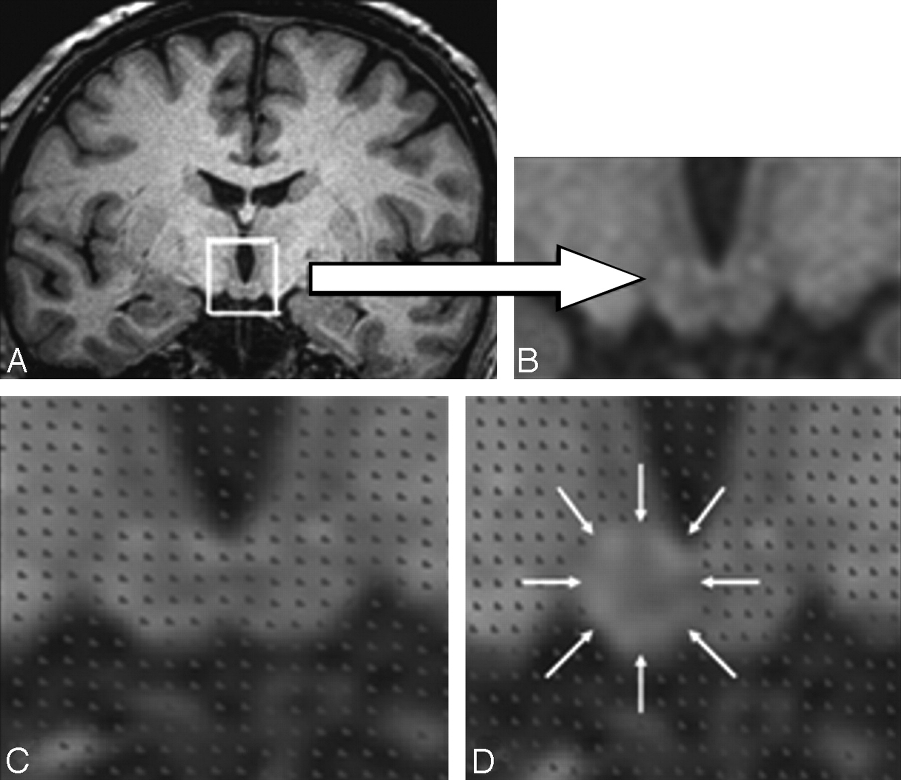
- Fig 1.
The stereologic procedure for measuring the mammillary bodies. A and B, Mammillary body location and detailed structure. C, Placement of the stereologic grid. D, Method of stereologic point counting. The mammillary body volume estimate is contained within the area indicated by the white arrows.
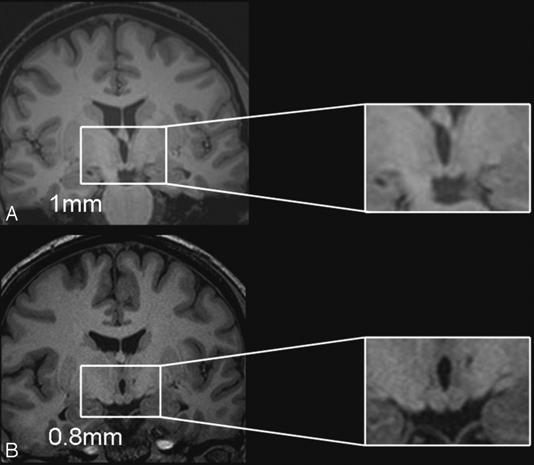
- Fig 2.
Comparison of the resolution of the 1 × 1 × 1 mm isotropic volume scan (A) with the 0.8-mm thin-section volume scan (B) in a patient with relatively normal mammillary body volumes. The 2 sections are taken from neighboring sections around the central region of the patient's mammillary bodies and show the higher resolution of the 0.8-mm scan and the resultant benefit for the identification of the mammillary body landmarks.
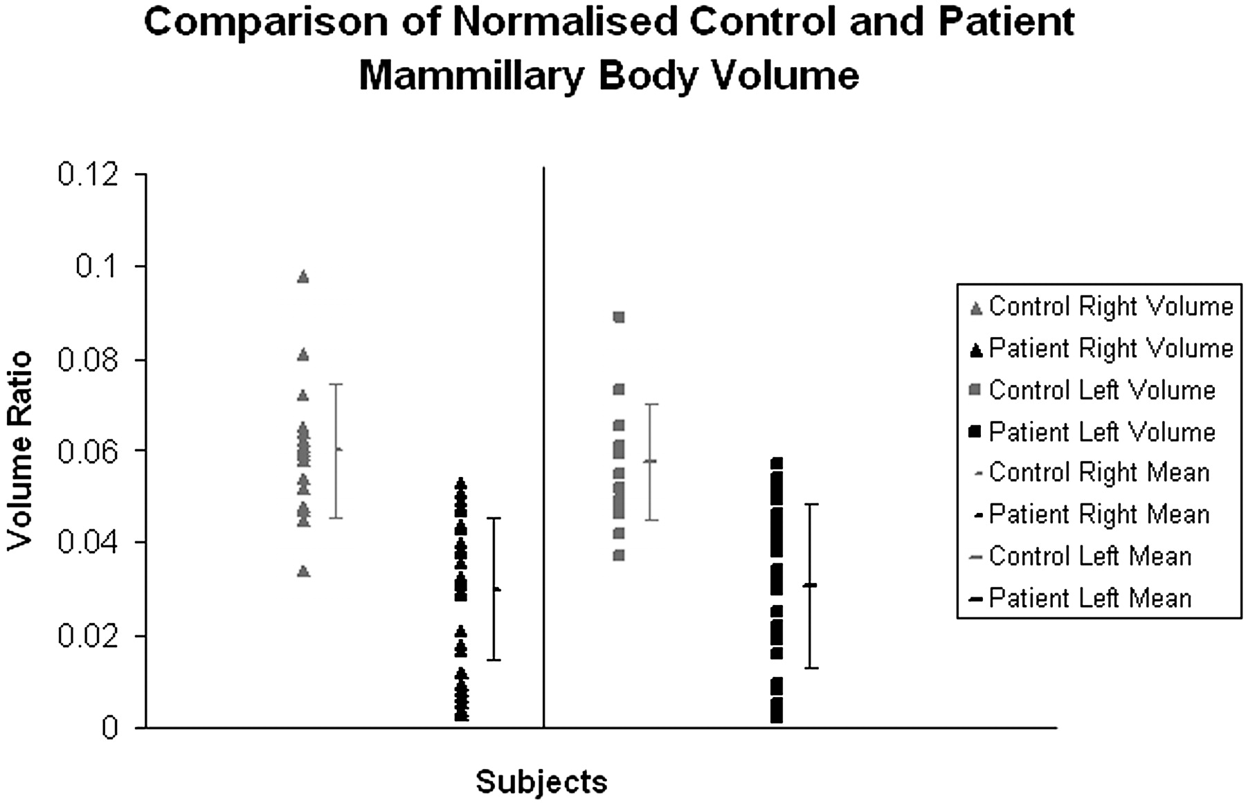
- Fig 3.
Distribution of intracranial normalized control and patient mammillary body volumes.
- Fig 4.
Cumulative classification of intracranial normalized patient mammillary body volumes according to the percentage of cases with atrophy below the control mean by using z-scores (1-sample t tests, 1-tailed).
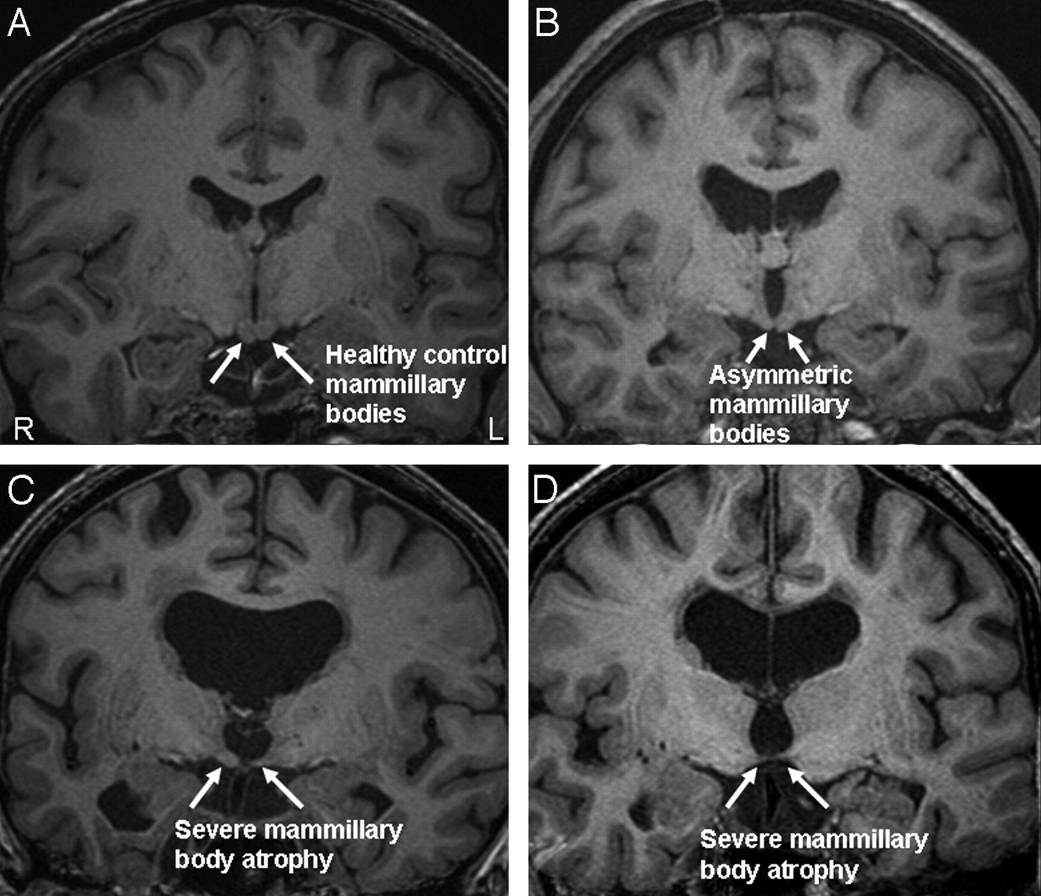
- Fig 5.
The range of atrophy across our study population. A, Healthy mammillary bodies from a control subject. B, Mild asymmetry in a patient with a colloid cyst. C and D, Severe mammillary body atrophy in the patient group. R indicates right; L, left.


Tables
- Table 1:
Mean absolute volume estimates, SDs, and volume ranges for the right and left mammillary bodies in the patients with colloid cysts and control subjects
Cyst Patients (n = 38) Controls (n = 20) Mean (SD) (mm3) Range Mean (SD) (mm3) Range 0.8 mm Right 38 (20) 4–67 69 (11) 44–86 Left 39 (22) 3–73 67 (10) 44–80 1.0 mm Right 38 (16) 5–72 56 (9) 41–83 Left 42 (19) 7–72 55 (16) 38–75 - Table 2:
Mean intracranial-volume normalized ratios, SDs, and volume ranges for the right and left mammillary bodies in patients with colloid cysts and controls
Cyst Patients (n = 38) Controls (n = 20) Mean (SD) Range Mean (SD) Range .08 mm Right 0.030 (0.015) 0.038–0.053 0.060 (0.014) 0.034–0.098 Left 0.031 (0.018) 0.020–0.057 0.057 (0.013) 0.042–0.09 1.0 mm Right 0.032 (0.016) 0.007–0.048 0.047 (0.080) 0.037–0.064 Left 0.035 (0.015) 0.007–0.059 0.047 (0.050) 0.038–0.054 - Table 3:
Number of patients and SDs below the control mean for the normalized mammillary body
SD below the Control Mean Normalized Mammillary Body Volume Right Left 0 to −1 7 10 −1 to −2 14 11 −2 to −3 9 7 −3 to −4 8 4 −4 to −5 0 6 Total 38 38
In this issue


Jump to section
Related Articles
Cited By...
- Impairments in the early consolidation of spatial memories via group II mGluR agonism in the mammillary bodies
- The Mammillary Bodies: A Review of Causes of Injury in Infants and Children
- Signal Change in the Mammillary Bodies after Perinatal Asphyxia
- Mammillothalamic Disconnection Alters Hippocampocortical Oscillatory Activity and Microstructure: Implications for Diencephalic Amnesia
- Mammillothalamic disconnection alters hippocampo-cortical oscillatory activity and microstructure: Implications for diencephalic amnesia
- Unraveling the contributions of the diencephalon to recognition memory: A review