
Article Figures & Data
Figures
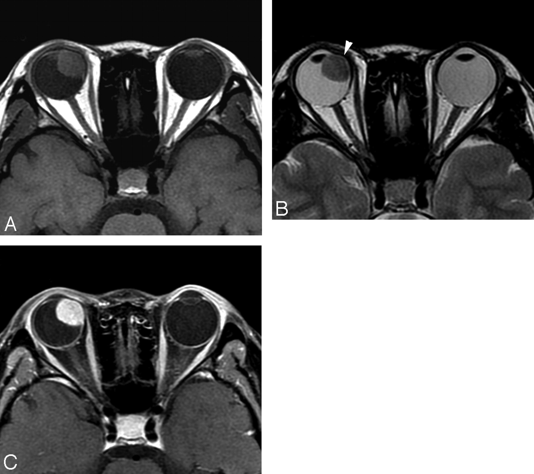
- Fig 1.
Case 3. Schwannoma in the right nasal ciliochoroidal region in a 22-year-old woman. Compared with the brain, the tumor demonstrates isointensity on SE T1WI (A), isointensity on FSE T2WI (B), and markedly heterogeneous enhancement on postcontrast SE T1WI (C). Displacement of the right lens is seen.
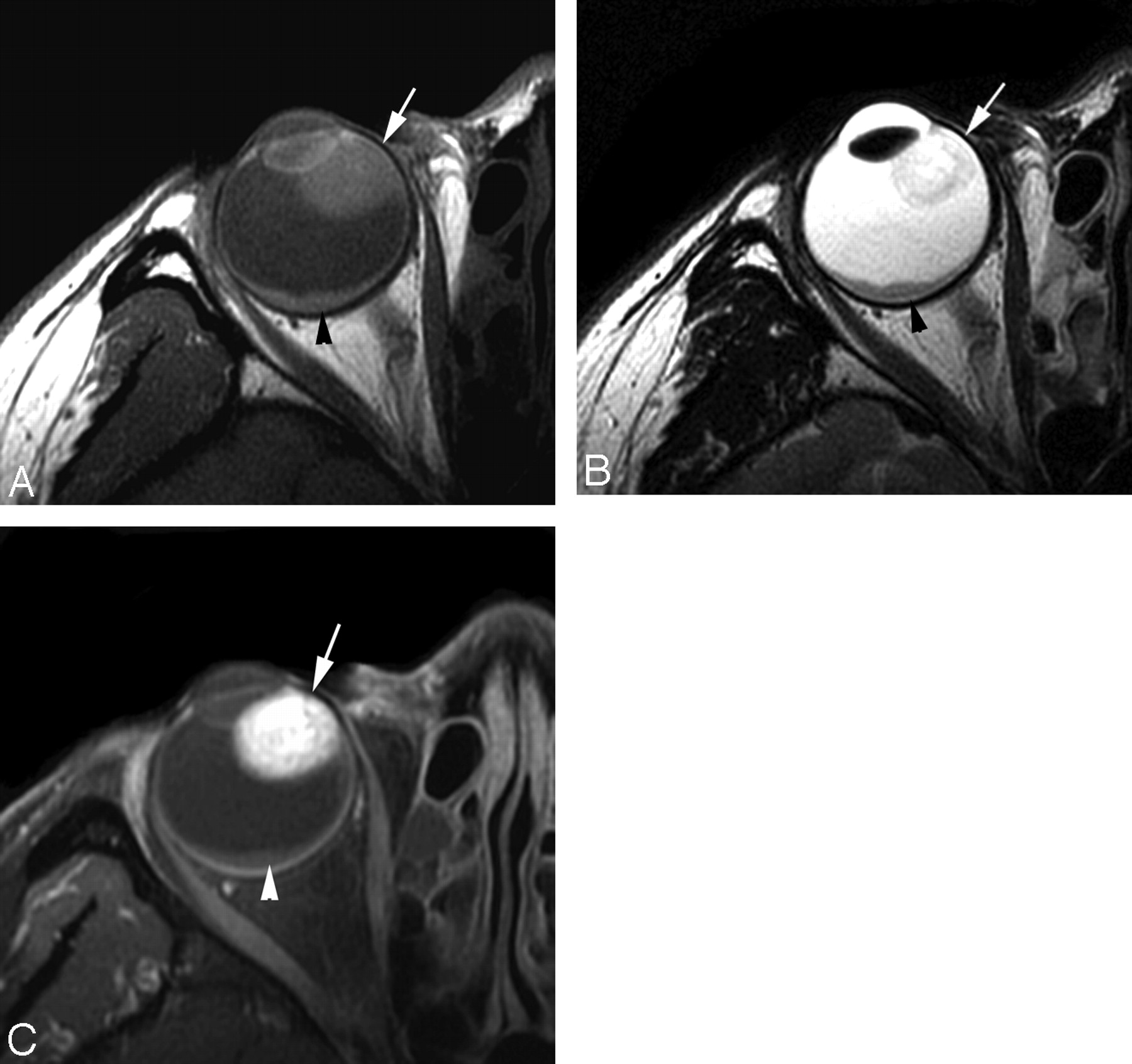
- Fig 2.
Case 5. Schwannoma in the right nasal ciliochoroidal region in a 38-year-old woman. With respect to the brain, the tumor (arrow) shows isointensity on SE T1WI (A), mixed hyperintensity and isointensity on FSE T2WI (B), and markedly heterogeneous enhancement on postcontrast SE T1WI (C). Crescent-shaped retinal detachment (arrowhead) without enhancement after contrast administration is found.
- Fig 3.
Dynamic contrast-enhancement curve shows patterns of enhancement in 5 cases of uveal schwannoma.


Tables
MR imaging findings in 6 patients with uveal schwannoma
No./Age (yr)* Location Shape Bulge of Globe Wall Displacement of Lens and Iris Size Margin Signal Intensity on MR Imaging Enhancement Other Findings Relative to Vitreus Relative to Brain T1WI T2WI T1WI T2WI 1/37 Right temporal ciliochoroidal region Oval No Yes 21 × 15 × 12 mm Well-defined Hyper, homo Mixed hypo, iso Iso, homo Mixed iso, hyper Hetero RD 2/19 Left nasal ciliochoroidal region Oval Yes Yes 15 × 13 × 12 mm Well-defined Hyper, homo Hypo, homo Iso, homo Iso, homo Homo 3/22 Right nasal ciliochoroidal region Oval Yes Yes 16 × 13 × 11 mm Well-defined Hyper, homo Hypo, homo Iso, homo Iso, homo Hetero 4/36 Left temporal choroid Oval Yes No 13 × 10 × 9 mm Well-defined Hyper, homo Mixed hypo, iso Iso, homo Mixed iso, hyper Homo RD, schwann of extremities 5/38 Right nasal ciliochoroidal region Round No Yes 10 × 10 × 10 mm Well-defined Hyper, homo Mixed hypo, iso Iso, homo Mixed hyper, iso Hetero RD 6/63 Left inferior ciliary body Oval No Yes 8 × 6 × 6 mm Well-defined Hyper, homo Hypo, homo Iso, homo Iso, homo Homo Note:—Hyper indicates hyperintense; hypo, hypointense; iso, isointense; homo, homogeneous; hetero, heterogeneous; RD, retinal detachment; schwann, schwannomas; T1WI, T1-weighted imaging; T2WI, T2-weighted imaging.
* All female sex.


