
Article Figures & Data
Figures
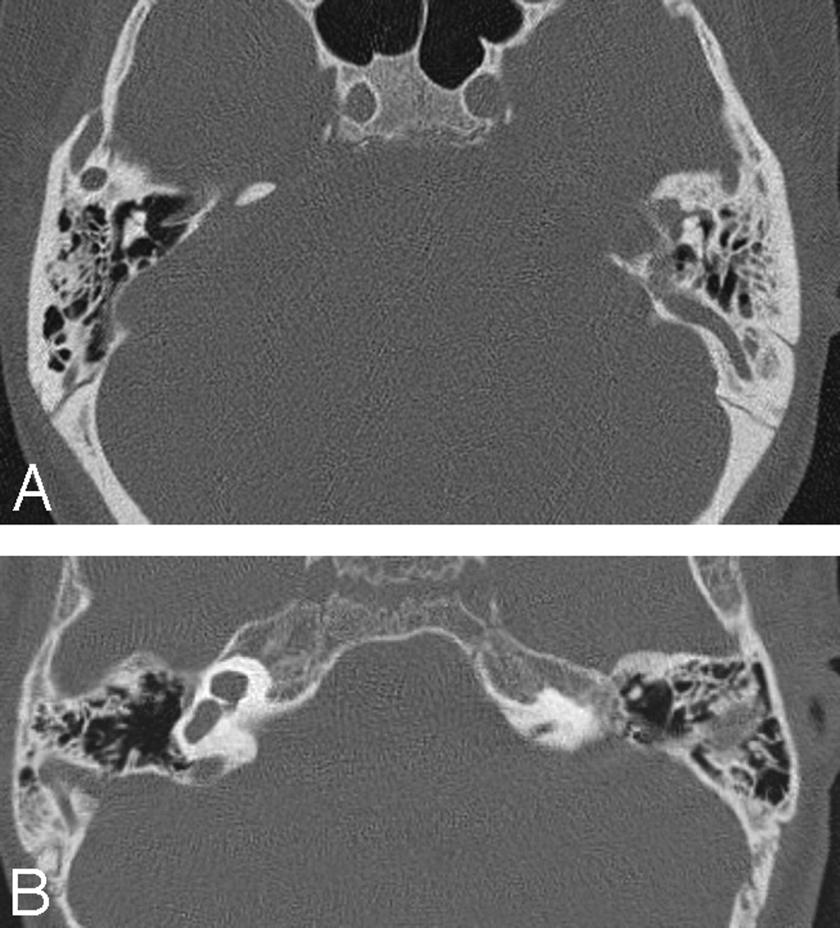
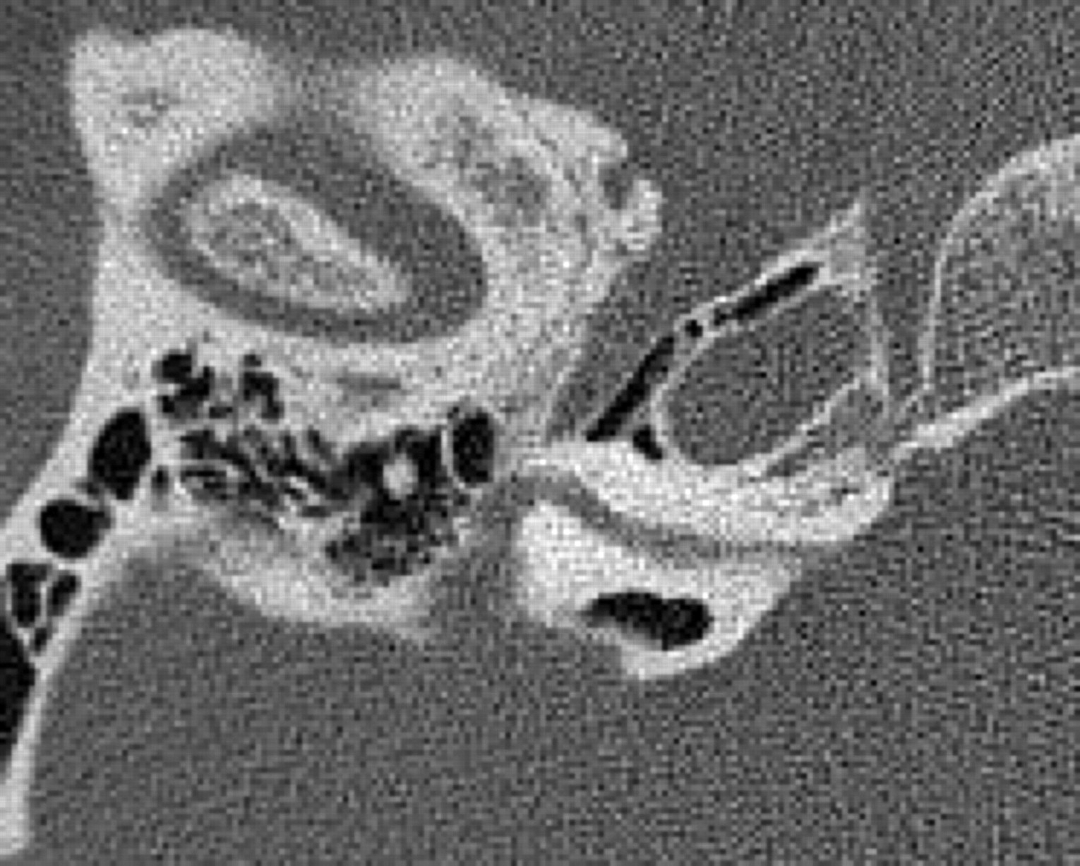
- Fig 1.
A, Axial CT scan of the temporal bones of patient 1 shows bilateral absence of total inner ear structures with aplasia of the otic capsules bilaterally. Note a large emissary vein on the left. B, CT scan of patient 4 reveals unilateral CLA on the left with a type 1 incomplete cochlear partition on the right. The petrous bone and the otic capsule are hypoplastic on the left.
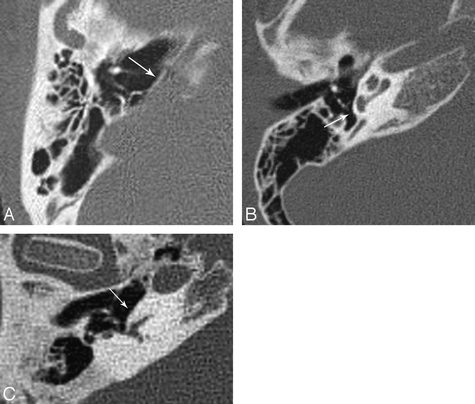
- Fig 2.
A, Flattening of the cochlear promontory (arrow) in CLA. B, Normal appearance of the cochlear promontory (arrow) and inner ear in a healthy patient. C, Normal appearance of the promontory (arrow) in a patient with severe LO. Note the normal size of the petrous bone in the patient with LO.
- Fig 3.
Narrowing of the middle ear and mastoid antrum.
- Fig 4.
A, Sagittal reformatted image of the temporal bone CT scan of an ear with CLA, demonstrating slight hypoplasia of the mastoid tip. B, Normal appearance of the mastoid tip in a patient of the same age.
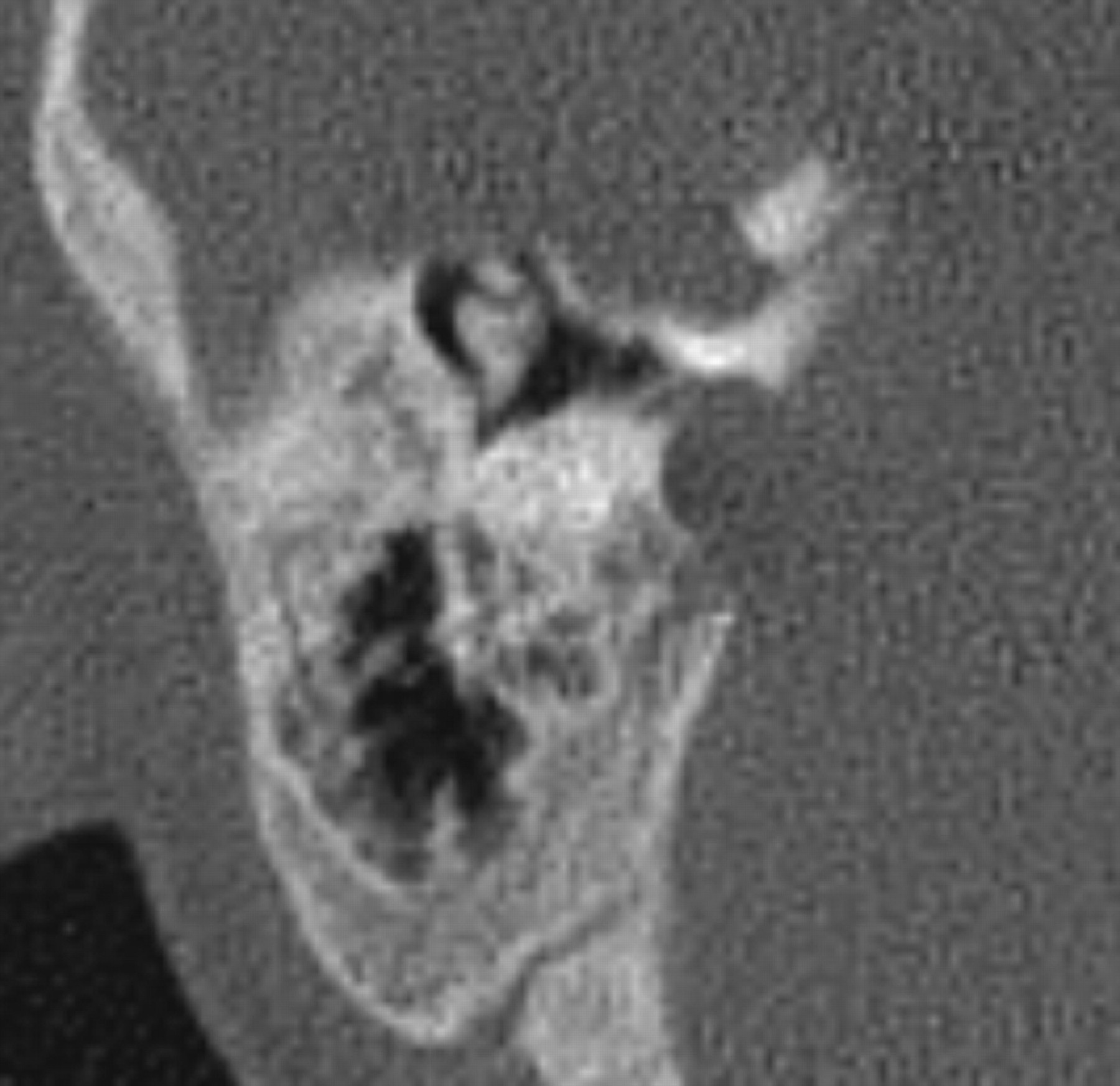
- Fig 5.
Anterolateral displacement and rotation of the stapes (white arrow) in an ear with CLA. Note the short process of the incus (black arrow) and head of the malleus (asterisk).
- Fig 6.
Coronal reformatted image of a patient with unilateral (left-sided) CLA reveals severe narrowing of the IAC on the left.
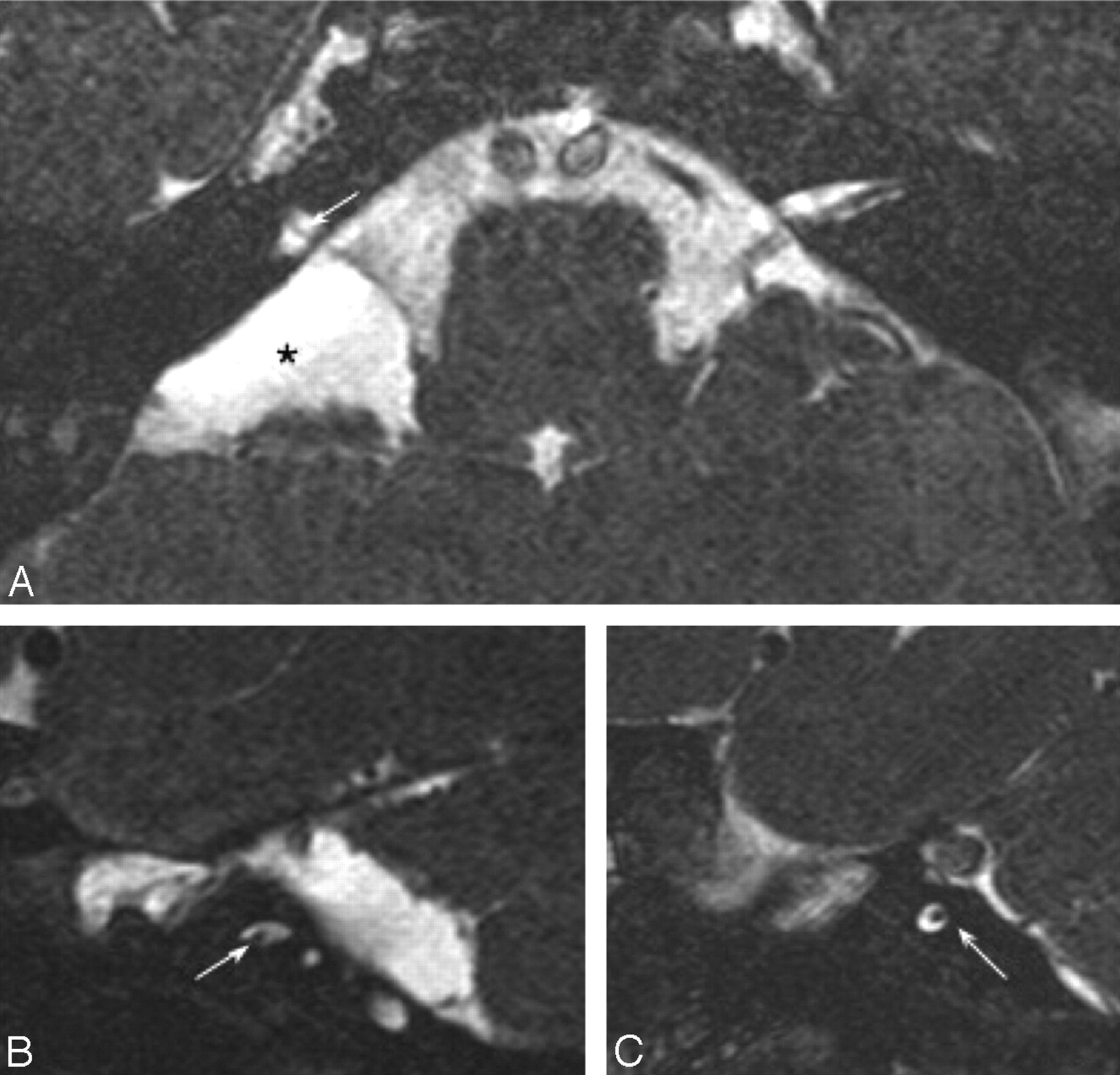
- Fig 7.
Axial (A) and bilateral sagittal oblique (B and C) 3D-CISS images of a patient with bilateral CLA demonstrates narrowing of the IACs bilaterally, containing only facial nerves (arrows). An arachnoid cyst (asterisk) is seen within the right cerebellopontine angle (A).
- Fig 8.
Axial CT scan of the temporal bones shows an aberrant facial canal with anterior displacement of the labyrinthine segment. The tympanic segment is short with a normal-appearing mastoid segment (not shown). The IAC is severely narrowed.
- Fig 9.
Axial CT image (A) and coronal reformatted image (B) demonstrate a high-riding jugular bulb (arrow), with an absent bony covering superiorly.
- Fig 10.
A, Coronal CT image shows a bony defect along the tegmen tympani with soft-tissue protrusion (arrow) into the middle ear. B, Coronal T2WI image of the same patient reveals inferior displacement of the temporal lobe (arrow) through this bony defect, consistent with a small encephalocele.


Tables
Imaging findings of the ears with labyrinthine aplasia
Patient CT/MRI CLA Otic Capsule Middle Ear Ossicle Anomaly IAC Cochlear Nerve Facial Nerve Course Jugular Anomaly Posterior Fossa Anomaly Skull Base−Other Bone Anomaly 1 +/+ R Apl Hypopl + Apl Apl Aberr + Large CPA Narrow clivus L Apl Hypopl + Apl Apl Aberr + 2 +/− L Hypopl Hypopl + Narrow ? Aberr + R CPA arachnoid cyst Low tegmen tympani, narrow clivus 3 +/+ R Apl Hypopl + Narrow Apl Aberr + – Tegmental defect (L), low tegmen tympani (R), narrow clivus L Hypopl Hypopl – Narrow Apl Aberr – 4 +/+ L Hypopl Hypopl + Narrow Apl Aberr + Split brain stem Narrow clivus 5 +/+ R Hypopl Hypopl ? Narrow Apl Aberr + Wide 4th v lateral recess Tegmental defect, narrow clivus 6 +/+ R Apl Hypopl ? Narrow Apl Aberr + Pontine malformation Encephalocele L Apl Hypopl + Apl Apl Aberr + 7 +/− R Hypopl N ? Apl ? ? + – – L Hypopl N ? Narrow ? Aberr – 8 +/+ R Hypopl Hypopl + Narrow Apl Aberr – – – 9 +/+ R Hypopl Hypopl – Narrow Apl Aberr – Chiari 1 – L Hypopl Hypopl + Narrow Apl Aberr + Note:—MRI indicates MR imaging; R, right; L, left; Apl, aplasia; Hypopl, hypoplasia; N, normal; ?, could not be evaluated in detail; Aberr, aberrant; CPA, cerebellopontine angle; 4th v, fourth ventricle; +/+, both present; +/−, MRI absent; –, not found; CLA, complete labyrinthine aplasia; IAC, internal auditory canal; +, present.


