
Article Figures & Data
Figures
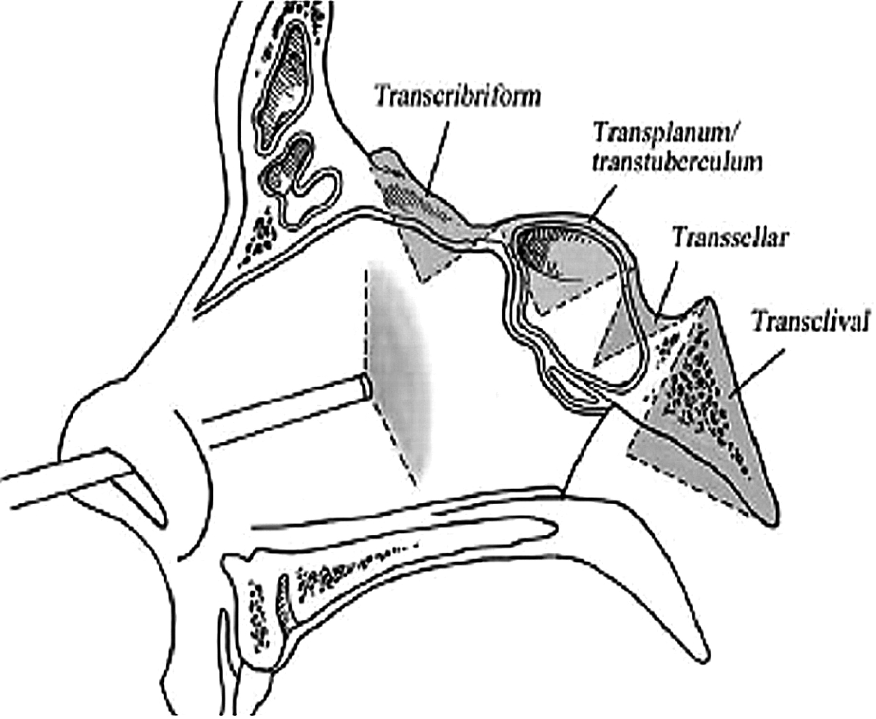
- Fig 1.
Drawing of the 4 corridors via an expanded endoscopic approach to the skull base in the sagittal plane. Reprinted with permission from the Journal of Neurosurgery Pediatric (2007:106:75–86).
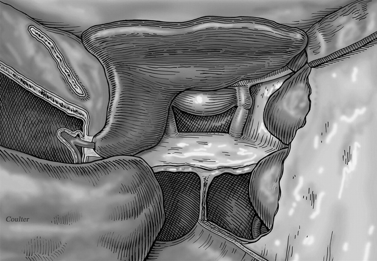
- Fig 2.
Coronal drawing of a vascular pedicled nasoseptal flap covering defect in the planum sphenoidale. The configuration is C shaped. There is an antrostomy defect (*) and the nasal septum (NS). Reprinted with permission from Neurosurgery (2008;63:ONS44-ONS53).
- Fig 3.
Sagittal drawing of the vascular pedicled nasoseptal flap covering the surgical defect with packing material and a Foley catheter balloon securing the flap in place. Reprinted with permission from Neurosurgery (2008;63:ONS44-ONS53).
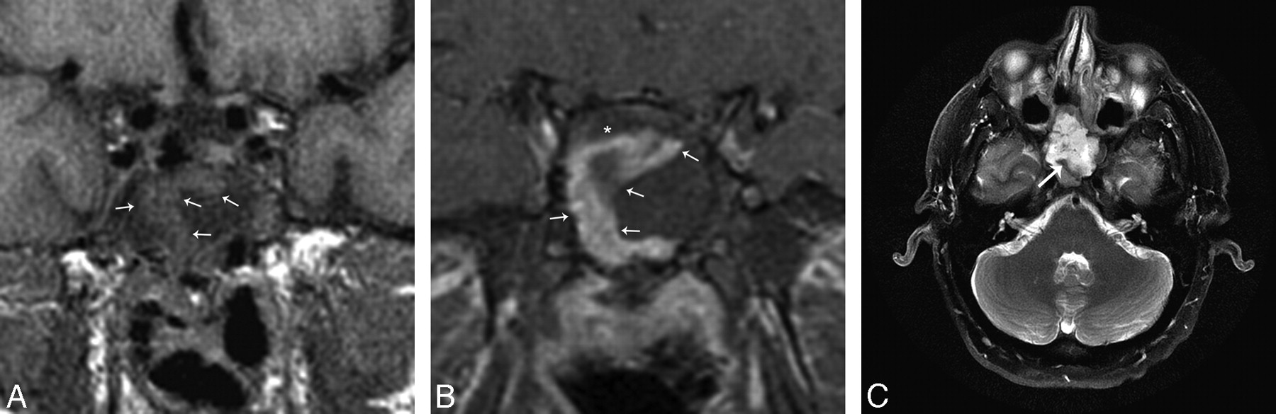
- Fig 4.
A, Immediate postoperative MR imaging. Coronal T1-weighted 3-mm precontrast image shows the nasoseptal flap (white arrows) subjacent to the surgical defect and is isointense. B, Immediate postoperative MR imaging. Coronal T1-weighted 3-mm postcontrast image with fat suppression shows a C-shaped enhancing nasoseptal flap (white arrows) underlying the surgical defect. There is linear hypointense, nonenhancing material deep to the flap, which represents the inlay and onlay graft material (*). C, Immediate postoperative MR imaging axial T2-weighted 5 mm sequence with fat suppression shows an isointense curvilinear flap (white arrow) in the surgical defect. There is slightly hyperintense material deep to the flap, which is multilayer reconstruction material (*). The T2 hyperintense material superficial to the flap is postoperative debris and fluid. This is a fat-suppressed image; as such, the hyperintense material is not fat packing.
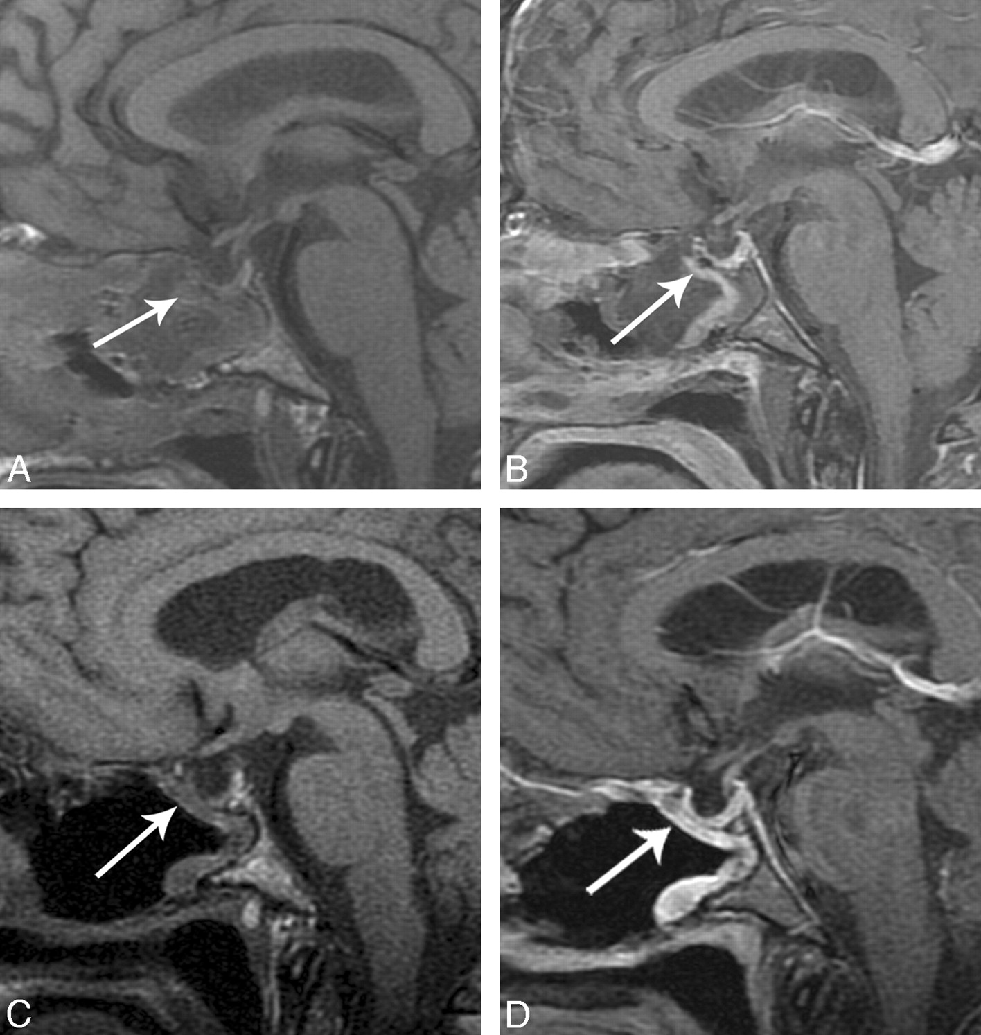
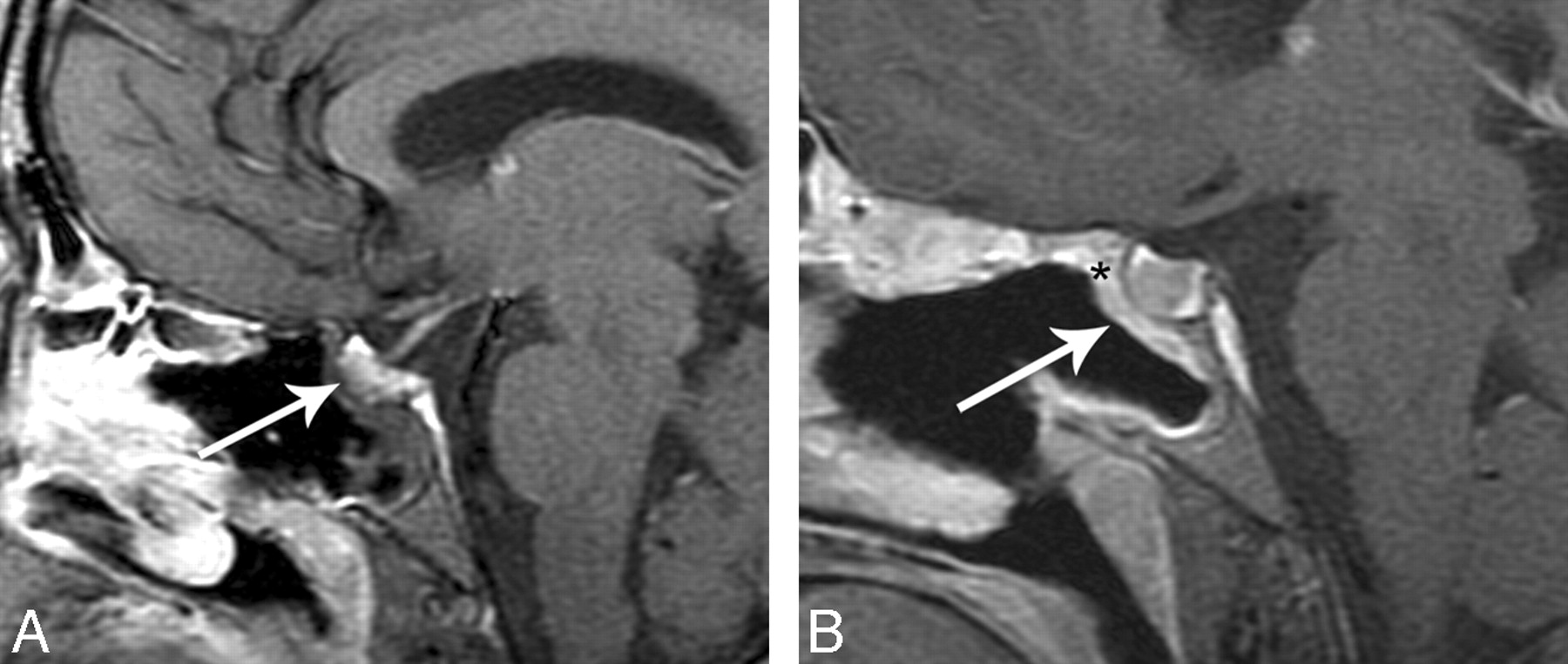
- Fig 5.
A, Immediate postoperative sagittal T1-weighted image shows a C-shaped flap underlying the operative defect (white arrow). B, Immediate postoperative sagittal T1-weighted image postcontrast with fat suppression shows a C-shaped flap underlying the operative defect (white arrow). C, Follow-up postoperative MR imaging scan sagittal unenhanced T1-weighted MR imaging shows decreased debris and removal of Foley catheter balloon in the sinonasal cavity and a C-shaped configuration of the flap, which is isointense (white arrow). D, Follow-up MR imaging sagittal T1-weighted postcontrast with fat suppression shows robust and thicker enhancement of the flap (white arrow).
- Fig 6.
This example is from another data group but illustrates how a flap may be displaced. A, Immediate postoperative sagittal T1-weighted MR imaging precontrast shows no enhancing nasoseptal flap in the expected region (white arrow). B, Immediate postoperative sagittal T1-weighted MR imaging postcontrast with fat suppression shows no enhancing C-shaped flap underlying the surgical defect (white arrowhead). There is linear soft tissue along the undersurface of the Foley balloon (small white arrow). This is presumed to represent a displaced enhancing flap. A CSF leak developed in this patient.
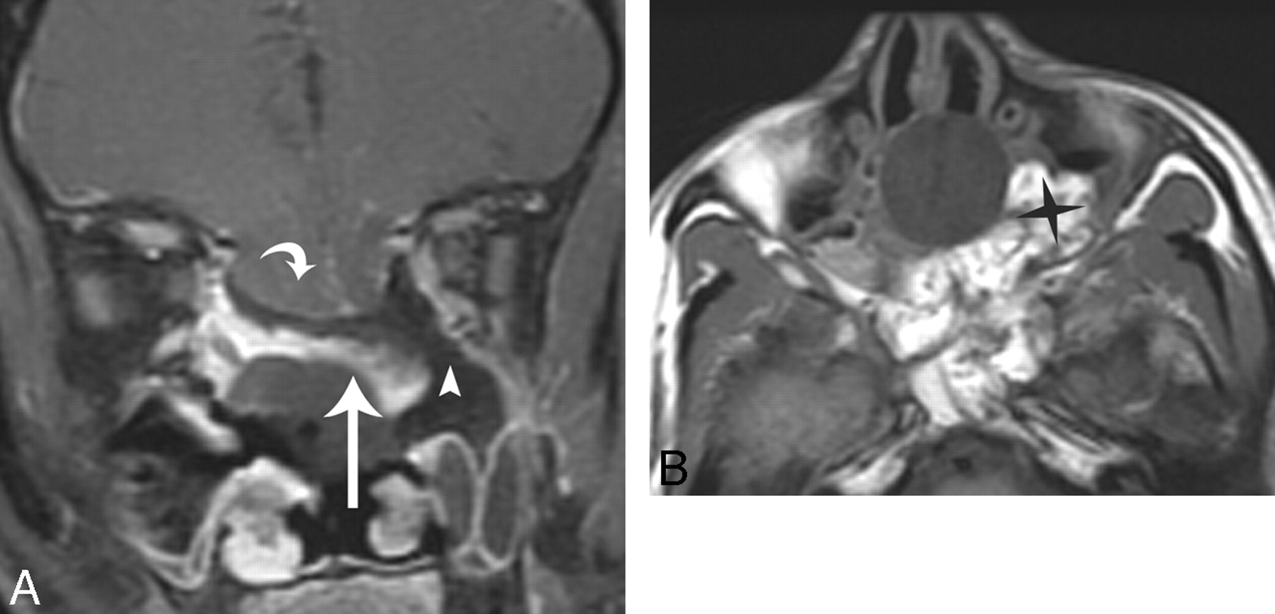
- Fig 7.
This example is from another data group but illustrates how flaps can be displaced and not adhere to the denuded sinonasal wall. A, Immediate postoperative coronal MR imaging T1-weighted postcontrast with fat suppression shows a defect (white arrowhead) left of the enhancing flap (white arrow). The flap (white arrow) is sloping inferiorly and is not in contact with the denuded sinonasal wall. A Foley catheter balloon is attempting to secure the flap. There is a large anterior skull base defect with herniation of brain parenchyma (white curved arrow). There is fat suppression making the defect (white arrowhead) look larger, which is a potential pitfall. The flap is displaced inferiorly on the left. This patient had a leak. B, Axial T1-weighted 5-mm MR imaging of no fat suppression shows the fat packing in the left defect (black star). This image demonstrates a potential pitfall when there is fat packing in overdiagnosing a defect on the fat-suppressed images.
- Fig 8.
This example is from another data group but illustrates how flaps can migrate and not adhere to the denuded sinonasal wall. A, Immediate postoperative coronal MR imaging T1-weighted postcontrast with fat suppression shows a defect (white arrowhead) left of the enhancing flap (white arrow). The flap (white arrow) is sloping inferiorly and is not in contact with the denuded sinonasal wall. A Foley catheter balloon is attempting to secure the flap. There is a large anterior skull base defect with herniation of brain parenchyma (white curved arrow). Fat suppression is making the defect (white arrowhead) look larger, which is a potential pitfall. The flap is displaced inferiorly on the left. This patient had a leak. B, Axial T1-weighted MR imaging of no fat suppression shows the fat packing in the left defect (black star). This image demonstrates a potential pitfall when there is fat packing in overdiagnosing a defect on the fat-suppressed images.


Tables
Pulse Sequence TE (ms) TR (ms) FOV (cm) Thickness (mm) Gap/Overlap (mm) Fat Suppression (Y/N) Contrast (Y/N) Precontrast Sag T1 FSE Min <600 20 3 0.5 No N Cor T1 FSE Min <600 16 3 0.5 N N Ax T2 FSE 102 3400 22 5 1 Y N Postcontrast Cor T1 FSE Min <600 16 3 0.5 Y Y Sag T1 FSE-XL Min <600 20 3 0.5 Y Y Ax 3D SPGR Min Min 25 1.5 – N Y Note:—Sag indicates sagittal; Cor, coronal; Ax, axial; FSE, fast spin-echo; N, no; Y, yes; SPGR, spoiled gradient-recalled echo.
- Table 2:
Enhancement patterns of the vascular pedicle nasoseptal flap on immediate and delayed postoperative MR images
Patient No. and Condition Flap Enhancement Immediate Postoperative MR Image (Y/N) Flap Enhancement Follow-up Postoperative MR Image (Y/N) Average Flap Thickness Immediate Postoperative MR Image (mm) Average Flap Thickness Follow-up Postoperative MR Image (mm) 1. Pituitary microadenoma Y Y 5 7 2. Pituitary apoplexy Y Y 5 3.5 3. Pituitary macroadenoma Y Y 6 3 4. Pituitary microadenoma Y Y 4 3 5. Pituitary macroadenoma with hemorrhage Y Y 3 4 6. Pituitary microadenoma Y N 2.5 – 7. Pituitary microadenoma N Y – 2 8. Pituitary macroadenoma N Y – 3 9. Pituitary microadenoma Y Y 6 4 10. Pituitary macroadenoma Y Y 4 3 Note:—N indicates no; Y, yes.


