
Article Figures & Data
Figures
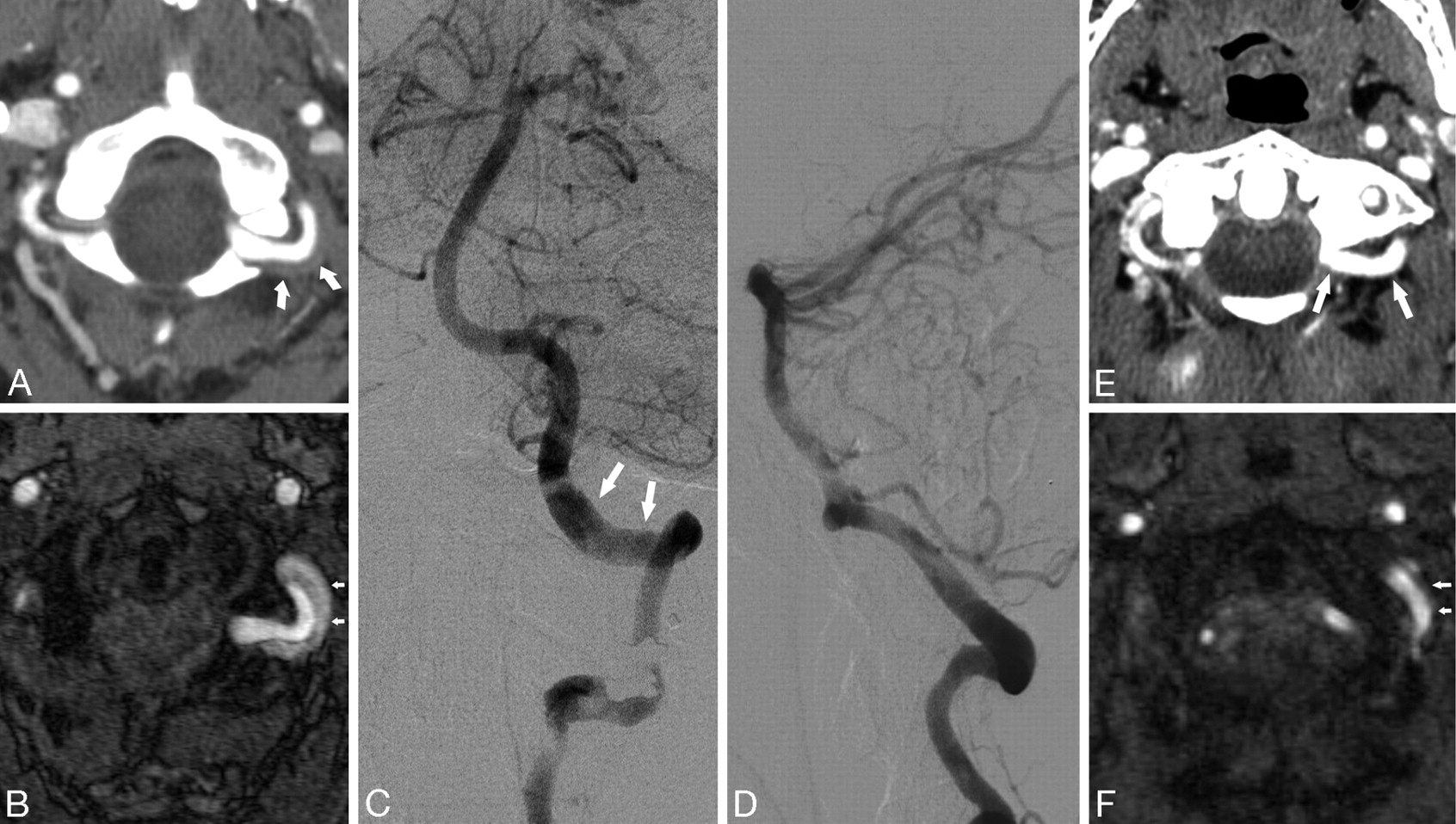
- Fig 1.
Patient 3. A 32-year-old woman who presented with headache. A, CTA demonstrates abnormal thickening of the wall of the vertebral artery, the suboccipital rind sign (arrows). B, Time-of-flight MRA source image shows subacute blood in the area of wall thickening. C and D, Anteroposterior and lateral catheter angiograms demonstrate relatively normal lumen with no significant narrowing of the vessel (arrows). E, Follow-up CTA 7 months later shows resolution, compared with A, of soft tissue around the VA and re-appearance of normal fat planes (arrows). F, Follow-up MRA shows resolution, compared with B, of the intramural hematoma (arrows).
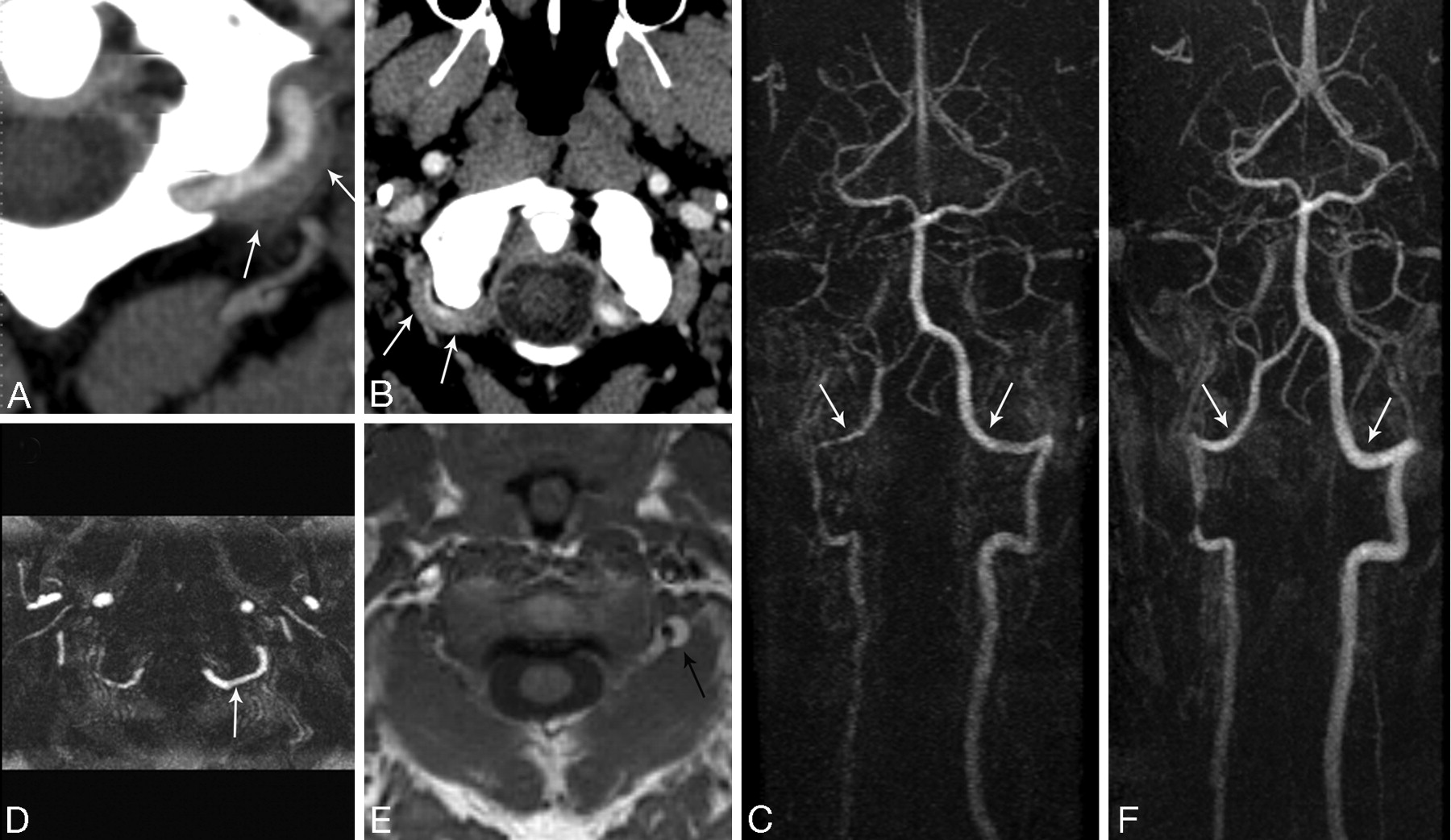
- Fig 2.
Patient 6. A 46-year-old woman with a history of cocaine abuse who presented with left neck pain, Horner syndrome, ataxia, and left facial droop. A, CTA image through the lower cervical spine level shows a hypoplastic left vertebral artery (arrow). B, CTA shows the abnormal suboccipital rind sign (arrows) and relatively normal caliber of a hypoplastic VA. C, Curved planar reformatted image demonstrates the entire length of a uniform-caliber hypoplastic left VA. D, Diffusion-weighted MR imaging shows an acute lateral medullary infarct (arrow). E, T2-weighted MR imaging image shows slow-flow or clot (arrow).
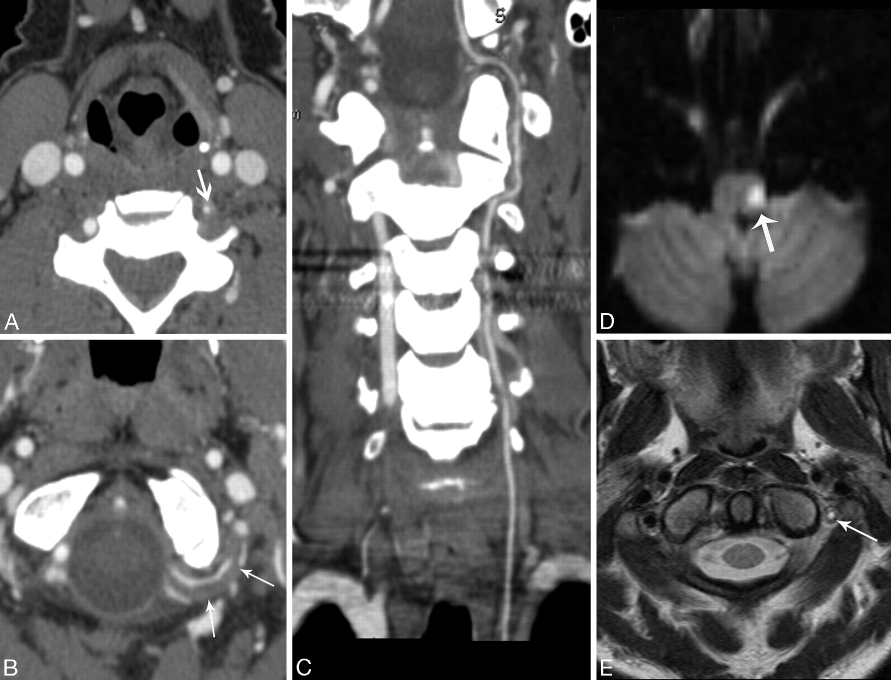
- Fig 3.
Patient 4. A 41-year-old man who presented with occipital headache. A and B, CTAs demonstrate a bilateral suboccipital rind sign (arrows). C, CEMRA demonstrates normal VA caliber in the suboccipital portion (arrows). More proximally, there is narrowing of the right VA at the C2 vertical portion. D, Axial maximum-intensity-projection image shows a normal caliber of the left vertebral artery (arrow). E, T1-weighted MR image demonstrates subacute blood within the mural hematoma (arrow). F, Follow-up CEMRA shows both vertebral arteries after resolution of the hematoma (arrows). Note the normal lumen diameter especially in the suboccipital portion. The focal narrowing at the C2 level on the right has persisted.
- Fig 4.
Diagram depicts a normal artery versus an artery with a rind sign. The lumen (x) in normal and dissected arteries is similar in caliber. The vessel wall (y) is thickened in patients with rind sign. The ratio of (x / x + y) will decrease in patients with rind sign as a result of wall thickening.
- Fig 5.
The wall thickness is plotted at 5 V3 segments. There is a difference in mean wall thickness between the patients with rind sign and controls (P < .001).


Tables
Baseline patient characteristics
Patient Sex Presenting Symptoms VAD Imaging Follow-up 1 M Trauma, neck pain Left Rind sign resolved, lumen normal 2 F Posterior circ. stroke Left Narrowing of VAs bilateral 3 F Headache, vertigo, occipital h/a Left Rind sign resolved, lumen normal 4 M Occipital headache Bilateral Rind sign resolved, lumens normal 5 M Posterior circ. stroke Right Rind sign resolved, lumen normal 6 F Horner syndrome, posterior circ. stroke, neck pain Left Occlusion LVA Note:—circ. indicates circulation; h/a, headache; LVA, left vertebral artery; VAD, vertebral artery dissection; VA, vertebral artery.
In this issue


Jump to section
Related Articles
Cited By...
- Cervical Arterial Dissections and Association With Cervical Manipulative Therapy: A Statement for Healthcare Professionals From the American Heart Association/American Stroke Association
- An atypical presentation of giant cell arteritis
- Added Value of High-Resolution MR Imaging in the Diagnosis of Vertebral Artery Dissection
- Dissection of the internal carotid artery causing Horner syndrome and palsy of cranial nerve XII