
Article Figures & Data
Figures
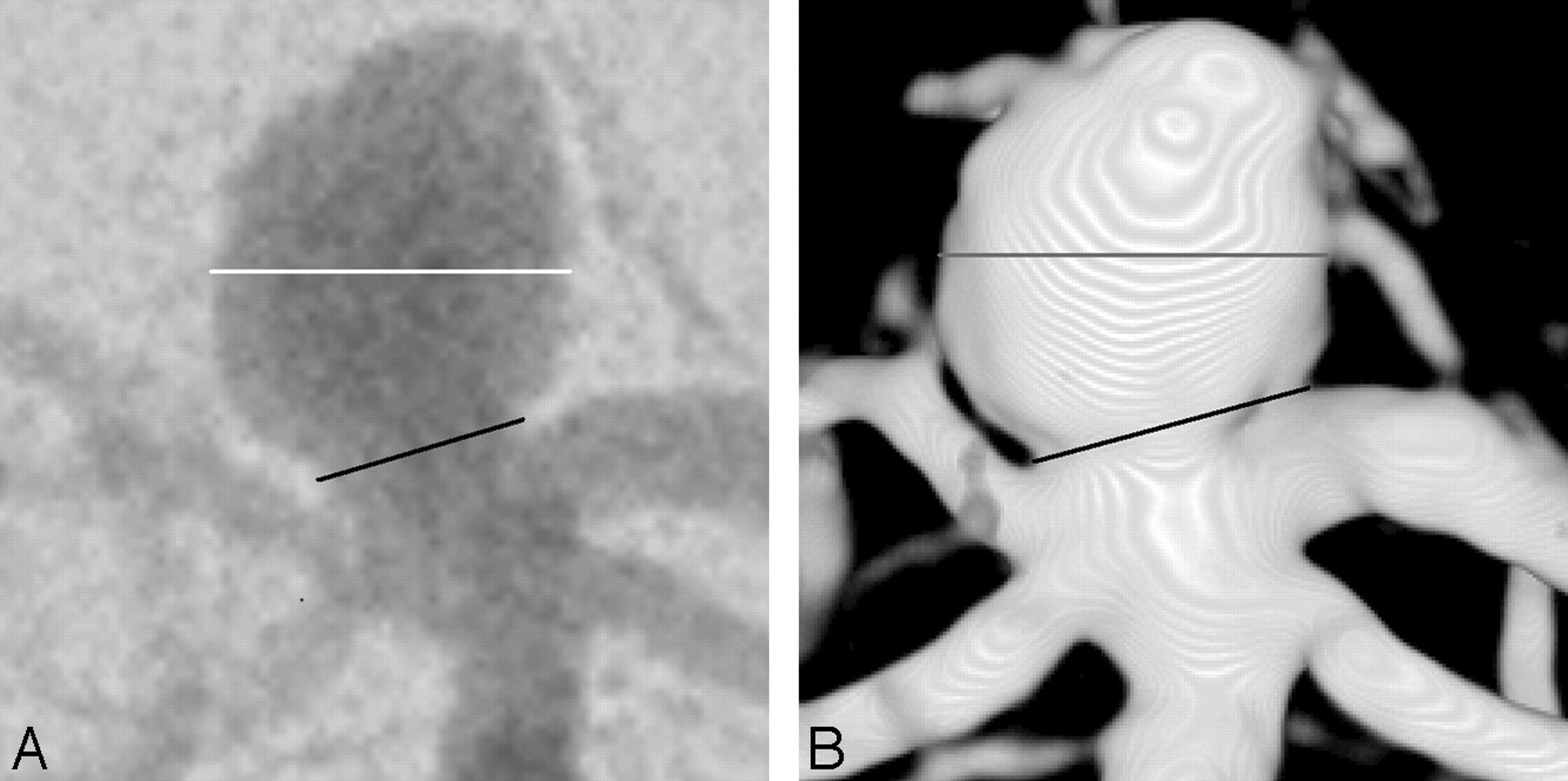
- Fig 1.
Images from a patient with a basilar tip aneurysm. A, Anteroposterior (AP) 2D DSA shows the measurement of dome diameter (white line) and neck width (black line). Dome-to-neck ratio for this 2D DSA image is 1.7. B, AP 3DRA from the same patient as in A shows dome diameter (gray line) and neck width (black line). Dome-to-neck ratio for this 3DRA image is 1.3.
- Fig 2.
Images from a patient with a superior hypophyseal aneurysm. A, Anteroposterior (AP) 2D DSA shows the measurement of dome diameter (white line) and neck width (black line). Dome-to-neck ratio for this 2D DSA image is 2.0. B, AP 3DRA from the same patient as in A shows dome diameter (gray line) and neck width (black line). Dome-to-neck ratio for this 3DRA image is 1.7.
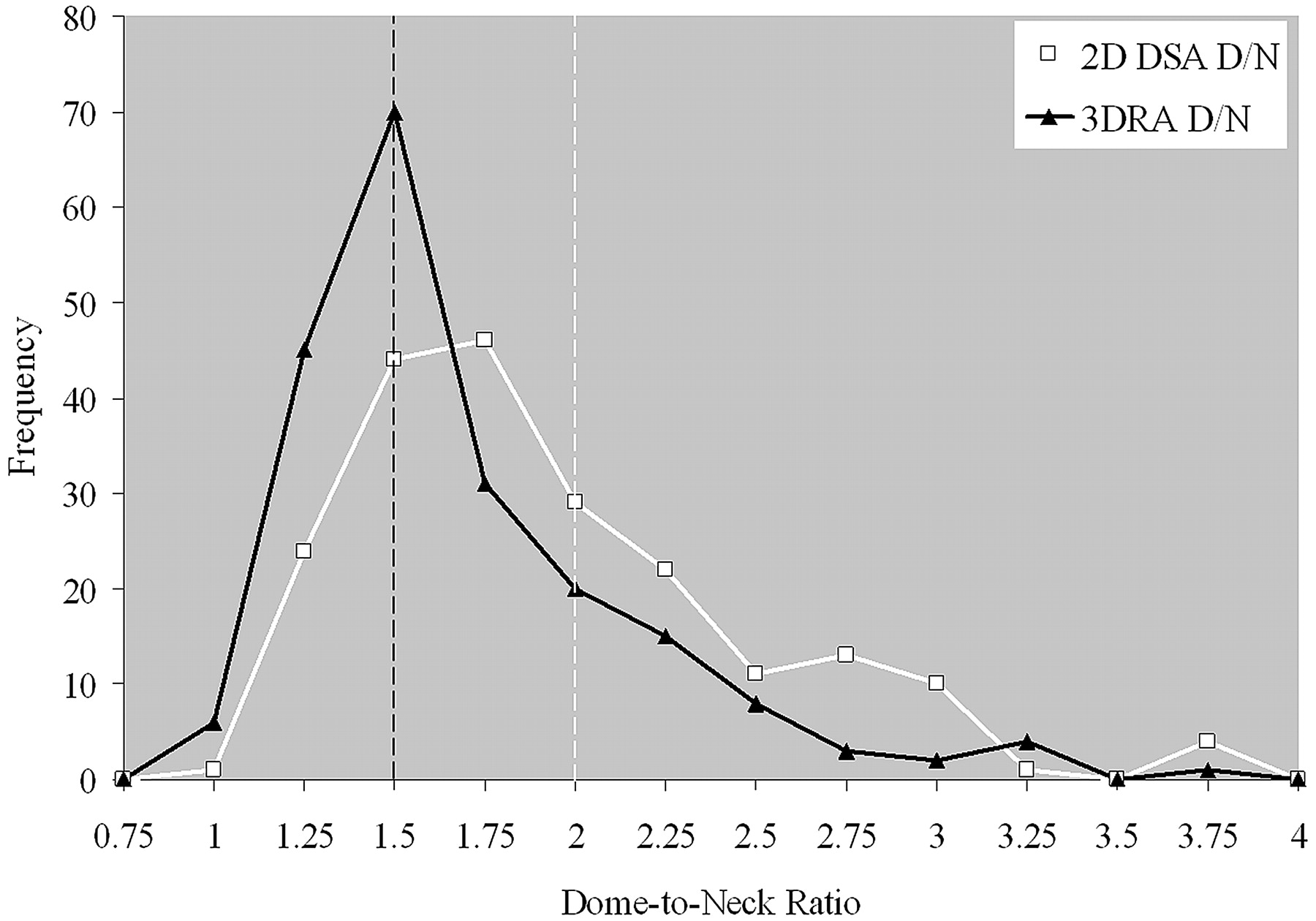
- Fig 3.
Comparison of the distribution of dome-to-neck ratios in 2D DSA and 3DRA. Histogram shows the frequency of aneurysms with various dome-to-neck ratios. Frequencies are calculated for dome-to-neck intervals of 0.25; for example, ratios from 2.0 to 2.25 are grouped into a single frequency number. The black line denotes 3DRA measurements, whereas the white line denotes 2D DSA measurements. Vertical dashed lines indicate previously published thresholds for wide-neck aneurysms (black line, 1.5; white line, 2.0). Thus, aneurysms to the left of a vertical line would be considered wide-neck.


Tables
Dome-to-neck ratios in 2D DSA versus 3DRA
2D DSA 3DRA P Value Mean D/N (SD) 1.81 (0.55) 1.56 (0.48) <.0001* D/N <1.5, No. (%) 69 (33.7) 119 (58.0) <.0001† D/N <2.0, No. (%) 142 (69.3) 173 (84.4) .0004† Note:—D/N indicates dome-to-neck ratio; DSA, digital subtraction angiography; 3DRA, 3D rotational angiography.
* Paired t test, Wilcoxon signed ranked test.
† Pearson χ2 test.
In this issue


Jump to section
Related Articles
Cited By...
- Improving visualization of three-dimensional aneurysm features via segmentation with upsampled resolution and gradient enhancement (SURGE)
- Does the DSA reconstruction kernel affect hemodynamic predictions in intracranial aneurysms? An analysis of geometry and blood flow variations
- Diagnostic Impact of Bone-Subtraction CT Angiography for Patients with Acute Subarachnoid Hemorrhage
- Interobserver variability of aneurysm morphology: discrimination of the daughter sac
- Mind the Gap: Impact of Computational Fluid Dynamics Solution Strategy on Prediction of Intracranial Aneurysm Hemodynamics and Rupture Status Indicators
- A carving method to determine an optimal working projection using three-dimensional volume rendering digital subtraction angiography in coil embolization of cerebral aneurysms
- Creation of Bifurcation-Type Elastase-Induced Aneurysms in Rabbits
- Intracranial Aneurysm Neck Size Overestimation with 3D Rotational Angiography: The Impact on Intra-Aneurysmal Hemodynamics Simulated with Computational Fluid Dynamics
- Aneurysm Ostium Angle: A Predictor of the Need for Stent as Assistance for Endovascular Aneurysm Coiling in Internal Carotid Artery Sidewall Aneurysms
- Reply:
- Identifying "Truth" in Computational Fluid Dynamics Research
- Difficult Aneurysms for Endovascular Treatment: Overwide or Undertall?