Advertisement

Main menu

  • Home
  • Content
    • Current Issue
    • Accepted Manuscripts
    • Article Preview
    • Past Issue Archive
    • AJNR Case Collection
    • Case of the Week Archive
    • Classic Case Archive
    • Case of the Month Archive
  • Special Collections
    • Spinal CSF Leak Articles (Jan 2020-June 2024)
    • 2024 AJNR Journal Awards
    • Most Impactful AJNR Articles
  • Multimedia
    • AJNR Podcast
    • AJNR Scantastics
    • Video Articles
  • For Authors
    • Submit a Manuscript
    • Author Policies
    • Fast publishing of Accepted Manuscripts
    • Graphical Abstract Preparation
    • Manuscript Submission Guidelines
    • Imaging Protocol Submission
    • Submit a Case for the Case Collection
  • About Us
    • About AJNR
    • Editorial Board
  • More
    • Become a Reviewer/Academy of Reviewers
    • Subscribers
    • Permissions
    • Alerts
    • Feedback
    • Advertisers
    • ASNR Home
  • Other Publications
    • ajnr

User menu

  • Alerts
  • Log in

Search

  • Advanced search
American Journal of Neuroradiology

American Journal of Neuroradiology

ASHNR American Society of Functional Neuroradiology ASHNR American Society of Pediatric Neuroradiology ASSR
  • Alerts
  • Log in

Advanced Search

  • Home
  • Content
    • Current Issue
    • Accepted Manuscripts
    • Article Preview
    • Past Issue Archive
    • AJNR Case Collection
    • Case of the Week Archive
    • Classic Case Archive
    • Case of the Month Archive
  • Special Collections
    • Spinal CSF Leak Articles (Jan 2020-June 2024)
    • 2024 AJNR Journal Awards
    • Most Impactful AJNR Articles
  • Multimedia
    • AJNR Podcast
    • AJNR Scantastics
    • Video Articles
  • For Authors
    • Submit a Manuscript
    • Author Policies
    • Fast publishing of Accepted Manuscripts
    • Graphical Abstract Preparation
    • Manuscript Submission Guidelines
    • Imaging Protocol Submission
    • Submit a Case for the Case Collection
  • About Us
    • About AJNR
    • Editorial Board
  • More
    • Become a Reviewer/Academy of Reviewers
    • Subscribers
    • Permissions
    • Alerts
    • Feedback
    • Advertisers
    • ASNR Home
  • Follow AJNR on Twitter
  • Visit AJNR on Facebook
  • Follow AJNR on Instagram
  • Join AJNR on LinkedIn
  • RSS Feeds

Welcome to the new AJNR, Updated Hall of Fame, and more. Read the full announcements.


AJNR is seeking candidates for the position of Associate Section Editor, AJNR Case Collection. Read the full announcement.


OtherPEDIATRICS

Salivary Gland Anlage Tumor in a Neonate Presenting with Respiratory Distress: Radiographic and Pathologic Correlation

M.A. Mogensen, A.C. Lin, K.W. Chang, G.J. Berry, P.D. Barnes and N.J. Fischbein
American Journal of Neuroradiology May 2009, 30 (5) 1022-1023; DOI: https://doi.org/10.3174/ajnr.A1364
M.A. Mogensen
  • Find this author on Google Scholar
  • Find this author on PubMed
  • Search for this author on this site
A.C. Lin
  • Find this author on Google Scholar
  • Find this author on PubMed
  • Search for this author on this site
K.W. Chang
  • Find this author on Google Scholar
  • Find this author on PubMed
  • Search for this author on this site
G.J. Berry
  • Find this author on Google Scholar
  • Find this author on PubMed
  • Search for this author on this site
P.D. Barnes
  • Find this author on Google Scholar
  • Find this author on PubMed
  • Search for this author on this site
N.J. Fischbein
  • Find this author on Google Scholar
  • Find this author on PubMed
  • Search for this author on this site
  • Article
  • Figures & Data
  • Info & Metrics
  • Responses
  • References
  • PDF
Loading

Abstract

SUMMARY: We present a case of congenital salivary gland anlage tumor (SGAT) of the nasal septum in a 2-week-old infant who had difficulty breathing through her nose since birth. CT and MR imaging demonstrated a circumscribed mass within the nasal cavity that did not communicate with the intracranial compartment. Differential diagnosis and clinical significance of recognizing this rare lesion are reviewed.

Nasal airway obstruction in the neonatal period is a potentially life-threatening condition that can be caused by a wide range of entities including congenital malformations, inflammatory conditions, hamartomas, and tumors. If a nasal mass is identified, then CT and MR imaging is often required to further characterize the mass and assess for intracranial involvement before intervention. Congenital salivary gland anlage tumor (SGAT) is a rare, benign lesion of the nasopharynx that presents with respiratory distress in the first days or weeks of life. Although this rare entity has been reported in the surgical and pediatric literature, its imaging features have not been reported in detail.

Case Report

A 2-week-old infant had trouble breathing through her nose since birth. The patient's pediatrician noted some bleeding when passing a catheter through the left naris, and the patient was referred to a pediatric otolaryngologist. A nasal endoscopy revealed a friable mass filling the left nasal cavity at the level of the middle turbinate. The right nasal cavity was narrowed because of a severe right septal deviation.

CT without contrast enhancement showed a soft tissue attenuation mass centered within the left nasal cavity with benign-appearing bony remodeling of the ethmoid labyrinth. A connection between the mass and the intracranial contents could not be excluded because of normal lack of ossification of the skull base at this age. An MR imaging examination was then performed and demonstrated a well-defined mass filling the posterior left nasal cavity that was T1 isointense and T2 iso-hyperintense to the brain parenchyma, with an intact skull base and no evidence of intracranial communication (Fig 1A–C).

Fig 1.
  • Download figure
  • Open in new tab
  • Download powerpoint
Fig 1.

Coronal (A) and sagittal (B) T2-weighted and sagittal T1-weighted (C) MR images show a well-circumscribed mass (white arrows) in the left nasal cavity and ethmoid labyrinth that is T1 isointense and heterogeneously T2 iso-hyperintense in relationship to brain parenchyma. The intact skull base is clearly visualized with no intracranial connection (black arrowhead).

The patient underwent CT-guided endoscopic resection of the mass, which was found to be pedicled to an approximately 4-mm area of the nasal septum. The choana and nasopharynx were clear.

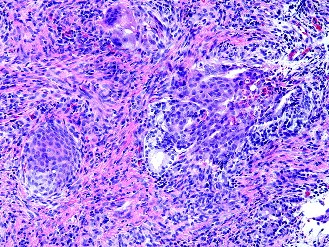
The fragmented tissue measured 3.0 × 2.3 × 0.8 cm in aggregate. Microscopic examination revealed an overlying nonkeratinized squamous cell lining with foci of mucosal erosions. Nests of epithelial cells arranged as cords, and ductal and solid structures extended from the mucosal layer into an edematous submucosa. The ductlike structures contained foci of squamous differentiation surrounded by round and spindled stromal cells (Fig 2). Cellular pleomorphism and mitotic figures were absent. No neural, glial, or rhabdomyomatous differentiation was present. These findings were compatible with an SGAT.

Fig 2.
  • Download figure
  • Open in new tab
  • Download powerpoint
Fig 2.

The pathologic features of SGAT: the biphasic nature of this hamartomatous lesion is demonstrated by the admixture of epithelial groups of ductlike and squamous structures and bland stromal cells (hematoxylin-eosin × 200).

Discussion

The term SGAT was first used in the literature by Dehner et al1 in 1994. They reported 9 cases of SGATs within the nasopharynx, all of which were midline and were attached to the posterior pharyngeal wall by a delicate pedicle. The tumors had a biphasic histologic pattern of squamous nests and ductlike structures along the periphery with solid, predominantly mesenchymal-appearing nodules centrally. Given the histologic similarity to the developing salivary gland, it was proposed that the SGAT is probably a hamartoma of minor salivary gland origin rather than a true neoplasm. Before this study, other similar lesions likely representing SGATs of the nasopharynx were discussed in the English-language literature, including a report in 1980 of a squamous cell proliferative lesion2 and a report in 1985 of a congenital pleomorphic adenoma.3

Congenital SGAT has a male predilection and is a rare cause of neonatal airway obstruction, with approximately 23 previous cases reported in the literature.4 The mass is midline and is typically attached to the posterior septum or the posterior nasopharyngeal wall by a thin pedicle. Excellent results have been obtained with simple excision, with no reported local recurrences. Two tumors were expelled during nasal manipulation (one during resuscitation and the other with nasal cannulation).1,5

Studies with CT and MR imaging are essential to evaluate anatomy, further define the characteristics and size of the mass, and assess for intracranial extension before surgical intervention. On CT examination, the homogeneous appearance of the mass with absence of bony destruction suggested a benign cause. Because of lack of ossification of the skull base, an MR imaging study was needed to assess for a nasoethmoidal encephalocele, which contains brain tissue of varied signal intensity on T2-weighted imaging.6 The intact skull base was clearly visualized on MR imaging. Although gadolinium contrast was not given in this case because of the absence of intravenous access, 1 previous case in the literature reported heterogeneous gadolinium enhancement in a SGAT.4 On the basis of the imaging appearance, the main consideration was a nasal glioma, a more common lesion that consists of disorganized, trapped brain tissue with no intracranial connection. As in the SGAT in our patient, nasal gliomas are typically T1 isointense and T2 iso-hyperintense to brain parenchyma.6

The causes of nasal or nasopharyngeal obstruction, unilateral or bilateral, in the neonate include developmental abnormalities such as nasal cavity or choanal stenosis or atresia.6 Masses in the nasal cavity and nasopharynx are rare in the neonate but are an important consideration in the setting of respiratory distress. The most common developmental masses are basal cephalocele, nasal glioma (neuroglial heterotopia), and nasolacrimal mucocele (dacryocystocele). Most neoplastic lesions involving the neonatal nasal and nasopharyngeal airway are benign such as dermoid, hamartoma, teratoma (epignathus), and hemangioma. Primary and secondary malignant neoplasms of this region are mainly seen in older infants and children and are extremely rare in the neonate. These lesions include neuroblastoma, Langerhans cell histiocytosis, chloroma, rhabdomyosarcoma, and lymphoma.

Congenital SGAT is usually not suggested as a possible cause in the setting of nasal obstruction from a mass in the neonatal period because it is uncommon. However, if this lesion is considered in the differential diagnosis on the basis of imaging studies, it is important to convey this to the referring physician because these lesions are attached by only a thin, delicate vascular pedicle that may be easily torn with gentle probing or nasal cannulation, resulting in dislodgement with possible unexpected complete airway obstruction.

References

  1. ↵
    Dehner LP, Valbuena L, Perez-Atayde A, et al. Salivary gland anlage tumor (“congenital pleomorphic adenoma”). A clinicopathologic, immunohistochemical and ultrastructural study of nine cases. Am J Surg Pathol 1994;18:25–36
    PubMed
  2. ↵
    Stillwater LB, Fee WE Jr. Squamous cell proliferative lesion of the nasopharynx in a newborn. Otolaryngol Head Neck Surg 1980;88:240–47
    PubMed
  3. ↵
    Har-El G, Zirkin HY, Tovi F, et al. Congenital pleomorphic adenoma of the nasopharynx (report of a case). J Laryngol Otol 1985;99:1281–87
    PubMed
  4. ↵
    Herrmann BW, Dehner LP, Lieu JE. Congenital salivary gland anlage tumor: a case series and review of the literature. Int J of Pediatr Otorhinolaryngol 2005;69:149–56
    CrossRef
  5. ↵
    Cohen EG, Yoder M, Thomas RM, et al. Congenital salivary gland anlage tumor of the nasopharynx. Pediatrics 2003;112:e66–e69
    Abstract/FREE Full Text
  6. ↵
    Valencia MP, Castillo M. Congenital and acquired lesions of the nasal septum: a practical guide for differential diagnosis. Radiographics 2008;28:205–24
    CrossRefPubMed
  • Received August 25, 2008.
  • Accepted after revision September 12, 2008.
  • Copyright © American Society of Neuroradiology
View Abstract
PreviousNext
Back to top

In this issue

American Journal of Neuroradiology: 30 (5)
American Journal of Neuroradiology
Vol. 30, Issue 5
May 2009
  • Table of Contents
  • Index by author
Advertisement
Print
Download PDF
Email Article

Thank you for your interest in spreading the word on American Journal of Neuroradiology.

NOTE: We only request your email address so that the person you are recommending the page to knows that you wanted them to see it, and that it is not junk mail. We do not capture any email address.

Enter multiple addresses on separate lines or separate them with commas.
Salivary Gland Anlage Tumor in a Neonate Presenting with Respiratory Distress: Radiographic and Pathologic Correlation
(Your Name) has sent you a message from American Journal of Neuroradiology
(Your Name) thought you would like to see the American Journal of Neuroradiology web site.
CAPTCHA
This question is for testing whether or not you are a human visitor and to prevent automated spam submissions.
Cite this article
M.A. Mogensen, A.C. Lin, K.W. Chang, G.J. Berry, P.D. Barnes, N.J. Fischbein
Salivary Gland Anlage Tumor in a Neonate Presenting with Respiratory Distress: Radiographic and Pathologic Correlation
American Journal of Neuroradiology May 2009, 30 (5) 1022-1023; DOI: 10.3174/ajnr.A1364

Citation Manager Formats

  • BibTeX
  • Bookends
  • EasyBib
  • EndNote (tagged)
  • EndNote 8 (xml)
  • Medlars
  • Mendeley
  • Papers
  • RefWorks Tagged
  • Ref Manager
  • RIS
  • Zotero
0 Responses
Respond to this article
Share
Bookmark this article
Salivary Gland Anlage Tumor in a Neonate Presenting with Respiratory Distress: Radiographic and Pathologic Correlation
M.A. Mogensen, A.C. Lin, K.W. Chang, G.J. Berry, P.D. Barnes, N.J. Fischbein
American Journal of Neuroradiology May 2009, 30 (5) 1022-1023; DOI: 10.3174/ajnr.A1364
del.icio.us logo Twitter logo Facebook logo Mendeley logo
  • Tweet Widget
  • Facebook Like
  • Google Plus One
Purchase

Jump to section

  • Article
    • Abstract
    • Case Report
    • Discussion
    • References
  • Figures & Data
  • Info & Metrics
  • Responses
  • References
  • PDF

Related Articles

  • No related articles found.
  • PubMed
  • Google Scholar

Cited By...

  • No citing articles found.
  • Crossref (11)
  • Google Scholar

This article has been cited by the following articles in journals that are participating in Crossref Cited-by Linking.

  • Salivary Gland Anlage Tumor: A Clinicopathological Study of Two Cases
    Guillaume Gauchotte, Laurent Coffinet, Emmanuelle Schmitt, Aude Bressenot, Virginie Hennequin, Jacqueline Champigneulle, Jean-Michel Vignaud
    Fetal and Pediatric Pathology 2011 30 2
  • Congenital salivary gland anlage tumor — in utero and postnatal imaging
    Rupa Radhakrishnan, Maria A. Calvo-Garcia, Foong-Yen Lim, Ravindhra G. Elluru, Bernadette L. Koch
    Pediatric Radiology 2015 45 3
  • Salivary gland anlage tumor: molecular profiling sheds light on a morphologic question
    Scott M. Peters, Andrew T. Turk
    Oral Surgery, Oral Medicine, Oral Pathology and Oral Radiology 2019 127 5
  • Newborn with congenital salivary gland anlage tumor presenting with respiratory distress
    Anil Kumar Swayampakula, Mariam Ischander, Craig W. Zuppan, Miguel Krishnan, Karen Tong, Sonea Qureshi
    International Journal of Pediatric Otorhinolaryngology Extra 2015 10 3
  • Pediatric Head and Neck Textbook
    Timothy J. Martin, Stephen F. Conley
    2021
  • Salivary anlage tumor of the nasopharynx: A case report and review of the literature
    Marcelo B. Antunes, Luv Ram Javia, Evan R. Ransom, Ken Kazahaya
    International Journal of Pediatric Otorhinolaryngology Extra 2011 6 2
  • Salivary gland anlage tumor: Evaluation and management of a rare pediatric condition
    Jessica Trac, Brigitte Routhier-Chevrier, Haiying Chen, Evan J. Propst, Nikolaus E. Wolter
    International Journal of Pediatric Otorhinolaryngology 2022 163
  • Atlas of Head and Neck Pathology
    Bruce M. Wenig
    2016
  • Diagnostic Pathology: Pediatric Neoplasms
    2018
  • Salivary gland anlage tumor
    Anuj Khurana, Saranya Singaravel, Urmi Mukherjee
    Indian Journal of Pathology and Microbiology 2016 59 3

More in this TOC Section

  • Comparison of Image Quality and Radiation Dose in Pediatric Temporal Bone CT Using Photon-Counting Detector CT and Energy-Integrating Detector CT
  • SyMRI & MR Fingerprinting in Brainstem Myelination
  • Pons&Vermis Localization on Fetal MRI Using U-Net
Show more Pediatrics

Similar Articles

Advertisement

Indexed Content

  • Current Issue
  • Accepted Manuscripts
  • Article Preview
  • Past Issues
  • Editorials
  • Editors Choice
  • Fellow Journal Club
  • Letters to the Editor

Cases

  • Case Collection
  • Archive - Case of the Week
  • Archive - Case of the Month
  • Archive - Classic Case

Special Collections

  • Special Collections

Resources

  • News and Updates
  • Turn around Times
  • Submit a Manuscript
  • Author Policies
  • Manuscript Submission Guidelines
  • Evidence-Based Medicine Level Guide
  • Publishing Checklists
  • Graphical Abstract Preparation
  • Imaging Protocol Submission
  • Submit a Case
  • Become a Reviewer/Academy of Reviewers
  • Get Peer Review Credit from Publons

Multimedia

  • AJNR Podcast
  • AJNR SCANtastic
  • Video Articles

About Us

  • About AJNR
  • Editorial Board
  • Not an AJNR Subscriber? Join Now
  • Alerts
  • Feedback
  • Advertise with us
  • Librarian Resources
  • Permissions
  • Terms and Conditions

American Society of Neuroradiology

  • Not an ASNR Member? Join Now

© 2025 by the American Society of Neuroradiology All rights, including for text and data mining, AI training, and similar technologies, are reserved.
Print ISSN: 0195-6108 Online ISSN: 1936-959X

Powered by HighWire