Advertisement

Main menu

  • Home
  • Content
    • Current Issue
    • Accepted Manuscripts
    • Article Preview
    • Past Issue Archive
    • AJNR Case Collection
    • Case of the Week Archive
    • Classic Case Archive
    • Case of the Month Archive
  • Special Collections
    • Spinal CSF Leak Articles (Jan 2020-June 2024)
    • 2024 AJNR Journal Awards
    • Most Impactful AJNR Articles
  • Multimedia
    • AJNR Podcast
    • AJNR Scantastics
    • Video Articles
  • For Authors
    • Submit a Manuscript
    • Author Policies
    • Fast publishing of Accepted Manuscripts
    • Graphical Abstract Preparation
    • Manuscript Submission Guidelines
    • Imaging Protocol Submission
    • Submit a Case for the Case Collection
  • About Us
    • About AJNR
    • Editorial Board
  • More
    • Become a Reviewer/Academy of Reviewers
    • Subscribers
    • Permissions
    • Alerts
    • Feedback
    • Advertisers
    • ASNR Home
  • Other Publications
    • ajnr

User menu

  • Alerts
  • Log in

Search

  • Advanced search
American Journal of Neuroradiology

American Journal of Neuroradiology

ASHNR American Society of Functional Neuroradiology ASHNR American Society of Pediatric Neuroradiology ASSR
  • Alerts
  • Log in

Advanced Search

  • Home
  • Content
    • Current Issue
    • Accepted Manuscripts
    • Article Preview
    • Past Issue Archive
    • AJNR Case Collection
    • Case of the Week Archive
    • Classic Case Archive
    • Case of the Month Archive
  • Special Collections
    • Spinal CSF Leak Articles (Jan 2020-June 2024)
    • 2024 AJNR Journal Awards
    • Most Impactful AJNR Articles
  • Multimedia
    • AJNR Podcast
    • AJNR Scantastics
    • Video Articles
  • For Authors
    • Submit a Manuscript
    • Author Policies
    • Fast publishing of Accepted Manuscripts
    • Graphical Abstract Preparation
    • Manuscript Submission Guidelines
    • Imaging Protocol Submission
    • Submit a Case for the Case Collection
  • About Us
    • About AJNR
    • Editorial Board
  • More
    • Become a Reviewer/Academy of Reviewers
    • Subscribers
    • Permissions
    • Alerts
    • Feedback
    • Advertisers
    • ASNR Home
  • Follow AJNR on Twitter
  • Visit AJNR on Facebook
  • Follow AJNR on Instagram
  • Join AJNR on LinkedIn
  • RSS Feeds

Welcome to the new AJNR, Updated Hall of Fame, and more. Read the full announcements.


AJNR is seeking candidates for the position of Associate Section Editor, AJNR Case Collection. Read the full announcement.


OtherHEAD & NECK

Pulsatile Tinnitus from Reversal of Flow in an Aberrant Occipital Artery: Resolved after Carotid Artery Stenting

P.O. Cowley, R. Jones, P. Tuch and W. McAuliffe
American Journal of Neuroradiology May 2009, 30 (5) 995-997; DOI: https://doi.org/10.3174/ajnr.A1393
P.O. Cowley
  • Find this author on Google Scholar
  • Find this author on PubMed
  • Search for this author on this site
R. Jones
  • Find this author on Google Scholar
  • Find this author on PubMed
  • Search for this author on this site
P. Tuch
  • Find this author on Google Scholar
  • Find this author on PubMed
  • Search for this author on this site
W. McAuliffe
  • Find this author on Google Scholar
  • Find this author on PubMed
  • Search for this author on this site
  • Article
  • Figures & Data
  • Info & Metrics
  • Responses
  • References
  • PDF
Loading

Abstract

SUMMARY: Carotid artery stent placement, performed for correction of an asymptomatic severe stenosis, leads to the resolution of persistent and troublesome pulsatile tinnitus. Tinnitus has been reported as a consequence of severe carotid stenoses on previous occasions. This case highlights how an aberrant occipital artery originating above a carotid artery stenosis can result in flow reversal and be a mechanism by which tinnitus may develop.

This case demonstrates that reversal of flow in the occipital artery may be an important cause of pulsatile tinnitus. Flow reversal in an aberrant occipital artery secondary to a carotid stenosis was rectified, leading to complete cessation of subjective and objective tinnitus.

Case Report

A 52-year-old woman was found to have a greater than 95% stenosis of the right common carotid artery on sonography and on 2D time-of-flight MR angiography. The patient elected to have internal carotid artery (ICA) stent placement for correction of the severe stenosis. At no time did she have an ischemic event in any territory.

Of note, she also complained of persistent right-sided pulsatile tinnitus. There was a prominent and extensive bruit radiating from the neck up to the angle of the jaw, higher up to the tragus of the ear, and around the temporomandibular joint on the right side. It was not audible anyplace else on the skull. Compression of the internal jugular vein had no effect, excluding a venous hum. She had a cholesteatoma removed on that side in 1975, on a background of frequent otitis media. Since then, there had been frequent fungal infections in that area until 4 years before presentation. The relationship between the tinnitus and the stenosis was not clinically certain.

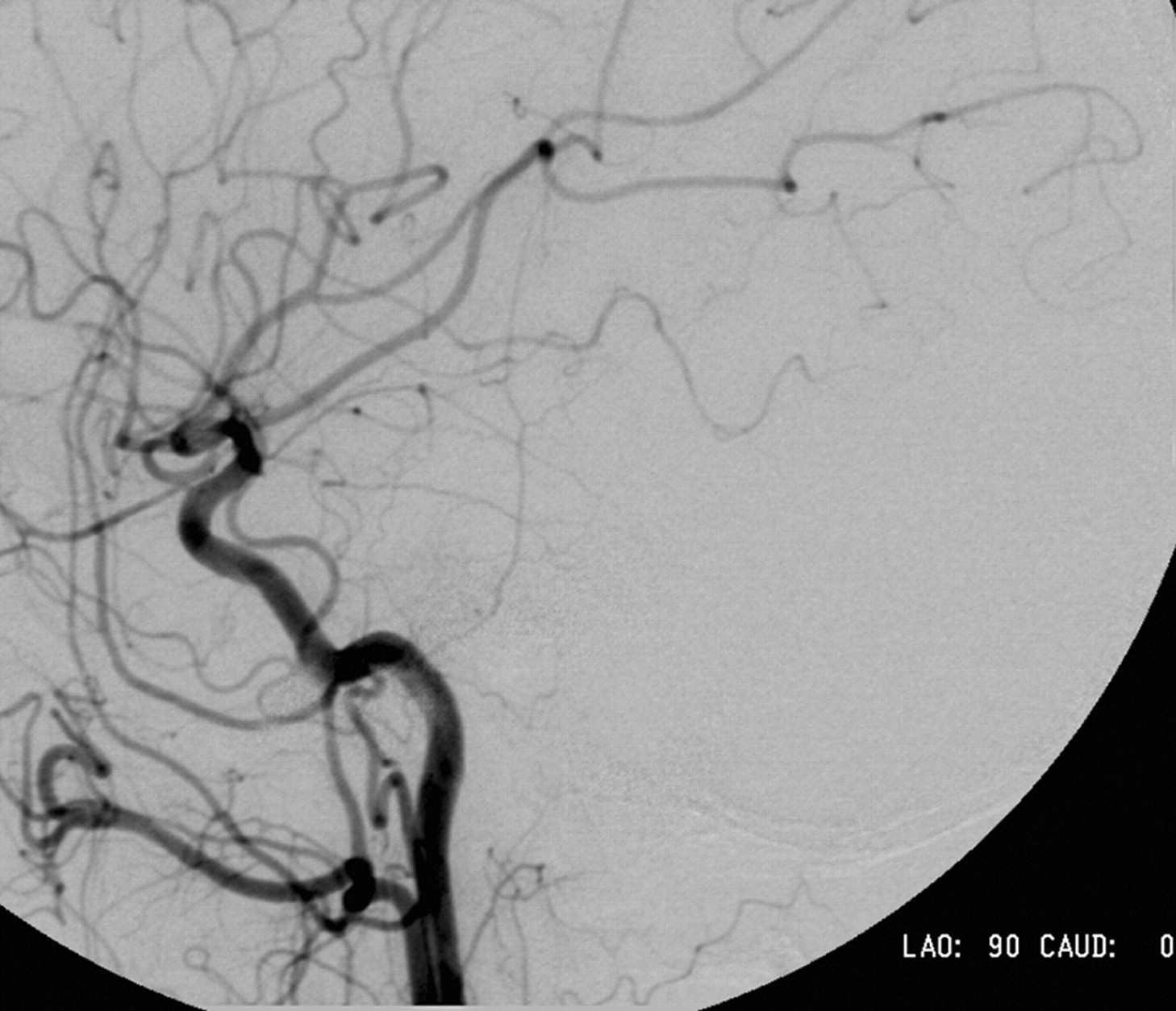
After antiplatelet medication (aspirin 150 mg once daily and clopidogrel 75 mg once daily), she then underwent angiography and proceeded directly to undergo endovascular stent placement. An apparent filling defect with streaming of unopacified blood in the ICA and absence of the occipital artery was noted on the preliminary images after common carotid artery injection (Figs 1 & 2).

Fig 1.
  • Download figure
  • Open in new tab
  • Download powerpoint
Fig 1.

Lateral view of the right common carotid artery injection demonstrates severe stenosis at the bifurcation. Note streaming of unopacified blood within the ICA and the absence of occipital artery branch from the posterior surface of the external carotid artery.

Fig 2.
  • Download figure
  • Open in new tab
  • Download powerpoint
Fig 2.

Lateral view of the right common carotid artery injection showing the apparent absence of the right occipital artery.

Using distal ICA protection (FilterWire EZ; Boston Scientific, Fremont, Calif), we placed an 8 × 30-mm nitinol self-expanding open-cell stent (Precise; Cordis, Miami Lakes, Fla) across the lesion followed by 5-mm angioplasty.

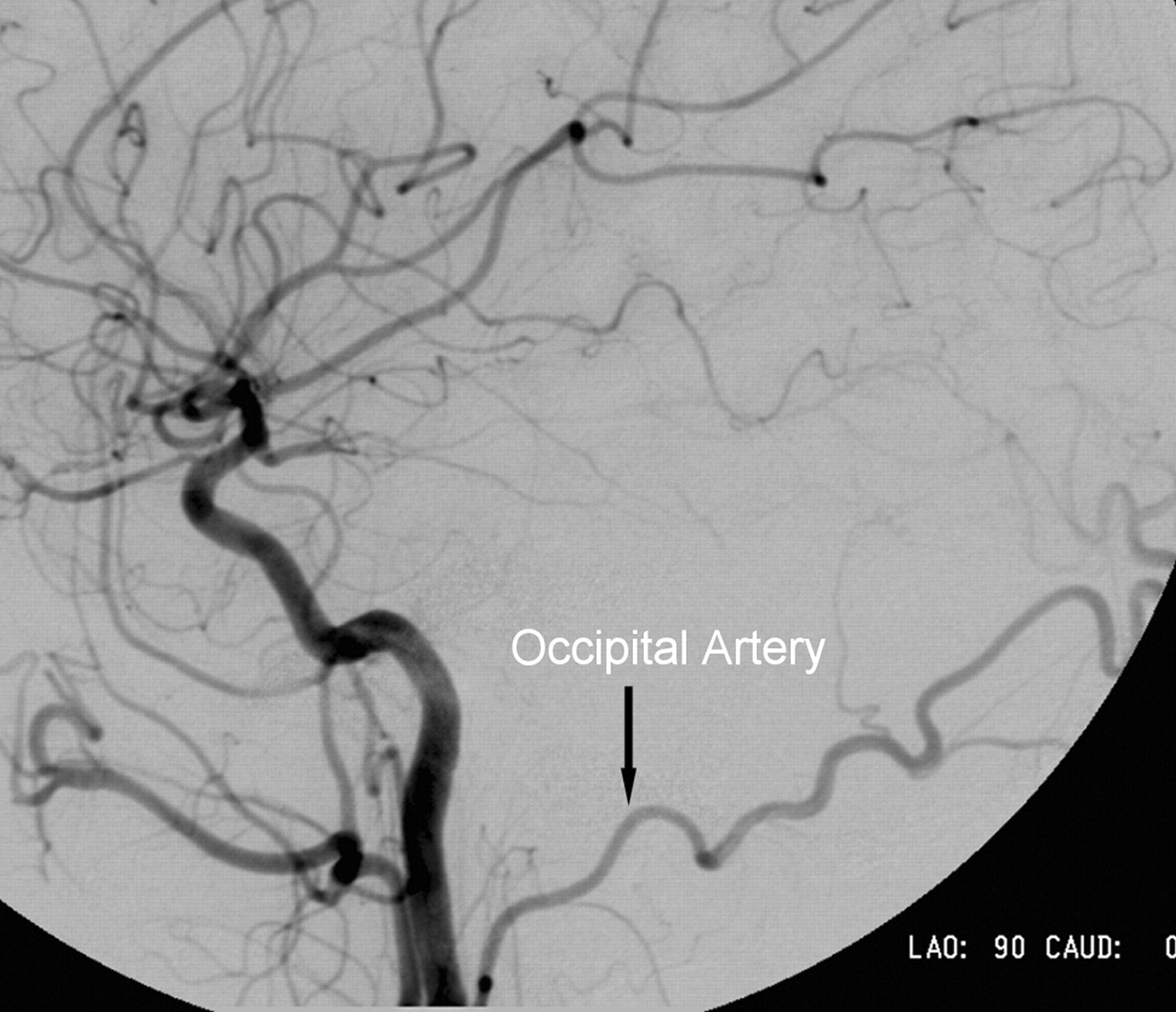
Repeated angiography after placement of the stent demonstrated filling of an aberrant large right occipital artery with its origin in the cervical right ICA (Figs 3 & 4). The patient stated that the tinnitus resolved at the time of stent placement.

Fig 3.
  • Download figure
  • Open in new tab
  • Download powerpoint
Fig 3.

Lateral view of the right common carotid artery injection after stent placement. The previously unseen occipital artery is now opacified, taking its origin from the cervical ICA at the level of C3.

Fig 4.
  • Download figure
  • Open in new tab
  • Download powerpoint
Fig 4.

Lateral view of the right common carotid artery injection confirms the presence of the hitherto unseen occipital artery.

Independent clinical auscultation confirmed complete resolution of the right-sided pulsatile tinnitus on the day of the procedure and demonstrated a sustained relief of symptoms 6 weeks after the procedure.

Discussion

Pulsatile tinnitus is a common presenting complaint that may arise from a host of possible underlying causes. The full list of differential diagnoses1 ranges from the benign to the impending lethal. A simplified scheme to review the causes of tinnitus considers 1) changes in flow velocity, 2) aberrant flow, and 3) turbulent flow. These 3 categories apply to both the arterial and the venous structures. Changes in flow velocity encompass causes such as dural arteriovenous fistulas and vascular tumors. Aberrant flow encompasses those such as high jugular bulb and persistent stapedial artery, and turbulence may be a contributing factor in any of the above causes and also in stenotic lesions of the carotid or transverse-sigmoid dural sinuses.

Reversal of flow and/or the possible changes in pulsatility and velocity within the occipital artery and its branches secondary to the carotid stenosis was the likely cause of the pulsatile tinnitus in the case of our patient. The close association of the occipital artery with the medial aspect of the mastoid process in the occipital groove makes it a good candidate for the propagation of abnormal pulsations to the structures of the middle and inner ear.

Pulsatile tinnitus has been attributed to many different vascular pathologic processes including stenotic lesions of the ICA.2,3 We cannot entirely exclude the possibility that the tinnitus was propagated directly from turbulence in the carotid stenosis. However, the character and distribution of the audible bruit that resolved after stent placement were not felt to be consistent with this explanation.

Many cases of pulsatile tinnitus secondary to dural arteriovenous fistulas involve abnormally high flow through the occipital artery. Furthermore, hypertrophy of the carotid to vertebral artery collateral vessels (the occipital artery in particular) has been linked to pulsatile tinnitus. Lehmann et al4 described such a case where subclavian stenosis lead to objective pulsatile tinnitus that resolved after subclavian artery stent placement.

Aberrant origins of the proximal branches of the external carotid artery have been described previously, including the ICA origin of the occipital artery.5-8 The exact incidence of this anatomic variant is unknown but is rare. Various series looking at the surgical anatomy of the carotid bifurcation have described the ICA origin of ascending pharyngeal, superior thyroid, lingual, and even facial arteries. Only 3 reports describing aberrant origin of the occipital artery have been found in the medical literature.5-7 The incidence of aberrant ICA origin of the occipital artery would seem to be considerably less than 1%.

Conclusions

We have presented a case that has never been described previously. In general, it highlights the potential role the occipital artery and, more specifically, reversal of flow within the occipital artery may play in the pathophysiology of pulsatile tinnitus.

References

  1. ↵
    Swartz JR, Harnsberger HR. Imaging of the Temporal Bone 3rd Ed. New York: Thieme;1998 :185
  2. ↵
    Waldvogel D, Mattle H, Sturzenegger M, et al. Pulsatile tinnitus –a review of 84 patients. J Neurol 1998;245:137–42
    CrossRefPubMed
  3. ↵
    Emery D, Ferguson R, Williams JS. Pulsatile tinnitus cured by angioplasty and stenting of petrous carotid artery stenosis. Arch Otolaryngol Head Neck Surg 1998;124:460–61
    PubMed
  4. ↵
    Lehmann M, Mounayer C, Benndorf G, et al. Pulsatile tinnitus: a symptom of chronic subclavian artery occlusion. AJNR Am J Neuroradiol 2005;26:1960–63
    Abstract/FREE Full Text
  5. ↵
    Aggarwal NR, Krishnamoorthy T, Devasia B, et al. Variant origin thyroid artery, occipital artery and ascending pharyngeal artery from a common trunk from the cervical segment of internal carotid artery. Surg Radiol Anat 2006;28:650–53
    CrossRefPubMed
  6. Hachem K, Slaba S, Nassar J, et al. Imaging of an aberrant occipital artery arising from the extracranial segment of the internal carotid artery. J Maladies Vasculaires 2004;29:205–09
    CrossRef
  7. ↵
    Ustunsoz B, Gumus B, Koksal A, et al. Missed total occlusion due to the occipital artery arising from the internal carotid artery. Cardiovasc Intervent Radiol 2007;30:116–17
    CrossRefPubMed
  8. ↵
    Hayashi N, Hori E, Ohtani Y, et al. Surgical anatomy of the cervical carotid artery for carotid endarterectomy. Neurol Med Chir (Tokyo) 2005;45:25–30
    CrossRefPubMed
  • Received August 5, 2008.
  • Accepted after revision September 21, 2008.
  • Copyright © American Society of Neuroradiology
View Abstract
PreviousNext
Back to top

In this issue

American Journal of Neuroradiology: 30 (5)
American Journal of Neuroradiology
Vol. 30, Issue 5
May 2009
  • Table of Contents
  • Index by author
Advertisement
Print
Download PDF
Email Article

Thank you for your interest in spreading the word on American Journal of Neuroradiology.

NOTE: We only request your email address so that the person you are recommending the page to knows that you wanted them to see it, and that it is not junk mail. We do not capture any email address.

Enter multiple addresses on separate lines or separate them with commas.
Pulsatile Tinnitus from Reversal of Flow in an Aberrant Occipital Artery: Resolved after Carotid Artery Stenting
(Your Name) has sent you a message from American Journal of Neuroradiology
(Your Name) thought you would like to see the American Journal of Neuroradiology web site.
CAPTCHA
This question is for testing whether or not you are a human visitor and to prevent automated spam submissions.
Cite this article
P.O. Cowley, R. Jones, P. Tuch, W. McAuliffe
Pulsatile Tinnitus from Reversal of Flow in an Aberrant Occipital Artery: Resolved after Carotid Artery Stenting
American Journal of Neuroradiology May 2009, 30 (5) 995-997; DOI: 10.3174/ajnr.A1393

Citation Manager Formats

  • BibTeX
  • Bookends
  • EasyBib
  • EndNote (tagged)
  • EndNote 8 (xml)
  • Medlars
  • Mendeley
  • Papers
  • RefWorks Tagged
  • Ref Manager
  • RIS
  • Zotero
0 Responses
Respond to this article
Share
Bookmark this article
Pulsatile Tinnitus from Reversal of Flow in an Aberrant Occipital Artery: Resolved after Carotid Artery Stenting
P.O. Cowley, R. Jones, P. Tuch, W. McAuliffe
American Journal of Neuroradiology May 2009, 30 (5) 995-997; DOI: 10.3174/ajnr.A1393
del.icio.us logo Twitter logo Facebook logo Mendeley logo
  • Tweet Widget
  • Facebook Like
  • Google Plus One
Purchase

Jump to section

  • Article
    • Abstract
    • Case Report
    • Discussion
    • Conclusions
    • References
  • Figures & Data
  • Info & Metrics
  • Responses
  • References
  • PDF

Related Articles

  • No related articles found.
  • PubMed
  • Google Scholar

Cited By...

  • Occipital artery anastomosis to vertebral artery causing pulsatile tinnitus
  • Occipital artery anastomosis to vertebral artery causing pulsatile tinnitus
  • Crossref (12)
  • Google Scholar

This article has been cited by the following articles in journals that are participating in Crossref Cited-by Linking.

  • Focal defect of mastoid bone shell in the region of the transverse-sigmoid junction: a new cause of pulsatile tinnitus
    J Xue, T Li, X Sun, Y Liu
    The Journal of Laryngology & Otology 2012 126 4
  • Occipital artery anastomosis to vertebral artery causing pulsatile tinnitus
    Matthew David Alexander, Joey English, Steven W Hetts
    Journal of NeuroInterventional Surgery 2014 6 2
  • Petrosquamosal sinus in the temporal bone as a cause of pulsatile tinnitus: a radiological detection
    Zhaohui Liu, Chengfang Chen, Zhenchang Wang, Shusheng Gong, Junfang Xian, Xihong Liang
    Clinical Imaging 2013 37 3
  • Vasopressin-Induced Constriction of the Isolated Rat Occipital Artery is Segment Dependent
    Stephen P. Chelko, Chad W. Schmiedt, Tristan H. Lewis, Stephen J. Lewis, Tom P. Robertson
    Journal of Vascular Research 2013 50 6
  • Pulsatile Tinnitus due to a Tortuous Siphon-Like Internal Carotid Artery Successfully Treated by Arterial Remodeling
    Dirk De Ridder, Sven Vanneste, Tomas Menovsky
    Case Reports in Otolaryngology 2013 2013
  • Textbook of Tinnitus
    Dirk De Ridder
    2011
  • An unexpected variation of occipital and ascending pharyngeal arteries: clinical implications
    Gabriella Roofeh, Nicholas Matthees, Manuel Cevallos
    Surgical and Radiologic Anatomy 2024 47 1
  • Effect of angioplasty and stent placement on sensorineural hearing loss and pulsatile tinnitus in a patient with severe internal carotid artery stenosis
    Navpreet K. Bains, Elizabeth Early, Brandi R. French, Farhan Siddiq, Camilo R. Gomez, Adnan I. Qureshi
    Journal of Stroke and Cerebrovascular Diseases 2022 31 11
  • Occipital artery anastomosis to vertebral artery causing pulsatile tinnitus
    Matthew David Alexander, Joey English, Steven W Hetts
    BMJ Case Reports 2013 2013
  • Current World Literature
    Current Opinion in Otolaryngology & Head & Neck Surgery 2010 18 5

More in this TOC Section

  • NI-RADS for HEAD&NECK Cancer Recurrence
  • WHO Classification Update: Nasal&Skull Base Tumors
  • Peritumoral Signal in Vestibular Schwannomas
Show more HEAD & NECK

Similar Articles

Advertisement

Indexed Content

  • Current Issue
  • Accepted Manuscripts
  • Article Preview
  • Past Issues
  • Editorials
  • Editors Choice
  • Fellow Journal Club
  • Letters to the Editor

Cases

  • Case Collection
  • Archive - Case of the Week
  • Archive - Case of the Month
  • Archive - Classic Case

Special Collections

  • Special Collections

Resources

  • News and Updates
  • Turn around Times
  • Submit a Manuscript
  • Author Policies
  • Manuscript Submission Guidelines
  • Evidence-Based Medicine Level Guide
  • Publishing Checklists
  • Graphical Abstract Preparation
  • Imaging Protocol Submission
  • Submit a Case
  • Become a Reviewer/Academy of Reviewers
  • Get Peer Review Credit from Publons

Multimedia

  • AJNR Podcast
  • AJNR SCANtastic
  • Video Articles

About Us

  • About AJNR
  • Editorial Board
  • Not an AJNR Subscriber? Join Now
  • Alerts
  • Feedback
  • Advertise with us
  • Librarian Resources
  • Permissions
  • Terms and Conditions

American Society of Neuroradiology

  • Not an ASNR Member? Join Now

© 2025 by the American Society of Neuroradiology All rights, including for text and data mining, AI training, and similar technologies, are reserved.
Print ISSN: 0195-6108 Online ISSN: 1936-959X

Powered by HighWire