Advertisement

Main menu

  • Home
  • Content
    • Current Issue
    • Accepted Manuscripts
    • Article Preview
    • Past Issue Archive
    • AJNR Case Collection
    • Case of the Week Archive
    • Classic Case Archive
    • Case of the Month Archive
  • Special Collections
    • Spinal CSF Leak Articles (Jan 2020-June 2024)
    • 2024 AJNR Journal Awards
    • Most Impactful AJNR Articles
  • Multimedia
    • AJNR Podcast
    • AJNR Scantastics
    • Video Articles
  • For Authors
    • Submit a Manuscript
    • Author Policies
    • Fast publishing of Accepted Manuscripts
    • Graphical Abstract Preparation
    • Manuscript Submission Guidelines
    • Imaging Protocol Submission
    • Submit a Case for the Case Collection
  • About Us
    • About AJNR
    • Editorial Board
  • More
    • Become a Reviewer/Academy of Reviewers
    • Subscribers
    • Permissions
    • Alerts
    • Feedback
    • Advertisers
    • ASNR Home
  • Other Publications
    • ajnr

User menu

  • Alerts
  • Log in

Search

  • Advanced search
American Journal of Neuroradiology

American Journal of Neuroradiology

ASHNR American Society of Functional Neuroradiology ASHNR American Society of Pediatric Neuroradiology ASSR
  • Alerts
  • Log in

Advanced Search

  • Home
  • Content
    • Current Issue
    • Accepted Manuscripts
    • Article Preview
    • Past Issue Archive
    • AJNR Case Collection
    • Case of the Week Archive
    • Classic Case Archive
    • Case of the Month Archive
  • Special Collections
    • Spinal CSF Leak Articles (Jan 2020-June 2024)
    • 2024 AJNR Journal Awards
    • Most Impactful AJNR Articles
  • Multimedia
    • AJNR Podcast
    • AJNR Scantastics
    • Video Articles
  • For Authors
    • Submit a Manuscript
    • Author Policies
    • Fast publishing of Accepted Manuscripts
    • Graphical Abstract Preparation
    • Manuscript Submission Guidelines
    • Imaging Protocol Submission
    • Submit a Case for the Case Collection
  • About Us
    • About AJNR
    • Editorial Board
  • More
    • Become a Reviewer/Academy of Reviewers
    • Subscribers
    • Permissions
    • Alerts
    • Feedback
    • Advertisers
    • ASNR Home
  • Follow AJNR on Twitter
  • Visit AJNR on Facebook
  • Follow AJNR on Instagram
  • Join AJNR on LinkedIn
  • RSS Feeds

Welcome to the new AJNR, Updated Hall of Fame, and more. Read the full announcements.


AJNR is seeking candidates for the position of Associate Section Editor, AJNR Case Collection. Read the full announcement.


LetterLetter

Isolated Reversible Thalamic Vasogenic Edema Following a Generalized Seizure

A. Aghaei Lasboo, M.T. Walker and T.A. Hijaz
American Journal of Neuroradiology May 2009, 30 (5) e72; DOI: https://doi.org/10.3174/ajnr.A1512
A. Aghaei Lasboo
  • Find this author on Google Scholar
  • Find this author on PubMed
  • Search for this author on this site
M.T. Walker
  • Find this author on Google Scholar
  • Find this author on PubMed
  • Search for this author on this site
T.A. Hijaz
  • Find this author on Google Scholar
  • Find this author on PubMed
  • Search for this author on this site
  • Article
  • Figures & Data
  • Info & Metrics
  • Responses
  • References
  • PDF
Loading

Transient MR imaging signal-intensity changes in the thalamus, usually accompanied by signal-intensity changes elsewhere in the brain, have been previously reported in the setting of seizures.1–3 We report a case in which an MR image obtained shortly after a seizure episode demonstrated abnormal signal intensity isolated to the left thalamus on T2-weighted and fluid-attenuated inversion recovery imaging sequences.

A 28-year-old man presented 5 hours after a witnessed seizure, which was characterized by sudden loss of consciousness and jerking movements of the extremities. The seizure lasted approximately 2 minutes, and it was followed by a period of lethargy. The patient had a history of a motor vehicle crash 2 years prior as a result of which he had cervical spine fractures and short-term memory deficits. There was no history of alcohol or recreational drug use. At the time of presentation to our institution, the patient was on medications with the potential to lower seizure threshold, including bupropion, oxybutinin, and duloxetine, none of which was newly prescribed.

Within 3 hours of presentation, the patient gradually became alert and oriented, and he had no memory of the seizure. Bupropion was held as a possible contributing factor to the patient's seizure, and levetiracetam therapy was initiated for seizure prophylaxis.

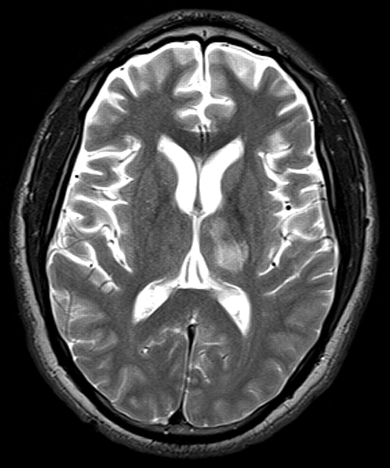
Electroencephalography (EEG), CT, and MR imaging were all performed on the day of admission. The EEG did not reveal any focal abnormality or epileptiform discharges. The CT demonstrated a nonspecific focus of hypoattenuation involving the left thalamus. The MR imaging revealed a nonenhancing T2-hyperintense focus predominantly isolated to the left thalamus (Fig. 1). There was no diffusion restriction, eliminating the possibility of cytotoxic edema from infarction. Differential diagnostic considerations included a low-grade glioma. The patient underwent a lumbar puncture, the findings of which were normal.

On day 7, a repeat MR imaging of the brain revealed complete resolution of the thalamic signal-intensity abnormality. During the remainder of the patient's admission, he remained seizure-free while on levetiracetam. On day 9, he was discharged in stable condition.

The thalamic nuclei act as relays between cortical and subcortical structures and are thought to be involved in regulation of seizure susceptibility and propagation.1 Thalamic signal-intensity abnormality, usually in conjunction with signal-intensity abnormality in other parts of the brain, has been documented by other researchers. Men et al1 reported a case with abnormal diffusion throughout the left hemispheric cortex, left thalamus, and right cerebellar hemisphere. Nagasaka et al2 reported “reversible” T2 signal-intensity abnormality involving the right temporoparietal cortex, contralateral cerebellum, and ipsilateral thalamus on an ictal MR imaging in a patient with a 1-month history of repetitive complex partial seizures. To the best of our knowledge, transient signal-intensity abnormality seen exclusively in the thalamus following a single episode of generalized seizure has not been reported previously. Our case reinforces the known role of the thalamus in seizure susceptibility and propagation. Involvement of the thalamus in our patient is strongly suggested by the development of transient thalamic signal-intensity abnormality immediately following a reported seizure.

It is not clear whether the location of the abnormal signal intensity in our patient means that the thalamus itself was the seizure focus or whether it was secondarily involved and inherently more susceptible to demonstrating signal-intensity changes. Roch et al4 studied the temporal evolution of lesions on MR imaging by using a rat epilepsy model. At 2 hours after status epilepticus, blood-brain barrier breakdown, which resolved in 6 hours, was observed only in the thalamus. It was followed by edema in the amygdala and piriform and entorhinal cortices with extensive neuronal loss. These findings resolved within 5 days.4

The rapid resolution of the signal-intensity abnormality in our patient raises the question of whether postseizure imaging should be performed more expeditiously to minimize the possibility of missing transient signal-intensity abnormality, which may help localize the seizure focus. Our case highlights the importance of follow-up imaging in a patient with abnormal imaging findings and a history of seizures before any potential medical or surgical interventions for other presumed etiologies, including a possible low-grade neoplasm.

  Fig 1.
  • Download figure
  • Open in new tab
  • Download powerpoint
Fig 1.

T2-weighted MR image shows a nonenhancing hyperintense focus predominantly isolated to the left thalamus with extension to a portion of the thalamocapsular junction.

References

  1. ↵
    Men S, Lee DH, Barron JR, et al. Selective neuronal necrosis associated with status epilepticus: MR findings. AJNR Am J Neuroradiol 2000;21:1837–40
    Abstract/FREE Full Text
  2. ↵
    Nagasaka T, Shindo K, Hiraide M, et al. Ipsilateral thalamic MRI abnormality in an epilepsy patient. Neurology 2002;58:641–44
    Abstract/FREE Full Text
  3. ↵
    Fazekas F, Kapeller P, Schmidt R, et al. Magnetic resonance imaging and spectroscopy findings after focal status epilepticus. Epilepsia 1995;36:946–49
    CrossRefPubMed
  4. ↵
    Roch C, Leroy C, Nehlig A, et al. Magnetic resonance imaging in the study of the lithium-pilocarpine model of temporal lobe epilepsy in adult rats. Epilepsia 2002;43:325–35
    CrossRefPubMed
  • Copyright © American Society of Neuroradiology
PreviousNext
Back to top

In this issue

American Journal of Neuroradiology: 30 (5)
American Journal of Neuroradiology
Vol. 30, Issue 5
May 2009
  • Table of Contents
  • Index by author
Advertisement
Print
Download PDF
Email Article

Thank you for your interest in spreading the word on American Journal of Neuroradiology.

NOTE: We only request your email address so that the person you are recommending the page to knows that you wanted them to see it, and that it is not junk mail. We do not capture any email address.

Enter multiple addresses on separate lines or separate them with commas.
Isolated Reversible Thalamic Vasogenic Edema Following a Generalized Seizure
(Your Name) has sent you a message from American Journal of Neuroradiology
(Your Name) thought you would like to see the American Journal of Neuroradiology web site.
CAPTCHA
This question is for testing whether or not you are a human visitor and to prevent automated spam submissions.
Cite this article
A. Aghaei Lasboo, M.T. Walker, T.A. Hijaz
Isolated Reversible Thalamic Vasogenic Edema Following a Generalized Seizure
American Journal of Neuroradiology May 2009, 30 (5) e72; DOI: 10.3174/ajnr.A1512

Citation Manager Formats

  • BibTeX
  • Bookends
  • EasyBib
  • EndNote (tagged)
  • EndNote 8 (xml)
  • Medlars
  • Mendeley
  • Papers
  • RefWorks Tagged
  • Ref Manager
  • RIS
  • Zotero
0 Responses
Respond to this article
Share
Bookmark this article
Isolated Reversible Thalamic Vasogenic Edema Following a Generalized Seizure
A. Aghaei Lasboo, M.T. Walker, T.A. Hijaz
American Journal of Neuroradiology May 2009, 30 (5) e72; DOI: 10.3174/ajnr.A1512
del.icio.us logo Twitter logo Facebook logo Mendeley logo
  • Tweet Widget
  • Facebook Like
  • Google Plus One
Purchase

Jump to section

  • Article
    • References
  • Figures & Data
  • Info & Metrics
  • Responses
  • References
  • PDF

Related Articles

  • No related articles found.
  • PubMed
  • Google Scholar

Cited By...

  • No citing articles found.
  • Crossref (2)
  • Google Scholar

This article has been cited by the following articles in journals that are participating in Crossref Cited-by Linking.

  • An exploration of the spectrum of peri -ictal MRI change; a comprehensive literature review
    Jennifer A Williams, Peter Bede, Colin P Doherty
    Seizure 2017 50
  • Thalamus lesions in chronic and acute seizure disorders
    Henriette J. Tschampa, Susanne Greschus, Robert Sassen, Christian G. Bien, Horst Urbach
    Neuroradiology 2011 53 4

More in this TOC Section

  • Brain AVM’s Nidus: What if We Hadn’t Understood Anything?
  • Letter to the Editor regarding “Automated Volumetric Software in Dementia: Help or Hindrance to the Neuroradiologist?”
  • Reply:
Show more Letters

Similar Articles

Advertisement

Indexed Content

  • Current Issue
  • Accepted Manuscripts
  • Article Preview
  • Past Issues
  • Editorials
  • Editors Choice
  • Fellow Journal Club
  • Letters to the Editor

Cases

  • Case Collection
  • Archive - Case of the Week
  • Archive - Case of the Month
  • Archive - Classic Case

Special Collections

  • Special Collections

Resources

  • News and Updates
  • Turn around Times
  • Submit a Manuscript
  • Author Policies
  • Manuscript Submission Guidelines
  • Evidence-Based Medicine Level Guide
  • Publishing Checklists
  • Graphical Abstract Preparation
  • Imaging Protocol Submission
  • Submit a Case
  • Become a Reviewer/Academy of Reviewers
  • Get Peer Review Credit from Publons

Multimedia

  • AJNR Podcast
  • AJNR SCANtastic
  • Video Articles

About Us

  • About AJNR
  • Editorial Board
  • Not an AJNR Subscriber? Join Now
  • Alerts
  • Feedback
  • Advertise with us
  • Librarian Resources
  • Permissions
  • Terms and Conditions

American Society of Neuroradiology

  • Not an ASNR Member? Join Now

© 2025 by the American Society of Neuroradiology All rights, including for text and data mining, AI training, and similar technologies, are reserved.
Print ISSN: 0195-6108 Online ISSN: 1936-959X

Powered by HighWire