
Article Figures & Data
Figures
- Fig 1.
Typical radiologic features of extra-axial CAPNON. A, Noncontrast CT scan shows a densely calcified mass in the right temporal horn. B, Axial T2-weighted MR image shows a uniform T2 hypointense mass centered in the right temporal horn. C, Axial T1-weighted postgadolinium sequence illustrates marked T1 hypointensity with scattered linear areas of enhancement that correspond to the strands of T2 hyperintensity. This appearance was seen in both of our larger lesions and may correspond to the vascular stromal elements seen within these lesions.
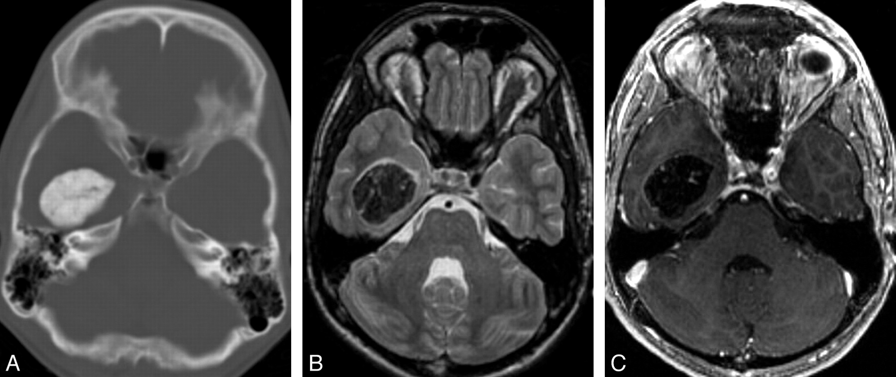
- Fig 2.
Typical radiologic features of intra-axial CAPNON. A, Noncontrast CT scan shows a left hippocampal mass with attenuated calcification. B, Coronal T2-weighted sequence demonstrates T2 hypointensity with a nodular border. C, Coronal T1-weighted sequence shows the typical T1 hypointensity. Preoperatively, this lesion was thought to represent a cavernous malformation. In retrospect, the nodular contour on T2 and lack of internal T2 hyperintensity would be atypical for a cavernous malformation of this size.
- Fig 3.
Typical histopathologic features of CAPNON. A, The typical chondromyxoid matrix of CAPNON (H&E, original magnification ×100). B, Focal osseous metaplasia is seen in all 4 cases (H&E, original magnification ×100). C, Medium-power magnification of the chondromyxoid matrix and the peripheral spindle cells (H&E, original magnification ×200). D, Immunohistochemical analysis for EMA demonstrating positive staining in the spindle cells surrounding the matrix (EMA immunohistochemistry, original magnification ×200).
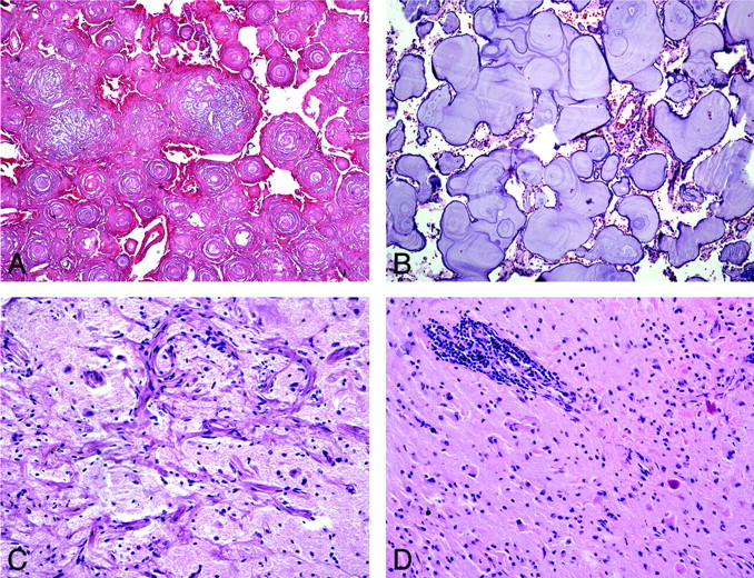
- Fig 4.
Atypical and unusual features of CAPNON. A, Areas of coalescent concentric lamellar calcifications without intervening chondromyxoid matrix or cells (H&E, original magnification ×100). B, More basophilic amorphous lamellar calcifications without intervening chondromyxoid matrix and with rare meningothelial cells (H&E, original magnification ×100). C, Adjacent cortical region showing meningioangiomatosis (H&E, original magnification ×200). D, Surrounding parenchyma with prominent perivascular lymphocytic infiltrates and Rosenthal fibers (H&E, original magnification ×200).


Tables
References Pt No. Age/Sex Location CT Performed Presentation Rhodes & Davis, 1978 1 27/F R frontal No HA 2 55/F Brain, dura No Autopsy finding 3 60/M L cerebellum No Autopsy finding 4 74/F Brain, dura No Autopsy finding 5 46/M Choroid No Autopsy finding 6 62/M Pineal No Autopsy finding 7 83/M Brain, dura No Autopsy finding Jun, 1984 8 55/M Corpus callosum Calc HA, N/V Garen, 1989 9 44/M Dura, Meckel cave Calc Atypical facial pain Bertoni, 1990 10 31/M Jugular foramen No HA, hoarseness 11 50/M Foramen magnum No Neck pain 12 48/M Skull base/cerebellum No R CN XI paralysis 13 23/M Spine, T10 No Back pain 14 58/M Spine, C2–3 No Back pain 15 32/M L frontal No Epilepsy 16 45/F Skull base No CN paralysis 17 58/M Skull base No Hoarseness 18 12/M Spine, C6 No Pain 19 32/M Spine, L4–5 No Back pain 20 33/F Spine, T9 No Back pain 21 68/F Spine, L4–5 No R hip pain 22 20/F Spine, C2 No Incidental 23 56/F Spine, L4–5 No Back pain Smith, 1994 24 48/M Spine, L2–3 No Sciatica Tsugu, 1999 25 22/F R parietal Calc Seizures Tatke, 2001 26 6/M L temporal Calc Seizures Qian, 1999 27 33/F L temporal Calc Developmental delay 28 49/M Spine, C1 & clivus No Weakness 29 59/M Spine, C1–2 No Shuffling gait 30 47/F Frontal lobe Calc Seizures Note:—CAPNON indicates calcifying pseudoneoplasms of the neuraxis; calc, densely calcified mass seen on CT; CN, cranial nerve; HA, headache; N/V, nausea and vomiting; L, left; R, right.
* This table includes an additional 30 patients from the literature with intracranial and intraspinal CAPNON.
Pt No. Age/Sex Presentation Location Size (cm) T1WI T2WI Enhancement 1 16/M Incidental Temporal horn, extra-axial 3.5 Hypo Hypo Internal linear C+ 2 35/M Seizures Temporal, intra-axial 2 Hypo Hypo Internal linear C+ 3 49/F Seizures Hippocampus, intra-axial 1 Hypo Hypo No C+ 4 59/M Left arm numbness Parietal, intra-axial 1 Hypo Hypo Rim C+ Shrier et al 32/F Incidental Temporal, intra-axial 0.8 Hypo Hypo Rim C+ Shrier et al 59/M Neck pain Foramen magnum, extra-axial 2 Hypo Hypo Heterogenous solid Note:—Hypo, indicates hypointense; C+, enhancement. All lesions showed dense calcification on CT; T1WI, T1-weighted imaging; T2WI, T2-weighted imaging.
* This table includes our 4 patients and 2 additional patients reported in the literature.
In this issue


Jump to section
Related Articles
Cited By...
- Calcified Pseudoneoplasm of the Neuraxis
- Calcified otogenic brain abscess
- Calcified pseudoneoplasm of the neuraxis
- Occipital calcified pseudoneoplasms of the neuraxis (CAPNON): understanding a rare pathology
- Clinical Reasoning: A 30-year-old woman with recurrent seizures and a cerebral lesion progressing over 2 decades
- Reply:
- Choroid Plexus Papilloma with Osseous Metaplasia as a Differential Diagnosis of Calcifying Pseudoneoplasms of the Neuraxis