Advertisement

Main menu

  • Home
  • Content
    • Current Issue
    • Accepted Manuscripts
    • Article Preview
    • Past Issue Archive
    • Video Articles
    • AJNR Case Collection
    • Case of the Week Archive
    • Case of the Month Archive
    • Classic Case Archive
  • Special Collections
    • AJNR Awards
    • Low-Field MRI
    • Alzheimer Disease
    • ASNR Foundation Special Collection
    • Photon-Counting CT
    • View All
  • Multimedia
    • AJNR Podcasts
    • AJNR SCANtastic
    • Trainee Corner
    • MRI Safety Corner
    • Imaging Protocols
  • For Authors
    • Submit a Manuscript
    • Submit a Video Article
    • Submit an eLetter to the Editor/Response
    • Manuscript Submission Guidelines
    • Statistical Tips
    • Fast Publishing of Accepted Manuscripts
    • Graphical Abstract Preparation
    • Imaging Protocol Submission
    • Author Policies
  • About Us
    • About AJNR
    • Editorial Board
    • Editorial Board Alumni
  • More
    • Become a Reviewer/Academy of Reviewers
    • Subscribers
    • Permissions
    • Alerts
    • Feedback
    • Advertisers
    • ASNR Home

User menu

  • Alerts
  • Log in

Search

  • Advanced search
American Journal of Neuroradiology

American Journal of Neuroradiology

ASHNR American Society of Functional Neuroradiology ASHNR American Society of Pediatric Neuroradiology ASSR
  • Alerts
  • Log in

Advanced Search

  • Home
  • Content
    • Current Issue
    • Accepted Manuscripts
    • Article Preview
    • Past Issue Archive
    • Video Articles
    • AJNR Case Collection
    • Case of the Week Archive
    • Case of the Month Archive
    • Classic Case Archive
  • Special Collections
    • AJNR Awards
    • Low-Field MRI
    • Alzheimer Disease
    • ASNR Foundation Special Collection
    • Photon-Counting CT
    • View All
  • Multimedia
    • AJNR Podcasts
    • AJNR SCANtastic
    • Trainee Corner
    • MRI Safety Corner
    • Imaging Protocols
  • For Authors
    • Submit a Manuscript
    • Submit a Video Article
    • Submit an eLetter to the Editor/Response
    • Manuscript Submission Guidelines
    • Statistical Tips
    • Fast Publishing of Accepted Manuscripts
    • Graphical Abstract Preparation
    • Imaging Protocol Submission
    • Author Policies
  • About Us
    • About AJNR
    • Editorial Board
    • Editorial Board Alumni
  • More
    • Become a Reviewer/Academy of Reviewers
    • Subscribers
    • Permissions
    • Alerts
    • Feedback
    • Advertisers
    • ASNR Home
  • Follow AJNR on Twitter
  • Visit AJNR on Facebook
  • Follow AJNR on Instagram
  • Join AJNR on LinkedIn
  • RSS Feeds

AJNR Awards, New Junior Editors, and more. Read the latest AJNR updates

Research ArticlePediatric Neuroimaging

Intracranial Aneurysms in Childhood: 27-Year Single-Institution Experience

S.W. Hetts, J. Narvid, N. Sanai, M.T. Lawton, N. Gupta, H.J. Fullerton, C.F. Dowd, R.T. Higashida and V.V. Halbach
American Journal of Neuroradiology August 2009, 30 (7) 1315-1324; DOI: https://doi.org/10.3174/ajnr.A1587
S.W. Hetts
  • Find this author on Google Scholar
  • Find this author on PubMed
  • Search for this author on this site
J. Narvid
  • Find this author on Google Scholar
  • Find this author on PubMed
  • Search for this author on this site
N. Sanai
  • Find this author on Google Scholar
  • Find this author on PubMed
  • Search for this author on this site
M.T. Lawton
  • Find this author on Google Scholar
  • Find this author on PubMed
  • Search for this author on this site
N. Gupta
  • Find this author on Google Scholar
  • Find this author on PubMed
  • Search for this author on this site
H.J. Fullerton
  • Find this author on Google Scholar
  • Find this author on PubMed
  • Search for this author on this site
C.F. Dowd
  • Find this author on Google Scholar
  • Find this author on PubMed
  • Search for this author on this site
R.T. Higashida
  • Find this author on Google Scholar
  • Find this author on PubMed
  • Search for this author on this site
V.V. Halbach
  • Find this author on Google Scholar
  • Find this author on PubMed
  • Search for this author on this site
  • Article
  • Figures & Data
  • Info & Metrics
  • Responses
  • References
  • PDF
Loading

Article Figures & Data

Figures

  • Tables
  • Fig 1.
    • Download figure
    • Open in new tab
    • Download powerpoint
    Fig 1.

    Clinical symptoms at presentation in relation to the type of aneurysm (percentages).

  • Fig 2.
    • Download figure
    • Open in new tab
    • Download powerpoint
    Fig 2.

    Vascular location of 103 aneurysms in relation to their type (percentages).
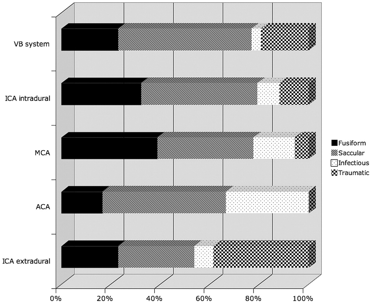
  • Fig 3.
    • Download figure
    • Open in new tab
    • Download powerpoint
    Fig 3.

    Aneurysm subtype expressed in relation to anatomic location (percentages).

  • Fig 4.
    • Download figure
    • Open in new tab
    • Download powerpoint
    Fig 4.

    Clinical management and treatment procedures in 77 children with intracranial aneurysms.

  • Fig 5.
    • Download figure
    • Open in new tab
    • Download powerpoint
    Fig 5.

    Progressive enlargement of a saccular paraophthalmic aneurysm superimposed on fusiform internal carotid vasculopathy. A, Right ICA angiogram of a patient at age 10, anteroposterior projection demonstrating a tortuous ICA with a superimposed 3-mm saccular paraophthalmic aneurysm (arrow). The patient underwent conservative observation with annual MR angiography which, 4 years later, suggested aneurysm progression. B, Right ICA angiogram of a patient at age 14, anteroposterior projection, demonstrating interval enlargement of the paraophthalmic aneurysm (arrow) to 13 mm, prompting a decision in favor of treatment. C and D, Right ICA angiogram, anteroposterior projection (C), and 3D rotational angiogram (D) of patient at age 14 immediately following successful stent-assisted coiling of the paraophthalmic aneurysm (arrowheads indicate coil mass).

  • Fig 6.
    • Download figure
    • Open in new tab
    • Download powerpoint
    Fig 6.

    Development of a de novo fusiform vertebrobasilar junction aneurysm 3 years after treatment of a fusiform ICA aneurysm with balloon takedown of the ICA in an adolescent male patient. A, Left ICA angiogram of the patient at age 11, lateral projection. B, Vertebrobasilar angiogram at age 11, anteroposterior projection. The left ICA was successfully occluded with 2 detachable balloons immediately following the angiograms shown in A and B. Three years later an MR angiogram (not shown) suggested a new aneurysm at the vertebrobasilar junction. Conventional angiography was performed, confirming this diagnosis (C and D). C, Vertebrobasilar angiogram at age 14, anteroposteror projection. D, Vertebrobasilar angiogram at age 14, lateral projection.

Tables

  • Figures
    • View popup
    Table 1:

    Comorbidities of patients in this series*

    CategorySpecific Comorbidity
    TraumaticMotor vehicle crash (3), Pedestrian-versus-automobile accident with skull base fractures, prior gunshot wound (2), prior skateboard accident, fall, head trauma not otherwise specified
    IschemicPrior insular infarct, prior external-carotid-to-internal carotid artery bypass grafting (2)
    VasculopathicFibromuscular dysplasia
    DermatologicPHACE, facial and retinal hemangiomas, vascular birthmark
    HormonalGrowth hormone deficiency with neuromigrational disorder and Moyamoya syndrome, Seckel syndrome (growth hormone deficiency), Majewski type II dwarfism
    HematologicVon Willebrand disease
    ImmunologicAIDS, congenital immunodeficiency, combined immunodeficiency syndrome with cerebral angiodysplasia
    CardiovascularCongenital aortic stenosis with endocarditis, tricuspid atresia with Fontan procedure, patent ductus arteriosus
    NeoplasticJuvenile pilocytic astrocytoma
    NeurologicSeizure disorder
    Other syndromicTuberous sclerosis (2), autosomal dominant polycystic kidney disease, trisomy 21 with endocarditis, hemiatrophy, mitochondrial disorder with optic atrophy, family history of Marfan syndrome
    • * Numbers in parentheses indicate comorbidities present in multiple patients. Note that infection is not listed as a comorbidity; however, patients with immunodeficiency syndromes and patients with endocarditis due to an identified underlying risk factor (eg, congenital aortic stenosis) are listed under their primary comorbid condition.

    • View popup
    Table 2:

    De novo aneurysms and enlarging untreated aneurysms in 6 children

    Age at First Presentation (yr)SexIndex AneurysmFirst TreatmentTime to De Novo or Enlarging AneurysmDe Novo or Enlarging Aneurysm LocationClinical ResultComorbidity
    11MICA fusiformICA balloon occlusion3 yrVB junction fusiformSuccessful clippingVascular birthmark
    7MICA fusiformICA balloon-coil occlusion3 yrIntracranial vertebral fusiformUntreatableHemiatrophy
    15FICA, MCA, ACA fusiform giantBypass, trapping12 yrAcomA fusiformWrapping, completion coilingNone
    8MACA giant mycoticCoiling20 moVB fusiformDied from SAHAIDS
    17F10 saccular aneurysmsClip 6 ipsilateral; coil 1 contralateral6 moEnlarging saccular PcomASuccessful coilingMajewski type II dwarfism
    10FLong-segment fusiform ICAObservation4 yrEnlarging saccular ICA aneurysmSuccessful stent coilingTricuspid atresia after Fontan procedure
    • Note:—VB indicates vertebrobasilar; ICA, internal carotid artery; MCA, middle cerebral artery; ACA, anterior cerebral artery; AcomA, anterior communicating artery; PcomA, posterior communicating artery; SAH, subarachnoid hemorrhage.

    • View popup
    Table 3:

    Aneurysm characteristics in various case series and meta-analyses

    SeriesCurrentLasjaunias et al18Huang et al22Agid et al29
    Patients (No.)77 (103 aneurysms)59 (75 aneurysms)70633 (37 aneurysms)
    Etiology/morphology
        Fusiform31%56%–19%
        Saccular46%27%–46%
        Infectious12%14%–8%
        Traumatic14%3%–14%
    Giant (>25 mm)11%–20%–
    Multiple16% of patients, 38% of aneurysms15%––
    Posterior circulation22%27%17%24%
    Age12 yr (3 mo to 18 y)7.6 yr (8 days to 15 yr)0–18 yr10.2 y (1 day to 17 yr)
    Sex (% male)48%59%63%48%
    Hemorrhage32%54%80%27%
    Mortality1.3%10.4%28%15%
    • Note:— –indicates not applicable.

PreviousNext
Back to top

In this issue

American Journal of Neuroradiology: 30 (7)
American Journal of Neuroradiology
Vol. 30, Issue 7
August 2009
  • Table of Contents
  • Index by author
Advertisement
Print
Download PDF
Email Article

Thank you for your interest in spreading the word on American Journal of Neuroradiology.

NOTE: We only request your email address so that the person you are recommending the page to knows that you wanted them to see it, and that it is not junk mail. We do not capture any email address.

Enter multiple addresses on separate lines or separate them with commas.
Intracranial Aneurysms in Childhood: 27-Year Single-Institution Experience
(Your Name) has sent you a message from American Journal of Neuroradiology
(Your Name) thought you would like to see the American Journal of Neuroradiology web site.
CAPTCHA
This question is for testing whether or not you are a human visitor and to prevent automated spam submissions.
Cite this article
S.W. Hetts, J. Narvid, N. Sanai, M.T. Lawton, N. Gupta, H.J. Fullerton, C.F. Dowd, R.T. Higashida, V.V. Halbach
Intracranial Aneurysms in Childhood: 27-Year Single-Institution Experience
American Journal of Neuroradiology Aug 2009, 30 (7) 1315-1324; DOI: 10.3174/ajnr.A1587

Citation Manager Formats

  • BibTeX
  • Bookends
  • EasyBib
  • EndNote (tagged)
  • EndNote 8 (xml)
  • Medlars
  • Mendeley
  • Papers
  • RefWorks Tagged
  • Ref Manager
  • RIS
  • Zotero
0 Responses
Respond to this article
Share
Bookmark this article
Intracranial Aneurysms in Childhood: 27-Year Single-Institution Experience
S.W. Hetts, J. Narvid, N. Sanai, M.T. Lawton, N. Gupta, H.J. Fullerton, C.F. Dowd, R.T. Higashida, V.V. Halbach
American Journal of Neuroradiology Aug 2009, 30 (7) 1315-1324; DOI: 10.3174/ajnr.A1587
del.icio.us logo Twitter logo Facebook logo Mendeley logo
  • Tweet Widget
  • Facebook Like
  • Google Plus One
Purchase

Jump to section

  • Article
    • Abstract
    • Materials and Methods
    • Results
    • Discussion
    • Conclusions
    • References
  • Figures & Data
  • Info & Metrics
  • Responses
  • References
  • PDF

Related Articles

  • No related articles found.
  • PubMed
  • Google Scholar

Cited By...

  • Pediatric infectious aneurysms: individual patient pooled analysis on presentation, management and outcomes
  • Pediatric infectious aneurysms: individual patient pooled analysis on presentation, management and outcomes
  • Intracranial aneurysms in microcephalic primordial dwarfism: a systematic review
  • Locations, associations and temporal evolution of intracranial arterial infundibular dilatations in children
  • Treatment of pediatric intracranial aneurysms: case series and meta-analysis
  • Flow Diverters in the Treatment of Pediatric Cerebrovascular Diseases
  • Endovascular treatment of pediatric intracranial aneurysms: a retrospective study of 35 aneurysms
  • De Novo and Recurrent Aneurysms in Pediatric Patients With Cerebral Aneurysms
  • Pediatric Intracranial Aneurysms: New and Enlarging Aneurysms after Index Aneurysm Treatment or Observation
  • MR Imaging of Partially Thrombosed Cerebral Aneurysms: Characteristics and Evolution
  • Crossref
  • Google Scholar

This article has not yet been cited by articles in journals that are participating in Crossref Cited-by Linking.

More in this TOC Section

  • FRACTURE MR in Congenital Vertebral Anomalies
  • Comparing MRI Perfusion in Pediatric Brain Tumors
  • Sodium MRI in Pediatric Brain Tumors
Show more Pediatric Neuroimaging

Similar Articles

Advertisement

Indexed Content

  • Current Issue
  • Accepted Manuscripts
  • Article Preview
  • Past Issues
  • Editorials
  • Editor's Choice
  • Fellows' Journal Club
  • Letters to the Editor
  • Video Articles

Cases

  • Case Collection
  • Archive - Case of the Week
  • Archive - Case of the Month
  • Archive - Classic Case

More from AJNR

  • Trainee Corner
  • Imaging Protocols
  • MRI Safety Corner
  • Book Reviews

Multimedia

  • AJNR Podcasts
  • AJNR Scantastics

Resources

  • Turnaround Time
  • Submit a Manuscript
  • Submit a Video Article
  • Submit an eLetter to the Editor/Response
  • Manuscript Submission Guidelines
  • Statistical Tips
  • Fast Publishing of Accepted Manuscripts
  • Graphical Abstract Preparation
  • Imaging Protocol Submission
  • Evidence-Based Medicine Level Guide
  • Publishing Checklists
  • Author Policies
  • Become a Reviewer/Academy of Reviewers
  • News and Updates

About Us

  • About AJNR
  • Editorial Board
  • Editorial Board Alumni
  • Alerts
  • Permissions
  • Not an AJNR Subscriber? Join Now
  • Advertise with Us
  • Librarian Resources
  • Feedback
  • Terms and Conditions
  • AJNR Editorial Board Alumni

American Society of Neuroradiology

  • Not an ASNR Member? Join Now

© 2025 by the American Society of Neuroradiology All rights, including for text and data mining, AI training, and similar technologies, are reserved.
Print ISSN: 0195-6108 Online ISSN: 1936-959X

Powered by HighWire