Advertisement

Main menu

  • Home
  • Content
    • Current Issue
    • Accepted Manuscripts
    • Article Preview
    • Past Issue Archive
    • AJNR Case Collection
    • Case of the Week Archive
    • Classic Case Archive
    • Case of the Month Archive
  • Special Collections
    • Spinal CSF Leak Articles (Jan 2020-June 2024)
    • 2024 AJNR Journal Awards
    • Most Impactful AJNR Articles
  • Multimedia
    • AJNR Podcast
    • AJNR Scantastics
    • Video Articles
  • For Authors
    • Submit a Manuscript
    • Author Policies
    • Fast publishing of Accepted Manuscripts
    • Graphical Abstract Preparation
    • Manuscript Submission Guidelines
    • Imaging Protocol Submission
    • Submit a Case for the Case Collection
  • About Us
    • About AJNR
    • Editorial Board
  • More
    • Become a Reviewer/Academy of Reviewers
    • Subscribers
    • Permissions
    • Alerts
    • Feedback
    • Advertisers
    • ASNR Home
  • Other Publications
    • ajnr

User menu

  • Alerts
  • Log in

Search

  • Advanced search
American Journal of Neuroradiology

American Journal of Neuroradiology

ASHNR American Society of Functional Neuroradiology ASHNR American Society of Pediatric Neuroradiology ASSR
  • Alerts
  • Log in

Advanced Search

  • Home
  • Content
    • Current Issue
    • Accepted Manuscripts
    • Article Preview
    • Past Issue Archive
    • AJNR Case Collection
    • Case of the Week Archive
    • Classic Case Archive
    • Case of the Month Archive
  • Special Collections
    • Spinal CSF Leak Articles (Jan 2020-June 2024)
    • 2024 AJNR Journal Awards
    • Most Impactful AJNR Articles
  • Multimedia
    • AJNR Podcast
    • AJNR Scantastics
    • Video Articles
  • For Authors
    • Submit a Manuscript
    • Author Policies
    • Fast publishing of Accepted Manuscripts
    • Graphical Abstract Preparation
    • Manuscript Submission Guidelines
    • Imaging Protocol Submission
    • Submit a Case for the Case Collection
  • About Us
    • About AJNR
    • Editorial Board
  • More
    • Become a Reviewer/Academy of Reviewers
    • Subscribers
    • Permissions
    • Alerts
    • Feedback
    • Advertisers
    • ASNR Home
  • Follow AJNR on Twitter
  • Visit AJNR on Facebook
  • Follow AJNR on Instagram
  • Join AJNR on LinkedIn
  • RSS Feeds

Welcome to the new AJNR, Updated Hall of Fame, and more. Read the full announcements.


AJNR is seeking candidates for the position of Associate Section Editor, AJNR Case Collection. Read the full announcement.


Getting new auth cookie, if you see this message a lot, tell someone!
Case ReportHead and Neck

Anterior-Segment Retinoblastoma Mimicking Pseudoinflammatory Angle-Closure Glaucoma: Review of the Literature and the Important Role of Imaging

R.R. Saket and M.F. Mafee
American Journal of Neuroradiology September 2009, 30 (8) 1607-1609; DOI: https://doi.org/10.3174/ajnr.A1565
R.R. Saket
  • Find this author on Google Scholar
  • Find this author on PubMed
  • Search for this author on this site
M.F. Mafee
  • Find this author on Google Scholar
  • Find this author on PubMed
  • Search for this author on this site
  • Article
  • Figures & Data
  • Info & Metrics
  • Responses
  • References
  • PDF
Loading

SUMMARY:

A 7-year-old boy presented with angle-closure glaucoma, initially presumed to be idiopathic. A ciliary body mass was later detected on MR imaging, suggestive of medulloepithelioma but pathologically proved to be diffuse infiltrating retinoblastoma. We discuss the patient management and review the literature, with emphasis on the role of CT and MR imaging in evaluating pediatric angle-closure glaucoma and in influencing the management of patients with retinoblastoma and medulloepithelioma.

Retinoblastoma is the most common intraocular tumor in children. Ninety-five percent of cases occur in patients younger than 5 years with a mean age of 18 months.(1) Medulloepithelioma occurs in older children, with mean age of 4 years.(2,3) As opposed to retinoblastoma, which typically arises in the sensory retina, medulloepithelioma almost always occurs in the ciliary body and is the primary consideration for an anterior segment mass in pediatric patients.(1–3)

The most frequent alternative diagnosis for a pediatric anterior segment mass is diffuse infiltrating retinoblastoma. Compared with conventional retinoblastomas, this variant typically occurs in older pediatric patients, overlapping the age group of medulloepithelioma.(2,4,5)

Given that both can elevate intraocular pressures, imaging plays an important role in identifying neoplastic etiologies in children with angle-closure glaucoma. Furthermore, MR imaging defines the local extent of such lesions, profoundly impacting management.

Case Report

A 7-year-old boy presented with left orbital pain, decreased vision, and findings of cataracts and angle-closure glaucoma with unrevealing funduscopic examination and orbital sonography. Six months after undergoing an Ahmed anterior chamber shunt surgery procedure for presumed idiopathic angle-closure glaucoma, he developed a white anterior chamber mass, initially believed to be a hypopyon from presumed granulomatous uveitis. He then underwent a nonconjunctival-disrupting procedure with fine-needle aspiration. Cytologic analysis revealed small malignant cells with scant cytoplasm, positive for synaptophysin and chromogranin immunostains, yielding a preliminary diagnosis of retinoblastoma.

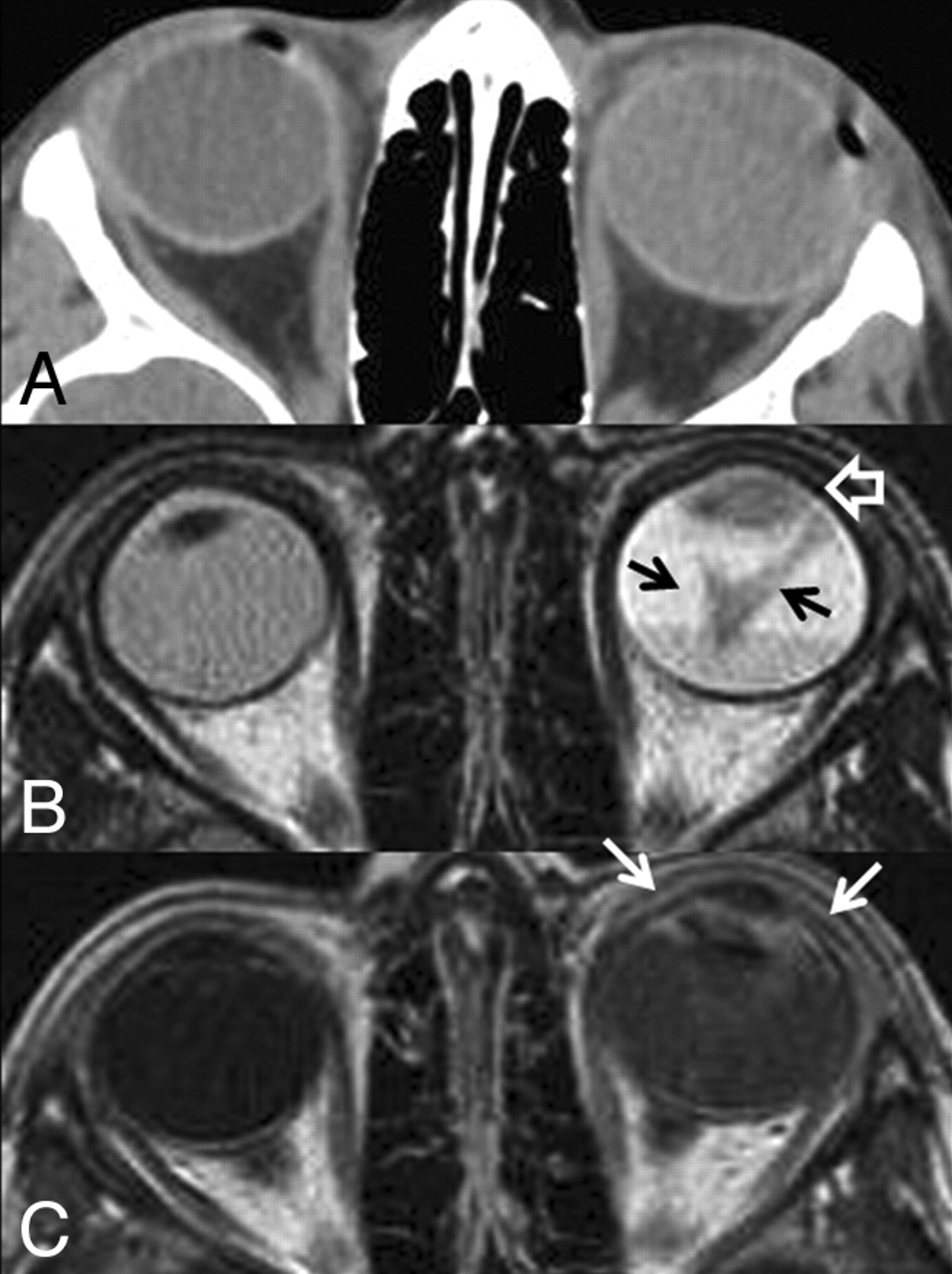
CT and MR imaging of the orbits demonstrated a noncalcified diffuse infiltrating mass involving a detached retina and anterior segment in the left globe (Fig 1), suggesting either advanced medulloepithelioma or diffuse infiltrating retinoblastoma. The patient subsequently underwent enucleation, providing a final pathologic diagnosis of retinoblastoma (Fig 2).

Fig 1.
  • Download figure
  • Open in new tab
  • Download powerpoint
Fig 1.

A, CT scan of the orbits. The left globe is asymmetrically enlarged and is hyperattenuating compared with the contralateral right globe, reflective of glaucoma and total retinal detachment, respectively. No calcifications are present. B, Axial T2-weighted MR image. There is total retinal detachment with a large fluid-fluid level. The detached retina is irregular and diffusely thickened (black arrows), and there is an irregular intermediate-signal-intensity mass involving the ciliary body and iris circumferentially (open white arrow). C, Axial T1-weighted postcontrast MR image. There is diffuse enhancement of the thickened detached retina and the anterior segment mass (white arrows). This constellation of findings is consistent with diffuse infiltrating retinal neoplasm extending into the anterior ocular segment. There is no evidence of optic nerve invasion or extrascleral extension.

Fig 2.
  • Download figure
  • Open in new tab
  • Download powerpoint
Fig 2.

Histologic section of the enucleated left globe. A, Photomicrograph (hematoxylin-eosin [HE] stain) of the whole-mount specimen at original magnification ×2. The ciliary body is diffusely infiltrated with neoplastic cells with round basophilic nuclei. The cells form rosettes around central lumens, known as Flexner-Wintersteiner rosettes, which are characteristic of retinoblastoma. B, Photomicrograph (HE stain) of the whole-mount specimen at original magnification ×40. The neoplastic cells have round basophilic nuclei with scant cytoplasm and are arranged in a rosette around a central lumen.

Discussion

Glaucoma in the first decade of life should be thoroughly evaluated for an ocular neoplasm. Up to 17%–22.8% of patients with retinoblastoma have increased intraocular pressures typically due to iris neovascularization.(6,7) However, with diffuse infiltrating retinoblastoma subtypes, glaucoma can result from neoplastic seeding of the trabecular meshwork in the anterior chamber, causing outflow obstruction of the aqueous humor.(6)

Retinoblastoma has 4 different growth patterns: endophytic growth toward the vitreous, exophytic growth toward the choroid, mixed exophytic and endophytic growth (most common), and diffuse infiltrating growth. Diffuse infiltrating retinoblastoma is rare, accounting for only 1%–2% of all cases,(1,8) and is characterized by plaquelike thickening of the retina without a discrete mass lesion. Patients often present when pseudoinflammatory complications occur once the lesion infiltrates into the anterior segment, and the condition may be misdiagnosed as uveitis and iritis.(5,8,9) When tumor involves the ciliary body, it can mimic a hypopyon, which can delay the diagnosis if anterior chamber paracentesis or tissue sampling is not performed.(5)

Secondary glaucoma from iris neovascularization actually occurs more frequently with medulloepithelioma, occurring in 60% of cases.(3) Medulloepitheliomas are typically malignant nonhereditary embryogenic neoplasms of the medullary epithelium almost always occurring in the ciliary body.(2,3) This neoplasm is divided into nonteratoid (60%) and teratoid (40%) histologic subtypes. Teratoid medulloepitheliomas are composed of heteroplastic tissue elements, which can include hyaline cartilage, skeletal muscle, and brain tissue.(2)

Anterior segment retinoblastoma and medulloepithelioma are nearly indistinguishable on imaging. Compared with conventional retinoblastomas, diffuse infiltrating retinoblastoma has an atypical imaging appearance, reflective of its growth pattern. On MR imaging, there is often retinal detachment without a discrete mass lesion. Diffuse irregular and nodular thickening of the detached retinal leaflets is evident, with abnormal enhancement that often extends to the anterior ocular segment (Fig 1).(8)

CT is not helpful in distinguishing these neoplasms because both may or may not calcify. Although 95% of conventional retinoblastomas calcify, diffusely infiltrating subtypes tend not to calcify.(1,5,8) Most medulloepitheliomas also do not calcify; however, calcifications do occur in 30% of teratoid subtypes (Fig 3).(2,3) Both neoplasms are hyperintense to the vitreous on T1-weighted images and hypointense on T2-weighted images and enhance either uniformly or heterogeneously.(2) The most reliable feature for distinguishing these 2 neoplasms is location. If the mass is confined to the ciliary body, then it is most likely a medulloepithelioma.(1) Anterior segment retinoblastomas typically have diffuse infiltration of the retina, iris, and ciliary body.(1,8) However, advanced cases of medulloepithelioma can infiltrate the retina and cause retinal detachment, so anterior-segment tumors that show diffuse infiltration cannot be reliably distinguished radiologically.(1)

Fig 3.
  • Download figure
  • Open in new tab
  • Download powerpoint
Fig 3.

Teratoid medulloepithelioma. A, Noncontrast axial CT scan shows a densely calcified mass of the ciliary body in the anterior segment of the right globe. Teratoid medulloepitheliomas can contain dystrophic calcifications due to the presence of hyaline cartilage tissue elements. B, Axial T1-weighted fat-saturated postcontrast MR image shows an enhancing mass of the ciliary body corresponding to the area of calcification on the CT scan. The mass is confined to the ciliary body; therefore, medulloepithelioma is more likely than diffuse infiltrating retinoblastoma. Alternative diagnoses for a ciliary body mass in pediatric patients include pyogenic granuloma, juvenile xanthogranuloma, mesoectodermal leiomyoma, and ciliary body cyst.

The role of imaging is not necessarily to make a specific pathologic diagnosis but rather to confirm the presence of an anterior segment mass and determine the local extent of the lesion. The presence of extrascleral or optic nerve invasion has significant prognostic and therapeutic implications for both retinoblastoma and medulloepithelioma. Tumors that are confined to the globe have excellent prognoses with 90%–95% 5-year survival after enucleation or focal therapy.(1,2,10) The globe is devoid of lymphatic tissue; therefore, metastatic disease is rare without episcleral or optic nerve involvement. When either neoplasm extends into the conjunctiva or extrascleral orbital soft tissues, the rate of metastatic disease and recurrence increases dramatically.(1,10–12) Metastatic disease for both neoplasms most commonly presents with diffuse subarachnoid seeding, resulting from optic nerve invasion, specifically with extension posterior to the lamina cribrosa.(12) On MR imaging, abnormal optic nerve enhancement that is contiguous with the tumor is indicative of postlaminar tumor infiltration.(10) Although limited in sensitivity for microscopic optic nerve invasion, this finding is 100% specific.(10) Patients with episcleral or optic nerve extension have poor overall prognoses and are often treated with orbital exenteration and chemotherapy as opposed to enucleation alone.(12)

References

  1. ↵
    1. Chung EM,
    2. Specht CS,
    3. Schroeder JW
    . Pediatric orbital tumors and tumorlike lesions: neuroepithelial lesions of the ocular globe and optic nerve. Radiographics 2007; 27: 1159– 86
    CrossRefPubMed
  2. ↵
    1. Vajaranant TS,
    2. Mafee MF,
    3. Kapur R,
    4. et al
    . Medulloepithelioma of the ciliary body and optic nerve: clinicopathologic, CT, and MR imaging features. Neuroimaging Clin N Am 2005; 15: 69– 83
    CrossRefPubMed
  3. ↵
    1. Shields JA,
    2. Eagle RC Jr.,
    3. Shields CL,
    4. et al
    . Congenital neoplasms of the nonpigmented ciliary epithelium (medulloepithelioma). Ophthalmology 1996; 103:
  4. ↵
    1. Karcioglu ZA,
    2. Abboud EB,
    3. Al-Mesfer SA,
    4. et al
    . Retinoblastoma in older children. J AAPOS 2002; 6: 26– 32
    CrossRefPubMed
  5. ↵
    1. Bhatnagar R,
    2. Vine AK
    . Diffuse infiltrating retinoblastoma. Ophthalmology 1991; 98: 1657– 61
    PubMed
  6. ↵
    1. de Leon JM,
    2. Walton DS,
    3. Latina MA,
    4. et al
    . Glaucoma in retinoblastoma. Semin Ophthalmol 2005; 20: 217– 22
    CrossRefPubMed
  7. ↵
    1. Shields CL,
    2. Shields JA,
    3. Shields MB,
    4. et al
    . Prevalence and mechanisms of secondary intraocular pressure elevation in eyes with intraocular tumors. Ophthalmology 1987; 94: 839– 46
    PubMed
  8. ↵
    1. Brisse HJ,
    2. Lumbroso L,
    3. Fréneaux PC,
    4. et al
    . Sonographic, CT, and MR imaging findings in diffuse infiltrative retinoblastoma: report of two cases with histologic comparison. AJNR Am J Neuroradiol 2001; 22: 499– 504
    Abstract/FREE Full Text
  9. ↵
    1. Foster BS,
    2. Mukai S
    . Intraocular retinoblastoma presenting as ocular and orbital inflammation. Int Ophthalmol Clin 1996; 36: 153– 60
    PubMed
  10. ↵
    1. de Graaf P,
    2. Barkhof F,
    3. Moll AC,
    4. et al
    . Retinoblastoma: MR imaging parameters in detection of tumor extent. Radiology 2005; 235: 197– 207
    CrossRefPubMed
  11. ↵
    1. Khelfaoui F,
    2. Validire P,
    3. Auperin A,
    4. et al
    . Histopathologic risk factors in retinoblastoma: a retrospective study of 172 patients treated in a single institution. Cancer 1996; 77: 1206– 13
    CrossRefPubMed
  12. ↵
    1. Chantada GL,
    2. Casco F,
    3. Fandiño AC,
    4. et al
    . Outcome of patients with retinoblastoma and postlaminar optic nerve invasion. Ophthalmology 2007; 114: 2083– 89
    CrossRefPubMed
  • Received December 27, 2008.
  • Accepted after revision January 28, 2009.
  • Copyright © American Society of Neuroradiology
View Abstract
PreviousNext
Back to top

In this issue

American Journal of Neuroradiology: 30 (8)
American Journal of Neuroradiology
Vol. 30, Issue 8
1 Sep 2009
  • Table of Contents
  • Index by author
Advertisement
Print
Download PDF
Email Article

Thank you for your interest in spreading the word on American Journal of Neuroradiology.

NOTE: We only request your email address so that the person you are recommending the page to knows that you wanted them to see it, and that it is not junk mail. We do not capture any email address.

Enter multiple addresses on separate lines or separate them with commas.
Anterior-Segment Retinoblastoma Mimicking Pseudoinflammatory Angle-Closure Glaucoma: Review of the Literature and the Important Role of Imaging
(Your Name) has sent you a message from American Journal of Neuroradiology
(Your Name) thought you would like to see the American Journal of Neuroradiology web site.
CAPTCHA
This question is for testing whether or not you are a human visitor and to prevent automated spam submissions.
Cite this article
R.R. Saket, M.F. Mafee
Anterior-Segment Retinoblastoma Mimicking Pseudoinflammatory Angle-Closure Glaucoma: Review of the Literature and the Important Role of Imaging
American Journal of Neuroradiology Sep 2009, 30 (8) 1607-1609; DOI: 10.3174/ajnr.A1565

Citation Manager Formats

  • BibTeX
  • Bookends
  • EasyBib
  • EndNote (tagged)
  • EndNote 8 (xml)
  • Medlars
  • Mendeley
  • Papers
  • RefWorks Tagged
  • Ref Manager
  • RIS
  • Zotero
0 Responses
Respond to this article
Share
Bookmark this article
Anterior-Segment Retinoblastoma Mimicking Pseudoinflammatory Angle-Closure Glaucoma: Review of the Literature and the Important Role of Imaging
R.R. Saket, M.F. Mafee
American Journal of Neuroradiology Sep 2009, 30 (8) 1607-1609; DOI: 10.3174/ajnr.A1565
del.icio.us logo Twitter logo Facebook logo Mendeley logo
  • Tweet Widget
  • Facebook Like
  • Google Plus One
Purchase

Jump to section

  • Article
    • SUMMARY:
    • Case Report
    • Discussion
    • References
  • Figures & Data
  • Info & Metrics
  • Responses
  • References
  • PDF

Related Articles

  • No related articles found.
  • PubMed
  • Google Scholar

Cited By...

  • Correlation of Apparent Diffusion Coefficient at 3T with Prognostic Parameters of Retinoblastoma
  • MRI of retinoblastoma
  • Crossref (12)
  • Google Scholar

This article has been cited by the following articles in journals that are participating in Crossref Cited-by Linking.

  • Guidelines for imaging retinoblastoma: imaging principles and MRI standardization
    Pim de Graaf, Sophia Göricke, Firazia Rodjan, Paolo Galluzzi, Philippe Maeder, Jonas A. Castelijns, Hervé J. Brisse
    Pediatric Radiology 2012 42 1
  • Correlation of Apparent Diffusion Coefficient at 3T with Prognostic Parameters of Retinoblastoma
    A.A.K. Abdel Razek, S. ElKhamary, S. Al-Mesfer, H.M. AlKatan
    American Journal of Neuroradiology 2012 33 5
  • MRI of retinoblastoma
    A A K A Razek, S Elkhamary
    The British Journal of Radiology 2011 84 1005
  • Clinical Presentation and Genetic Paradigm of Diffuse Infiltrating Retinoblastoma: A Review
    Peter G. Traine, Katharina J. Schedler, Eduardo B. Rodrigues
    Ocular Oncology and Pathology 2016 2 3
  • Camouflage Nanoparticles Enable in Situ Bioluminescence-Driven Optogenetic Therapy of Retinoblastoma
    Jiali Ding, Jianping Lu, Qian Zhang, Yanan Xu, Bin Song, Yuqi Wu, Haoliang Shi, Binbin Chu, Houyu Wang, Yao He
    ACS Nano 2023 17 8
  • A typical anterior retinoblastoma: diagnosis by aqueous humor cell-free DNA analysis
    Stephanie N. Kletke, Sameh Soliman, Hilary Racher, Ashwin Mallipatna, Furqan Shaikh, Kamiar Mireskandari, Brenda L. Gallie
    Ophthalmic Genetics 2022 43 6
  • Masquerade syndrome: A review of uveitic imposters
    Parthopratim Dutta Majumder, Vikas Khetan, Jyotirmay Biswas
    Asia-Pacific Journal of Ophthalmology 2024 13 2
  • Vibrational Spectral Biodiagnosis of Ocular Calcification
    Shan-Yang Lin
    Applied Spectroscopy Reviews 2014 49 1
  • A case of retinoblastoma masquerading as endophthalmitis: Unusual presentation and clinicopathological correlation
    Ghadah M.S. AlQahtani, Hind M. Alkatan, Saleh AlMesfer, Sahar Elkhamary, Azza M.Y. Maktabi
    International Journal of Surgery Case Reports 2024 123
  • La tomografía computarizada y la resonancia magnética en patologías frecuentes de órbita y vía óptica. Una revisión
    Lina Marcela Acuña Bedoya, Sara Viviana Angulo Sánchez
    Ciencia y Tecnología para la Salud Visual y Ocular 2013 11 1

More in this TOC Section

  • Correlation of Apparent Diffusion Coefficient at 3T with Prognostic Parameters of Retinoblastoma
  • Parathyroid Lesions: Characterization with Dual-Phase Arterial and Venous Enhanced CT of the Neck
  • Efficacy of Diffusion-Weighted Imaging for the Differentiation between Lymphomas and Carcinomas of the Nasopharynx and Oropharynx: Correlations of Apparent Diffusion Coefficients and Histologic Features
Show more Head and Neck

Similar Articles

Advertisement

Indexed Content

  • Current Issue
  • Accepted Manuscripts
  • Article Preview
  • Past Issues
  • Editorials
  • Editors Choice
  • Fellow Journal Club
  • Letters to the Editor

Cases

  • Case Collection
  • Archive - Case of the Week
  • Archive - Case of the Month
  • Archive - Classic Case

Special Collections

  • Special Collections

Resources

  • News and Updates
  • Turn around Times
  • Submit a Manuscript
  • Author Policies
  • Manuscript Submission Guidelines
  • Evidence-Based Medicine Level Guide
  • Publishing Checklists
  • Graphical Abstract Preparation
  • Imaging Protocol Submission
  • Submit a Case
  • Become a Reviewer/Academy of Reviewers
  • Get Peer Review Credit from Publons

Multimedia

  • AJNR Podcast
  • AJNR SCANtastic
  • Video Articles

About Us

  • About AJNR
  • Editorial Board
  • Not an AJNR Subscriber? Join Now
  • Alerts
  • Feedback
  • Advertise with us
  • Librarian Resources
  • Permissions
  • Terms and Conditions

American Society of Neuroradiology

  • Not an ASNR Member? Join Now

© 2025 by the American Society of Neuroradiology All rights, including for text and data mining, AI training, and similar technologies, are reserved.
Print ISSN: 0195-6108 Online ISSN: 1936-959X

Powered by HighWire