
Article Figures & Data
Figures
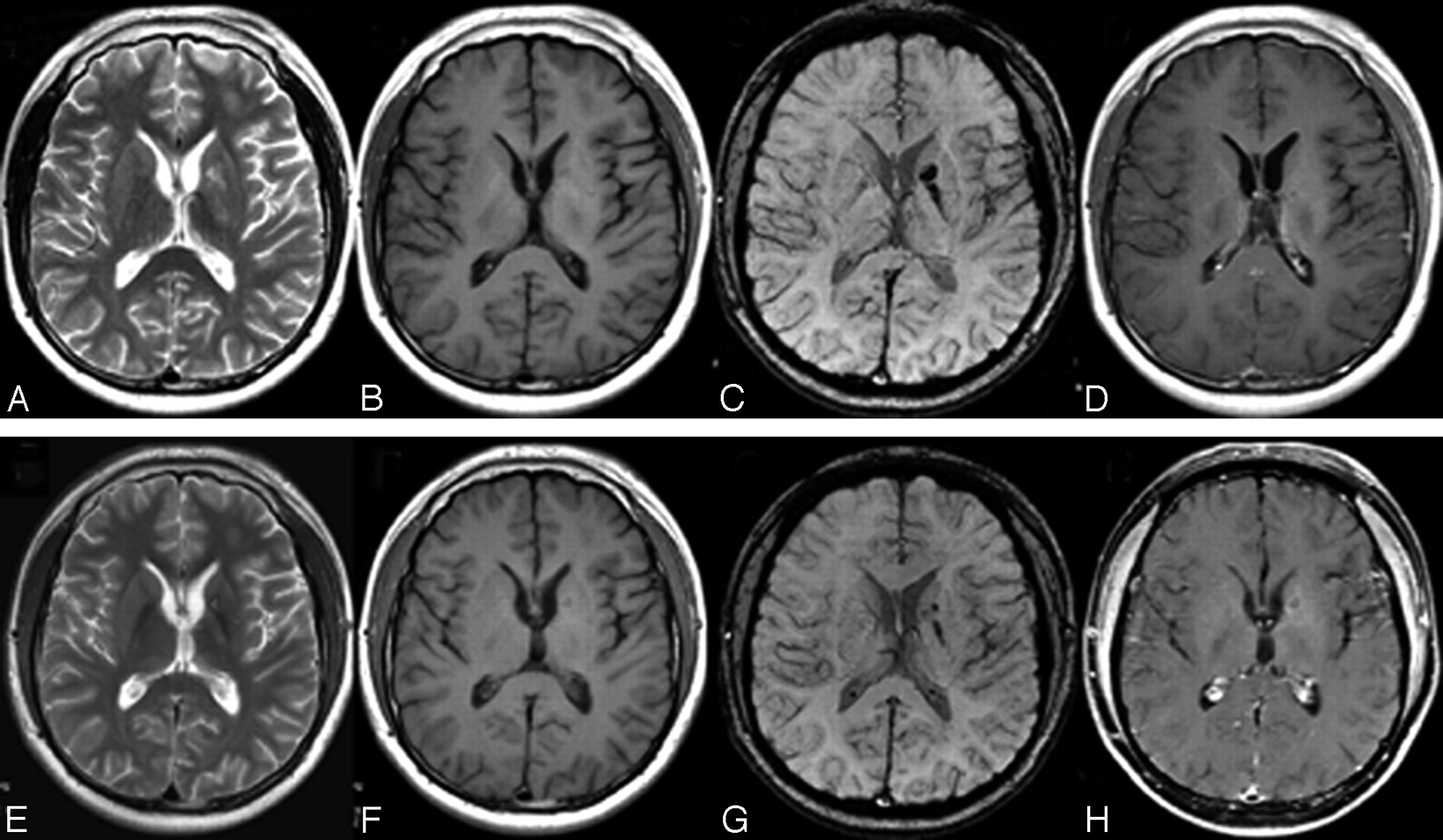
- Fig 1.
MR imaging of case 1. A, Axial T2WI shows patchy hyperintensity at the left BG and the thalamus. B, T1WI shows slight hyperintensity at the left BG without obvious mass effect. C, SWI shows obvious hypointensity at the left posterior limb of internal capsule, globus pallidus, and thalamus. D, Postcontrast T1WI shows no enhancement. E and F, Repeat MR imaging 66 days later shows increased tumor size and hyperintensity on both T2WI (E) and T1WI (F), indicating intratumoral bleeding. G–I, Twenty-seven days after RT, MR images show shrinkage of the tumor on T2WI (G), T1WI (H), and postcontrast T1WI (I). Follow-up SWI was not performed in this case.
- Fig 2.
MR imaging of case 2. A, Axial T2WI shows patchy hyperintensity at the left BG. B, The tumor is invisible on T1WI. C, SWI shows obvious hypointensity at the globus pallidus, the genu, and the posterior limb of the internal capsule on the left side. D, Postcontrast T1WI shows no enhancement. E–H, Twenty-seven days after RT, the tumor markedly decreases on follow-up T2WI (E), T1WI (F) (still invisible), and SWI (G) but shows slight ringlike enhancement (H).
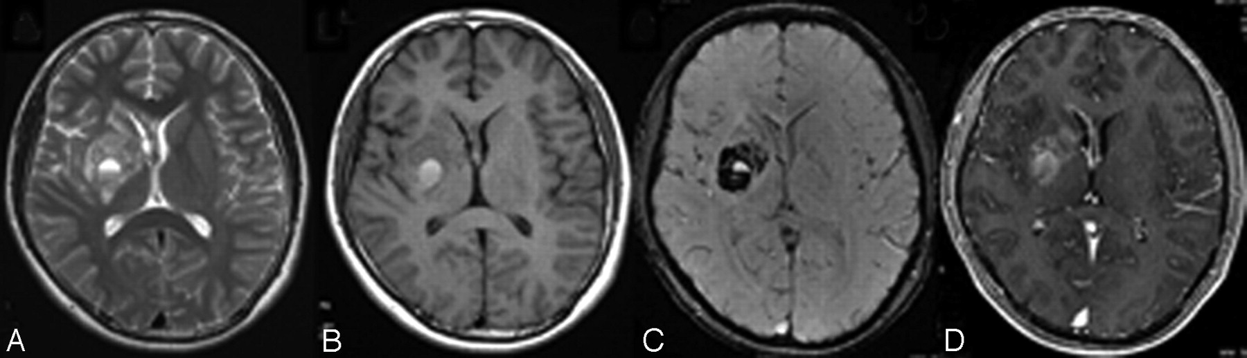
- Fig 3.
MR imaging of case 4. A and B, Axial T2WI (A) and T1WI (B) show heterogeneous signal-intensity changes at the right BG, involving the genu of corpus callosum and septum pellucidum, and the right thalamus is also involved (not shown). C, SWI demonstrates obvious heterogeneous hypointensity together with fluid-fluid level. D, Heterogeneous enhancement in the BG and subependymal spread are noted on the postcontrast T1WI.
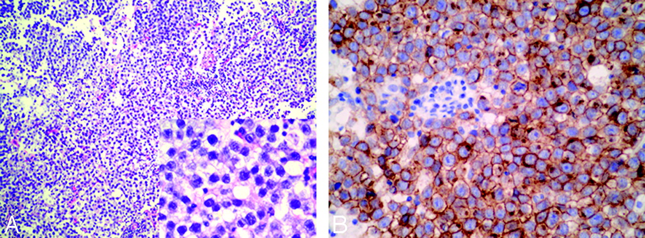
- Fig 4.
Photomicrographs of histopathology of the right BG germinoma in case 4. A, Standard HE coloration (original magnification × 100; insert figure: original magnification ×400) shows that the tumor is composed of large cells with vacuolated cytoplasm and round or ovoid nuclei with prominent nucleoli. Lymphocytic element admixes with tumor cells focally. B, Immunolabeling of germ cells with CD117a (c-kit) is positive (original magnification ×400).


Tables
Case No. Age (yr) Sex Symptom Duration (mo) Weakness of Limbs Diabetes Insipidus Increased ICP Diagnosis Tumor Marker (AFP, HCG) Tumor Location T1WI T2WI Contrast Enhancement SWI 1 14 M 2 Y N N HP Neg BG/TH Slight hyper Hyper N Hypo 2 16 M 3 Y Y N HP Neg BG Iso Hyper N Hypo 3 14 M 6 Y N Y HP Neg BG Hetero/hyper Hetero Y (hetero) Hetero/Hypo 4 12 M 16 Y N Y HP N/A BG/TH Hetero/hyper Hetero Y (hetero) Hetero/Hypo 5 13 M 8 Y Y N HP Neg BG/TH Hetero Hetero Y (hetero) Hetero/Hypo 6 15 M 2 Y N N HP Neg BG Iso Hyper N Hypo Note:—Y indicates yes; N, no; ICP, intracranial pressure; HP, histologically proven; AFP, α-fetoprotein; HCG, human chorionic gonadotropin; N/A, not available; Neg, negative; BG, basal ganglia; TH, thalamus; Hetero, heterogeneous; Hyper, hyperintense; Hypo, hypointense; Iso, isointense; SWI, susceptibility-weighted imaging; T1WI, T1-weighted imaging; T2WI, T2-weighted imaging.
Case No. T1WI (mm) T2WI (mm) SWI (mm) 1 4 7 18 2 N/A 8 21 3 38 38 32 4 45 45 35 5 15 15 12 6 N/A 8 20 Case No. T1WI T2WI SWI 1 22.58 32.29 −80.43 2 13.30 67.48 −100.29 3 152.46 16.62 −10.74 4 25.60 130.10 −15.90 5 -18.19 197.00 −8.64 6 18.94 45.13 −70.98 Reference Total No. Hemorrhage Mass Effect Contrast Enhancement SWI Radiologic Features on T1WI Radiologic Features on T2WI Kim et al1 14 7 4 14 None Hyper in 7, hetero in 7 Hyper in 7, hetero in 7 Klein et al2 1 0 1 1 None Hetero Hetero Wong et al3 6 (3 performed MR imaging) No data 2 3 None Hetero in 3 Hetero in 3 Moon et al5 10 7 No data 10 None Hyper in 5, hetero in 5 Hyper in 5, hetero in 5 Higano et al6 6 1 3 No data None hyper in 2, hetero in 4 Hyper in 2, hetero in 4 Liu et al7 1 0 0 1 None Hypo Hetero Ozelame et al8 4 2 2 4 None Hyper in 2, hetero in 2 Hyper in 2, hetero in 2 Rushing et al12 2 1 1 No data None Hypo in 2 Hyper in 1, hetero in 1 Nagata et al13 1 0 0 1 None Hypo Hyper Fujii et al18 1 0 0 1 Yes Hypo Hyper Current series 6 6 3 3 Yes Iso in 2, hyper in 1, hetero in 3 Hyper in 3, hetero in 3
In this issue


Jump to section
Related Articles
Cited By...
- No citing articles found.