
Article Figures & Data
Figures
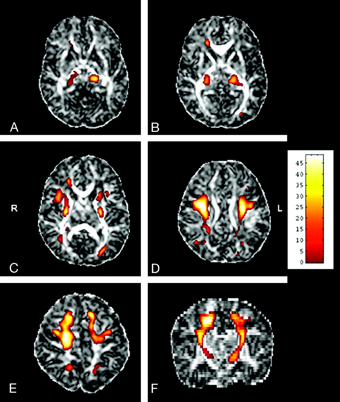
- Fig 1.
Areas with significant FA increase with age are located in the thalami (A, B), the anterior and posterior arms of the internal capsules, and the optic radiations (C), the centrum semiovale (D), and the subcortical projection pathways of the frontal and parietal cortices (E) in the axial (A–E) and coronal (F) planes. The anatomic underlay is the FA image of the template. The color maps represent the F-scores. Only regions of more than 50 voxels attaining a corrected P value of less than .05 for the voxel-level of statistical inference were considered significant.
- Fig 2.
Regression plots derived from gestational age in relationship to FA values at the most statistically significant voxels located in the CST (F = 59.63), the thalamus (F = 31.23), the STR (F = 30.54), the ATR (F = 29.61), the CR (F = 29.44), and the PTR (F = 25.27) on the right side of the brain. The positive slopes of the regression lines indicate that FA increases with age.
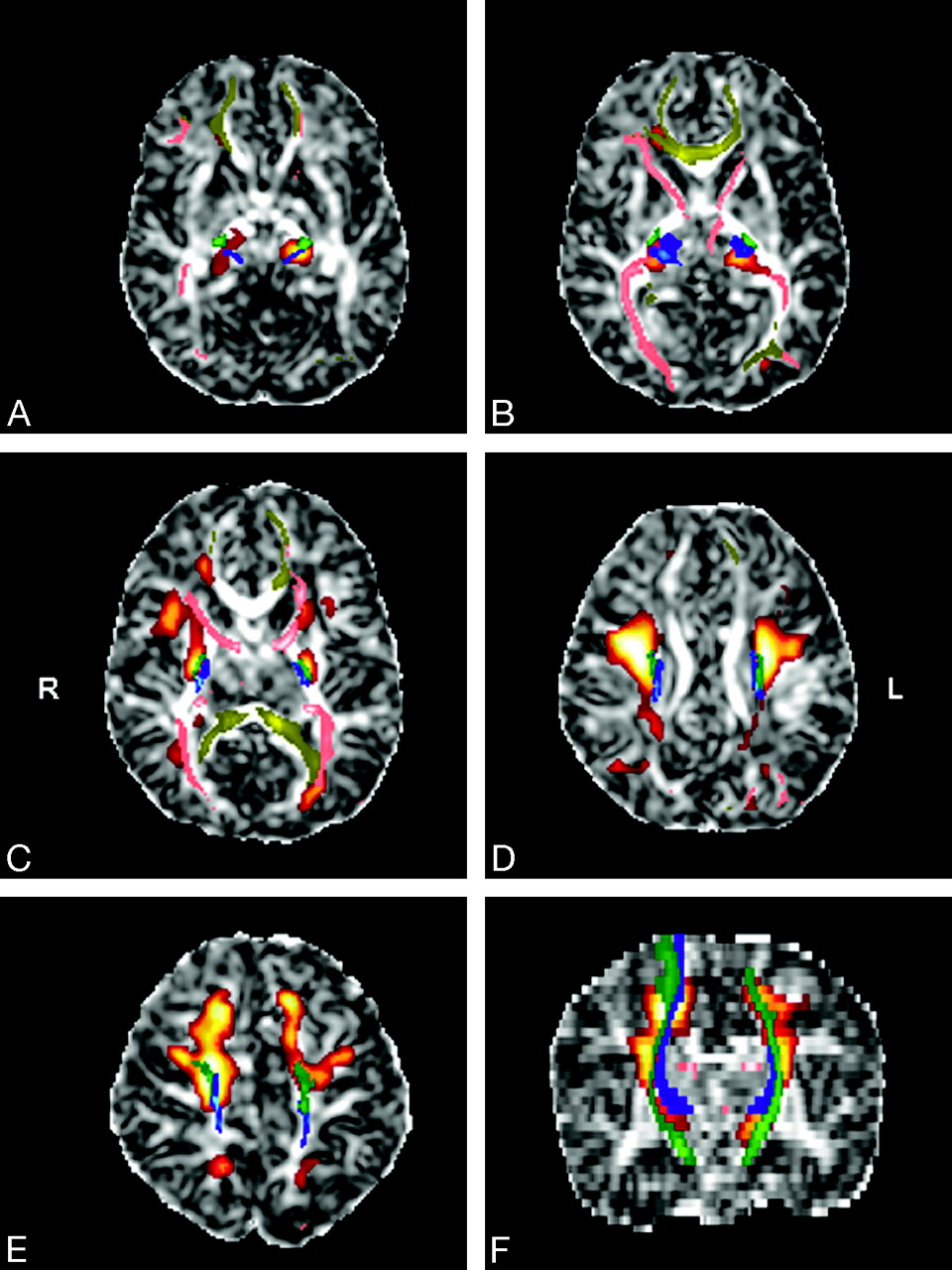
- Fig 3.
Superimposition of statistical parametric maps (red-orange), probabilistic tractography images (blue, green, pink, yellow), and FA images of the template in the axial (A–E) and coronal (F) planes. The CST (green), the STR (blue), the ATR, the PTR (pink), and the CR (yellow) are included in the significant voxels of the parametric analysis.


In this issue


Jump to section
Related Articles
Cited By...
- Effect of number of diffusion encoding directions in Neonatal Diffusion Tensor Imaging using Tract-Based Spatial Statistical analysis
- Structural network maturation of the preterm human brain
- Comparison of Spin-Echo T1- and T2-Weighted and Gradient-Echo T1-Weighted Images at 3T in Evaluating Very Preterm Neonates at Term-Equivalent Age
- White Matter Abnormalities Are Related to Microstructural Changes in Preterm Neonates at Term-Equivalent Age: A Diffusion Tensor Imaging and Probabilistic Tractography Study
- Gender Differences in Language and Motor-Related Fibers in a Population of Healthy Preterm Neonates at Term-Equivalent Age: A Diffusion Tensor and Probabilistic Tractography Study