
Article Figures & Data
Figures
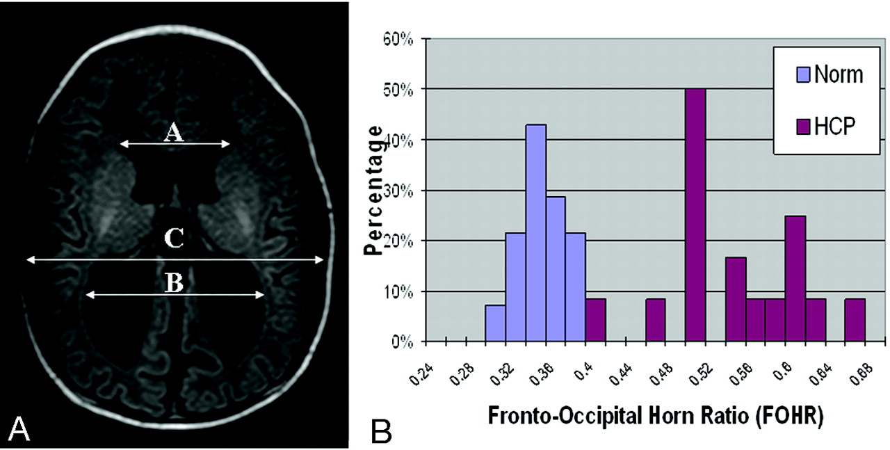
- Fig 1.
A, The measurement and calculation of the FOHR are demonstrated on an axial section of T1-weighted MR imaging study from a child with hydrocephalus in the preshunt group. B, Histogram showing that children in the control group demonstrate a narrow range (0.29∼0.37) in the FOHR, whereas children in the patient group have a distinct range of distribution of the ratio (0.40∼0.65).
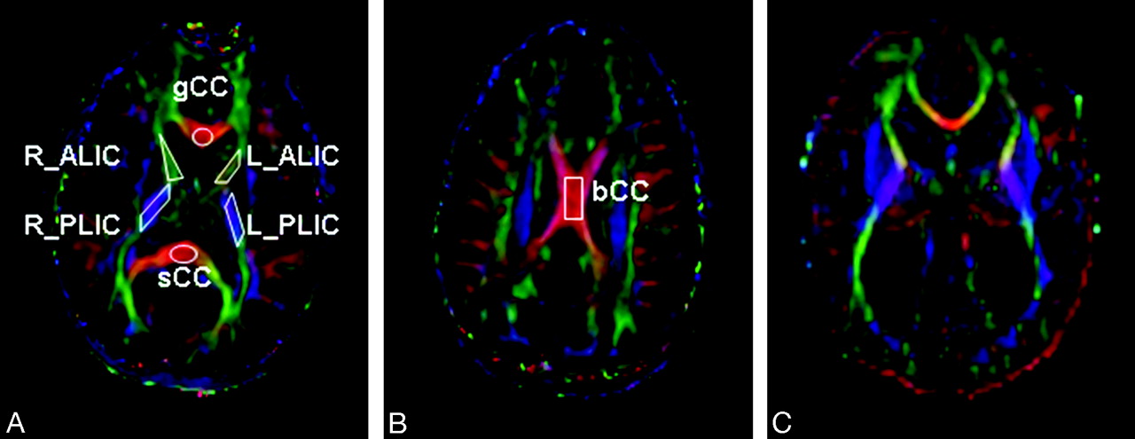
- Fig 2.
Color-coded FA maps for a child in the healthy control group (A and B) and a child with hydrocephalus in the preshunt group (C). The orientation of white matter tracts is coded with different colors with red, green, and blue indicating left-right, anteroposterior, and superior-inferior, respectively. The ROIs used in the study are outlined for the gCC, L_ALIC and R_ALIC, L_PLIC and R_PLIC and sCC in (A), and the bCC in (B). In both ALIC and PLIC, because there were no statistically significant differences between the left and the right side, the average values of DTI measurements were used in the analysis.
- Fig 3.
A, sCC. B, PLIC. Scatterplots of FA fitted to a linear model (solid line) in healthy children and in children with hydrocephalus (regression line not shown). The 95% prediction intervals are shown in dashed lines representing age-specific range in which a normal value is supposed to be at 95% certainty.
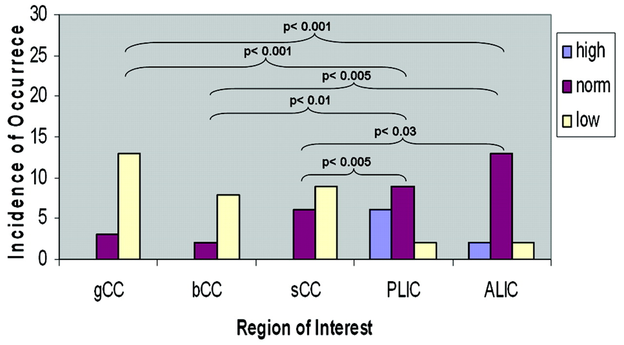
- Fig 4.
Comparisons of frequency of occurrence of FA categorized as abnormally high, normal, and abnormally low in the 5 ROIs. Statistics were conducted on the basis of the Fisher exact test with Freeman-Halton extension.


Tables
Pt. No. Sex Age Preop DTI (mo) Age Surg (mo) Clinical Signs/Symptoms HCP Diagnosis (Type/Cause) Clinical Outcome 1 M 0.03 0.13 Macrocephaly, accelerated head growth Congenital Developmentally appropriate for age 2 F 1.81 3.04 Macrocephaly, accelerated head growth Congenital Stable with developmental delay 3 F 16.14 17.28 Accelerated head growth after repair of myelomeningocele Myelomeningocele Stable with decreased lower extremity movement 4 M 6.25 6.75 Accelerated head growth Peter plus syndrome Stable with developmental delay 5 M 9.60 10.30 Progressive ventriculomegaly Posttraumatic Stable with hemiparesis 6 F 4.90 5.10 Accelerated head growth after repair of myelomeningocele Myelomeningocele Stable with decreased lower extremity movement 7 M 1.71 2.61 Macrocephaly, accelerated head growth Communicating Stable with developmental delay 8 M 0.07 0.70 Accelerated head growth Communicating Normal development 9 M 8.02 8.96 Accelerated head growth Postmeningitis Stable with developmental delay 10 M 6.84 6.94 Accelerated head growth Dandy-Walker variant Stable neurologically 11 M 1.61 2.38 Accelerated head growth Aqueduct stenosis Stable neurologically 12 M 3.32 5.85 Downward gaze Aqueduct stenosis Stable neurologically 13 F 6.12 6.18 Accelerated head growth Quadrigeminal plate arachnoid cyst Normal development 14 F 3.16 3.25 Accelerated head growth Prematurity, meningitis Stable neurologically 15 M 0.69 1.61 Accelerated head growth Prematurity, IVH Stable neurologically 16 F 0.79 0.99 Apnea/brady, bulging fontanel Prematurity, IVH Stable neurologically 17 F 8.05 8.94 Papilledema, enlarged ventricles Achondroplasia Resolved papilledema, stable Note:—HCP indicates hydrocephalus; DTI, diffusion tensor imaging; IVH, intraventricular hemorrhage; Preop, preoperative; Surg, surgery.
- Table 2:
Comparison of DTI parameters between children with HCP and age-matched control subjects
ROI DTI Parameter n HCP Norm Paired t test df P gCC FA 16 0.341 ± 0.101 0.525 ± 0.092 6.66 15 .002* MD† 16 1.724 ± 0.848 1.355 ± 0.398 2.66 15 .049* Axial diffusivity† 16 2.368 ± 1.100 2.201 ± 0.559 0.58 15 NS Radial diffusivity† 16 1.406 ± 0.741 0.929 ± 0.341 4.08 15 .010* bCC FA 7 0.270 ± 0.113 0.513 ± 0.088 4.20 6 .023* MD† 7 1.750 ± 0.634 1.334 ± 0.443 3.50 6 .043* Axial diffusivity† 7 2.206 ± 0.64 2.181 ± 0.681 0.135 6 NS Radial diffusivity† 7 1.526 ± 0.647 0.907 ± 0.335 4.22 6 .028* sCC FA 15 0.403 ± 0.164 0.564 ± 0.088 3.42 14 .027* MD† 15 1.722 ± 0.757 1.393 ± 0.454 1.92 14 NS Axial diffusivity† 15 2.426 ± 0.876 2.383 ± 0.717 0.19 14 NS Radial diffusivity† 15 1.373 ± 0.722 0.946 ± 0.408 2.62 14 .050* ALIC FA 17 0.400 ± 0.112 0.402 ± 0.082 0.07 16 NS MD† 17 1.099 ± 0.473 1.048 ± 0.367 0.64 16 NS Axial diffusivity† 17 1.596 ± 0.586 1.531 ± 0.457 0.58 16 NS Radial diffusivity† 17 0.854 ± 0.427 0.811 ± 0.331 0.60 16 NS PLIC FA 17 0.530 ± 0.123 0.501 ± 0.068 1.71 16 NS MD† 17 1.060 ± 0.475 1.062 ± 0.312 0.02 16 NS Axial diffusivity† 17 1.701 ± 0.628 1.697 ± 0.435 0.03 16 NS Radial diffusivity† 17 0.741 ± 0.417 0.749 ± 0.266 0.12 16 NS Note:—ROI indicates region of interest; df, degrees of freedom; gCC, genu of the corpus callosum; FA, fractional anisotropy; MD, mean diffusivity; bCC, body of the corpus callosum; sCC, splenium of the corpus callosum; ALIC, anterior limb of the internal capsule; PLIC, posterior limb of the internal capsule; NS, not significant.
* P value is adjusted for multiple testing with the false discovery rate method.
† The units of MD, axial diffusivity, and radial diffusivity are all in 10 −3mm2/s.
ROI Group Linear Regression R2 P Error Mean Error SD gCC Norm Y = 0.019X+0.438 0.79 .0010* 0 0.04 HCP Y = 0.010X+0.294 0.21 NS 0 0.09 bCC Norm Y = 0.016X+0.446 0.67 .0005* 0 0.05 HCP Y = 0.005X+0.310 0.03 NS 0 0.15 sCC Norm Y = 0.017X+0.486 0.69 .0003* 0 0.05 HCP Y = 0.008X+0.365 0.05 NS 0 0.16 ALIC Norm Y = 0.016X+0.326 0.66 .0003* 0 0.05 HCP Y = 0.014X+0.333 0.30 .0314* 0 0.09 PLIC Norm Y = 0.014X+0.437 0.72 .0002* 0 0.04 HCP Y = 0.021X+0.431 0.55 .0017* 0 0.08 * P value is adjusted for multiple testing with the false discovery rate method.
In this issue


Jump to section
Related Articles
Cited By...
- Evidence of supratentorial white matter injury prior to treatment in children with posterior fossa tumours using diffusion MRI
- The Construction of a Predictive Composite Index for Decision-Making of CSF Diversion Surgery in Pediatric Patients following Prenatal Myelomeningocele Repair
- Diffusion Tensor Imaging and Fiber Tractography in Children with Craniosynostosis Syndromes
- White Matter Microstructural Abnormality in Children with Hydrocephalus Detected by Probabilistic Diffusion Tractography
- DTI Values in Key White Matter Tracts from Infancy through Adolescence
- Diffusion Tensor Imaging Properties and Neurobehavioral Outcomes in Children with Hydrocephalus
- Differences in Microstructural Alterations of the Hippocampus in Alzheimer Disease and Idiopathic Normal Pressure Hydrocephalus: A Diffusion Tensor Imaging Study