Advertisement

Main menu

  • Home
  • Content
    • Current Issue
    • Accepted Manuscripts
    • Article Preview
    • Past Issue Archive
    • AJNR Case Collection
    • Case of the Week Archive
    • Classic Case Archive
    • Case of the Month Archive
  • Special Collections
    • Spinal CSF Leak Articles (Jan 2020-June 2024)
    • 2024 AJNR Journal Awards
    • Most Impactful AJNR Articles
  • Multimedia
    • AJNR Podcast
    • AJNR Scantastics
    • Video Articles
  • For Authors
    • Submit a Manuscript
    • Author Policies
    • Fast publishing of Accepted Manuscripts
    • Graphical Abstract Preparation
    • Manuscript Submission Guidelines
    • Imaging Protocol Submission
    • Submit a Case for the Case Collection
  • About Us
    • About AJNR
    • Editorial Board
  • More
    • Become a Reviewer/Academy of Reviewers
    • Subscribers
    • Permissions
    • Alerts
    • Feedback
    • Advertisers
    • ASNR Home
  • Other Publications
    • ajnr

User menu

  • Alerts
  • Log in

Search

  • Advanced search
American Journal of Neuroradiology

American Journal of Neuroradiology

ASHNR American Society of Functional Neuroradiology ASHNR American Society of Pediatric Neuroradiology ASSR
  • Alerts
  • Log in

Advanced Search

  • Home
  • Content
    • Current Issue
    • Accepted Manuscripts
    • Article Preview
    • Past Issue Archive
    • AJNR Case Collection
    • Case of the Week Archive
    • Classic Case Archive
    • Case of the Month Archive
  • Special Collections
    • Spinal CSF Leak Articles (Jan 2020-June 2024)
    • 2024 AJNR Journal Awards
    • Most Impactful AJNR Articles
  • Multimedia
    • AJNR Podcast
    • AJNR Scantastics
    • Video Articles
  • For Authors
    • Submit a Manuscript
    • Author Policies
    • Fast publishing of Accepted Manuscripts
    • Graphical Abstract Preparation
    • Manuscript Submission Guidelines
    • Imaging Protocol Submission
    • Submit a Case for the Case Collection
  • About Us
    • About AJNR
    • Editorial Board
  • More
    • Become a Reviewer/Academy of Reviewers
    • Subscribers
    • Permissions
    • Alerts
    • Feedback
    • Advertisers
    • ASNR Home
  • Follow AJNR on Twitter
  • Visit AJNR on Facebook
  • Follow AJNR on Instagram
  • Join AJNR on LinkedIn
  • RSS Feeds

Welcome to the new AJNR, Updated Hall of Fame, and more. Read the full announcements.


AJNR is seeking candidates for the position of Associate Section Editor, AJNR Case Collection. Read the full announcement.


Case ReportPediatrics

The Rare Third Branchial Cleft Cyst

M.J. Joshi, M.J. Provenzano, R.J.H. Smith, Y. Sato and W.R.K. Smoker
American Journal of Neuroradiology October 2009, 30 (9) 1804-1806; DOI: https://doi.org/10.3174/ajnr.A1627
M.J. Joshi
  • Find this author on Google Scholar
  • Find this author on PubMed
  • Search for this author on this site
M.J. Provenzano
  • Find this author on Google Scholar
  • Find this author on PubMed
  • Search for this author on this site
R.J.H. Smith
  • Find this author on Google Scholar
  • Find this author on PubMed
  • Search for this author on this site
Y. Sato
  • Find this author on Google Scholar
  • Find this author on PubMed
  • Search for this author on this site
W.R.K. Smoker
  • Find this author on Google Scholar
  • Find this author on PubMed
  • Search for this author on this site
  • Article
  • Figures & Data
  • Info & Metrics
  • Responses
  • References
  • PDF
Loading

Article Figures & Data

Figures

  • Fig 1.
    • Download figure
    • Open in new tab
    • Download powerpoint
    Fig 1.

    Sagittal (A) T1 and axial (B) T1-and (C) T2-weighted MR images. A paraglottic and retropharyngeal cystic mass with an air-fluid level is present, displacing the left carotid sheath structures laterally and anteriorly.
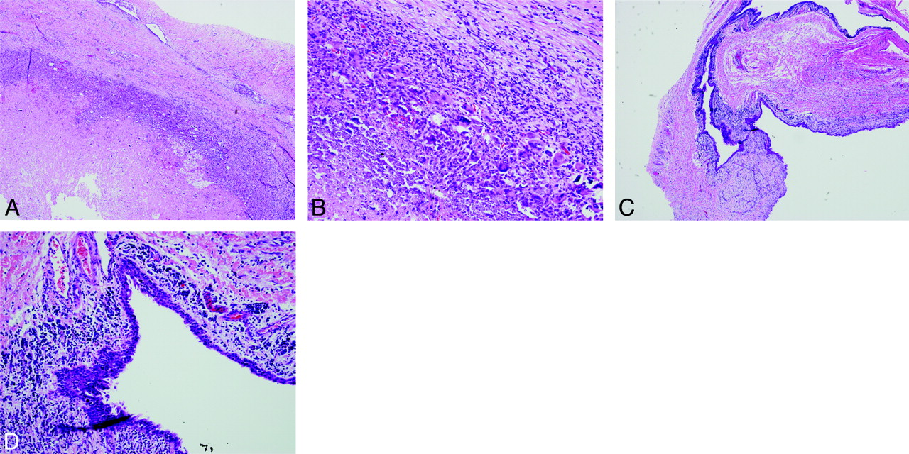
  • Fig 2.
    • Download figure
    • Open in new tab
    • Download powerpoint
    Fig 2.

    Photomicrographs (A, low-powered) and (B, high-powered) show rupture of the cyst wall with granulation tissue and foreign-body giant-cell reaction. Photomicrographs (C, low-powered and D, high-powered) demonstrate a cyst lining composed of inflamed squamous mucosa with surrounding lymphoid tissue.

PreviousNext
Back to top

In this issue

American Journal of Neuroradiology: 30 (9)
American Journal of Neuroradiology
Vol. 30, Issue 9
1 Oct 2009
  • Table of Contents
  • Index by author
Advertisement
Print
Download PDF
Email Article

Thank you for your interest in spreading the word on American Journal of Neuroradiology.

NOTE: We only request your email address so that the person you are recommending the page to knows that you wanted them to see it, and that it is not junk mail. We do not capture any email address.

Enter multiple addresses on separate lines or separate them with commas.
The Rare Third Branchial Cleft Cyst
(Your Name) has sent you a message from American Journal of Neuroradiology
(Your Name) thought you would like to see the American Journal of Neuroradiology web site.
CAPTCHA
This question is for testing whether or not you are a human visitor and to prevent automated spam submissions.
Cite this article
M.J. Joshi, M.J. Provenzano, R.J.H. Smith, Y. Sato, W.R.K. Smoker
The Rare Third Branchial Cleft Cyst
American Journal of Neuroradiology Oct 2009, 30 (9) 1804-1806; DOI: 10.3174/ajnr.A1627

Citation Manager Formats

  • BibTeX
  • Bookends
  • EasyBib
  • EndNote (tagged)
  • EndNote 8 (xml)
  • Medlars
  • Mendeley
  • Papers
  • RefWorks Tagged
  • Ref Manager
  • RIS
  • Zotero
0 Responses
Respond to this article
Share
Bookmark this article
The Rare Third Branchial Cleft Cyst
M.J. Joshi, M.J. Provenzano, R.J.H. Smith, Y. Sato, W.R.K. Smoker
American Journal of Neuroradiology Oct 2009, 30 (9) 1804-1806; DOI: 10.3174/ajnr.A1627
del.icio.us logo Twitter logo Facebook logo Mendeley logo
  • Tweet Widget
  • Facebook Like
  • Google Plus One
Purchase

Jump to section

  • Article
    • Abstract
    • Case Report
    • Discussion
    • References
  • Figures & Data
  • Info & Metrics
  • Responses
  • References
  • PDF

Related Articles

  • Erratum
  • PubMed
  • Google Scholar

Cited By...

  • Neck Mass in a Full-term Infant with Hypoxic-Ischemic Encephalopathy
  • Crossref (19)
  • Google Scholar

This article has been cited by the following articles in journals that are participating in Crossref Cited-by Linking.

  • Imaging of Pediatric Neck Masses
    Elliott R. Friedman, Susan D. John
    Radiologic Clinics of North America 2011 49 4
  • Pyriform Sinus Fistula in the Fetus and Neonate: A Systematic Review of Published Cases
    Tong Chen, Jianglong Chen, Qingfeng Sheng, Linlin Zhu, Zhibao Lv
    Frontiers in Pediatrics 2020 8
  • Atypical imaging observations of branchial cleft cysts
    SU HU, CHUN-HONG HU, LING YANG, JIAN-MING XING, JIAN-HUA CHEN, ZI-LI GE, JI-SHENG LIU
    Oncology Letters 2014 7 1
  • Congenital cystic lesions of the neck
    Mahmud Mossa-Basha, David M. Yousem
    Applied Radiology 2013
  • Features of Diagnostics, Clinical Course and Treatment of the Branchial Cleft Cysts
    Journal of Diagnostics and Treatment of Oral and Maxillofacial Pathology 2017 1 1
  • Lumps and Bumps of the Neck in Children—Neuroimaging of Congenital and Acquired Lesions
    Marjolein H.G. Dremmen, Aylin Tekes, Samantha Mueller, Donna Seyfert, David E. Tunkel, Thierry A.G.M. Huisman
    Journal of Neuroimaging 2016 26 6
  • Cysts in the Posterior Triangle of the Neck in Adults
    Beatriz Brea-Álvarez, Amaya Roldán-Fidalgo
    Acta Otorrinolaringologica (English Edition) 2015 66 2
  • Third branchial cleft anomalies
    Janet W. Lee, Jamie L. Funamura
    Operative Techniques in Otolaryngology-Head and Neck Surgery 2017 28 3
  • Quistes en el triángulo cervical posterior en adultos
    Beatriz Brea-Álvarez, Amaya Roldán-Fidalgo
    Acta Otorrinolaringológica Española 2015 66 2
  • Diagnostic Imaging: Head and Neck
    2017

More in this TOC Section

  • Comparison of Image Quality and Radiation Dose in Pediatric Temporal Bone CT Using Photon-Counting Detector CT and Energy-Integrating Detector CT
  • SyMRI & MR Fingerprinting in Brainstem Myelination
  • ASL in PEDS Nontraumatic Orbital Lesions
Show more Pediatrics

Similar Articles

Advertisement

Indexed Content

  • Current Issue
  • Accepted Manuscripts
  • Article Preview
  • Past Issues
  • Editorials
  • Editors Choice
  • Fellow Journal Club
  • Letters to the Editor

Cases

  • Case Collection
  • Archive - Case of the Week
  • Archive - Case of the Month
  • Archive - Classic Case

Special Collections

  • Special Collections

Resources

  • News and Updates
  • Turn around Times
  • Submit a Manuscript
  • Author Policies
  • Manuscript Submission Guidelines
  • Evidence-Based Medicine Level Guide
  • Publishing Checklists
  • Graphical Abstract Preparation
  • Imaging Protocol Submission
  • Submit a Case
  • Become a Reviewer/Academy of Reviewers
  • Get Peer Review Credit from Publons

Multimedia

  • AJNR Podcast
  • AJNR SCANtastic
  • Video Articles

About Us

  • About AJNR
  • Editorial Board
  • Not an AJNR Subscriber? Join Now
  • Alerts
  • Feedback
  • Advertise with us
  • Librarian Resources
  • Permissions
  • Terms and Conditions

American Society of Neuroradiology

  • Not an ASNR Member? Join Now

© 2025 by the American Society of Neuroradiology All rights, including for text and data mining, AI training, and similar technologies, are reserved.
Print ISSN: 0195-6108 Online ISSN: 1936-959X

Powered by HighWire