Advertisement

Main menu

  • Home
  • Content
    • Current Issue
    • Accepted Manuscripts
    • Article Preview
    • Past Issue Archive
    • Video Articles
    • AJNR Case Collection
    • Case of the Week Archive
    • Case of the Month Archive
    • Classic Case Archive
  • Special Collections
    • AJNR Awards
    • Low-Field MRI
    • Alzheimer Disease
    • ASNR Foundation Special Collection
    • Photon-Counting CT
    • View All
  • Multimedia
    • AJNR Podcasts
    • AJNR SCANtastic
    • Trainee Corner
    • MRI Safety Corner
    • Imaging Protocols
  • For Authors
    • Submit a Manuscript
    • Submit a Video Article
    • Submit an eLetter to the Editor/Response
    • Manuscript Submission Guidelines
    • Statistical Tips
    • Fast Publishing of Accepted Manuscripts
    • Graphical Abstract Preparation
    • Imaging Protocol Submission
    • Author Policies
  • About Us
    • About AJNR
    • Editorial Board
    • Editorial Board Alumni
  • More
    • Become a Reviewer/Academy of Reviewers
    • Subscribers
    • Permissions
    • Alerts
    • Feedback
    • Advertisers
    • ASNR Home

User menu

  • Alerts
  • Log in

Search

  • Advanced search
American Journal of Neuroradiology

American Journal of Neuroradiology

ASHNR American Society of Functional Neuroradiology ASHNR American Society of Pediatric Neuroradiology ASSR
  • Alerts
  • Log in

Advanced Search

  • Home
  • Content
    • Current Issue
    • Accepted Manuscripts
    • Article Preview
    • Past Issue Archive
    • Video Articles
    • AJNR Case Collection
    • Case of the Week Archive
    • Case of the Month Archive
    • Classic Case Archive
  • Special Collections
    • AJNR Awards
    • Low-Field MRI
    • Alzheimer Disease
    • ASNR Foundation Special Collection
    • Photon-Counting CT
    • View All
  • Multimedia
    • AJNR Podcasts
    • AJNR SCANtastic
    • Trainee Corner
    • MRI Safety Corner
    • Imaging Protocols
  • For Authors
    • Submit a Manuscript
    • Submit a Video Article
    • Submit an eLetter to the Editor/Response
    • Manuscript Submission Guidelines
    • Statistical Tips
    • Fast Publishing of Accepted Manuscripts
    • Graphical Abstract Preparation
    • Imaging Protocol Submission
    • Author Policies
  • About Us
    • About AJNR
    • Editorial Board
    • Editorial Board Alumni
  • More
    • Become a Reviewer/Academy of Reviewers
    • Subscribers
    • Permissions
    • Alerts
    • Feedback
    • Advertisers
    • ASNR Home
  • Follow AJNR on Twitter
  • Visit AJNR on Facebook
  • Follow AJNR on Instagram
  • Join AJNR on LinkedIn
  • RSS Feeds

AJNR Awards, New Junior Editors, and more. Read the latest AJNR updates

Research ArticleBrain
Open Access

Enhanced Detection of Diffusion Reductions in Creutzfeldt-Jakob Disease at a Higher B Factor

H. Lee, C. Hoffman, P.B. Kingsley, A. Degnan, O. Cohen and I. Prohovnik
American Journal of Neuroradiology January 2010, 31 (1) 49-54; DOI: https://doi.org/10.3174/ajnr.A1756
H. Lee
  • Find this author on Google Scholar
  • Find this author on PubMed
  • Search for this author on this site
C. Hoffman
  • Find this author on Google Scholar
  • Find this author on PubMed
  • Search for this author on this site
P.B. Kingsley
  • Find this author on Google Scholar
  • Find this author on PubMed
  • Search for this author on this site
A. Degnan
  • Find this author on Google Scholar
  • Find this author on PubMed
  • Search for this author on this site
O. Cohen
  • Find this author on Google Scholar
  • Find this author on PubMed
  • Search for this author on this site
I. Prohovnik
  • Find this author on Google Scholar
  • Find this author on PubMed
  • Search for this author on this site
  • Article
  • Figures & Data
  • Info & Metrics
  • Responses
  • References
  • PDF
Loading

Article Figures & Data

Figures

  • Tables
  • Fig 1.
    • Download figure
    • Open in new tab
    • Download powerpoint
    Fig 1.

    Clusters in which the apparent diffusion coefficient (ADC) was found to be significantly reduced in patients with Creutzfeldt-Jakob disease (CJD) compared with healthy controls at P < .001 and K > 5. Clusters were overlaid onto averaged T1-weighted images. A, Result at b = 1000 s/mm2. B, Result at b = 2000 s/mm2. The left and right hemispheres are identified as L and R.

  • Fig 2.
    • Download figure
    • Open in new tab
    • Download powerpoint
    Fig 2.

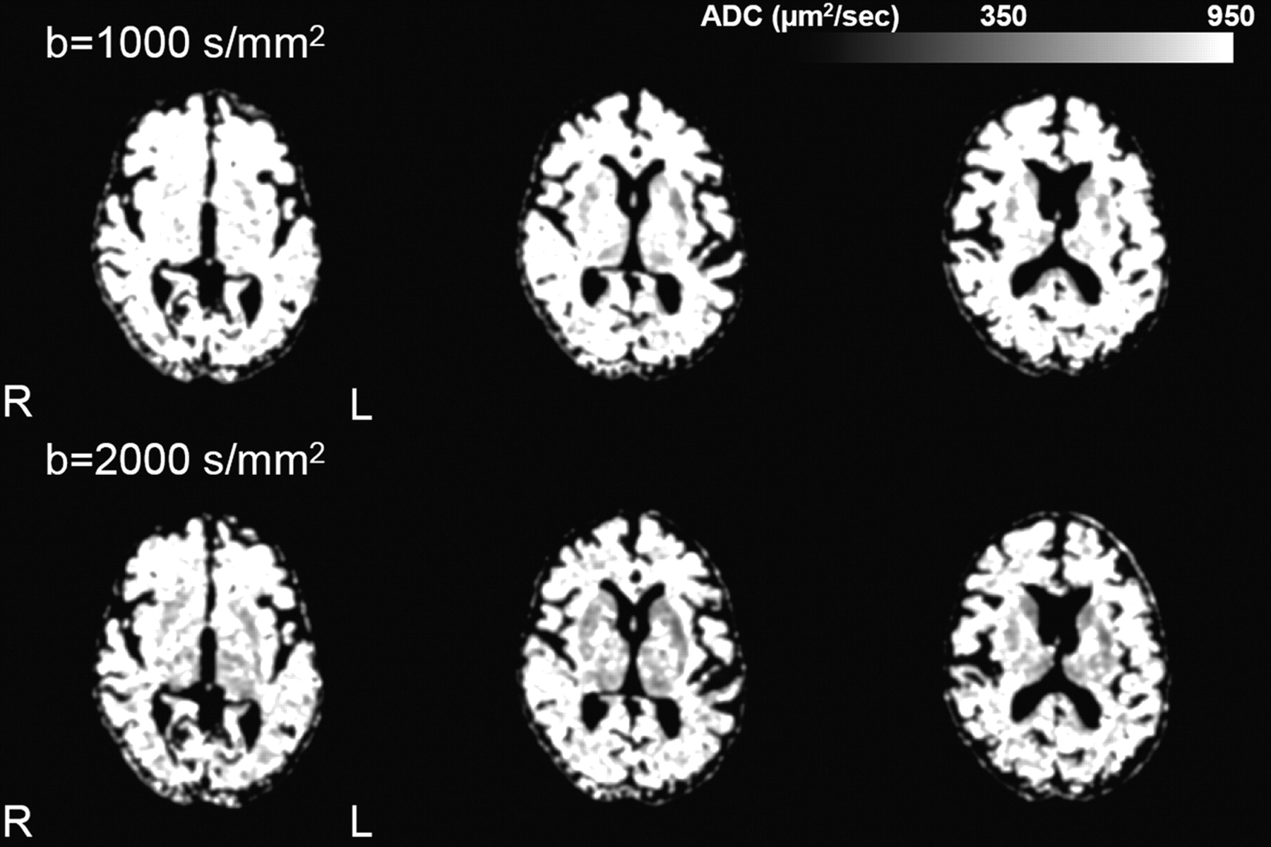
    Representative ADC maps of a patient with sporadic CJD (sCJD) (case 2018) at b = 1000 s/mm2 and b = 2000 s/mm2. For illustration purposes, images were smoothed with a 3-mm isotropic smoothing kernel. Signal-intensity abnormality involves the caudate, putamen, and thalamus bilaterally and becomes more extensive at b = 2000 s/mm2 compared with b= 1000 s/mm2. The left and right hemispheres are identified as L and R.

  • Fig 3.
    • Download figure
    • Open in new tab
    • Download powerpoint
    Fig 3.

    Bar graphs (b = 1000 s/mm2 in black, b = 2000 s/mm2 in gray) depict the patient ADC as a percentage of the control ADC. Mean ± SD ADC values are listed in the table for each structure.

  • Fig 4.
    • Download figure
    • Open in new tab
    • Download powerpoint
    Fig 4.

    Representative imaging findings on diffusion-weighted imaging at b = 1000 s/mm2 and b = 2000 s/mm2 sequences in 4 symptomatic patients (2 with familial CJD [fCJD] and 2 with sCJD). Signal-intensity abnormalities involve the caudate nucleus, putamen, thalamus, and cortex. The left and right hemispheres are identified as L and R.

Tables

  • Figures
    • View popup
    Table 1:

    Subject characteristicsa

    PatientsControlsP Value
    N1315
    Age59.72 ± 7.7257.45 ± 10.34NS
    Sex (% male)6247NS
    CNS9.85 ± 3.720.33 ± 0.62<.0001
    MMSE25.22 ± 3.1128.50 ± 1.62<.005
    • Note:—CNS indicates Clinical Neurological Scale; MMS, Mini-Mental State Examination; NS, not significant.

    • a Significant differences between the groups were assessed by t test for continuous measures and χ2 for gender distributions. Symptom severity was quantified by the CNS12 and MMS.11

    • View popup
    Table 2:

    Summary of clusters in which ADC was found to be significantly reduced (P < .001) among patients compared with normal controlsa

    MNI (x,y,z)KtControlsPatients% DifferenceAnatomical LocationsSide
    MeanSDMeanSD
    b = 1000
        (−12,−16,8)204.96777456846312Thalamus (medial dorsal nucleus, ventral lateral nucleus, ventral posterior lateral nucleus)L
        (10,−12,0)304.90769406836011Thalamus (medial dorsal nucleus, ventral lateral nucleus)R
        (18,12,−2)64.47782686707614Caudate body, caudate head, putamenR
        (24,4, −4)314.24725466138015PutamenR
        (16,10,10)64.12746586329615Caudate bodyR
    b = 2000
        (16,−16,8)1216.37722406247013Thalamus (medial dorsal nucleus, ventral lateral nucleus, ventral posterior lateral nucleus, pulvinar)R
        (26,6,2)1955.67706355827717Caudate body, caudate head, putamenR
        (28,−14,2)385.27702375938215PutamenR
        (−6,−16,6)435.05700356076713Thalamus (medial dorsal nucleus, ventral lateral nucleus, ventral posterior lateral nucleus, ventral posterior medial nucleus)L
        (−16,−26,8)64.27737596418013PulvinarL
        (−24,6,4)203.877124058512318PutamenL
    • Note:—ADC indicates apparent diffusion coefficient; MNI (x,y,z), Montreal Neurological Institute coordinates of voxel at a peak t value; K, size of clusters in the unit of voxels; t, corresponding t value of a peak height.

    • a Mean and SD were computed within each cluster. Percentage difference is defined by taking the ratio between mean ADC of patients and controls. The left and right hemisphere are identified as L and R.

PreviousNext
Back to top

In this issue

American Journal of Neuroradiology: 31 (1)
American Journal of Neuroradiology
Vol. 31, Issue 1
1 Jan 2010
  • Table of Contents
  • Index by author
Advertisement
Print
Download PDF
Email Article

Thank you for your interest in spreading the word on American Journal of Neuroradiology.

NOTE: We only request your email address so that the person you are recommending the page to knows that you wanted them to see it, and that it is not junk mail. We do not capture any email address.

Enter multiple addresses on separate lines or separate them with commas.
Enhanced Detection of Diffusion Reductions in Creutzfeldt-Jakob Disease at a Higher B Factor
(Your Name) has sent you a message from American Journal of Neuroradiology
(Your Name) thought you would like to see the American Journal of Neuroradiology web site.
CAPTCHA
This question is for testing whether or not you are a human visitor and to prevent automated spam submissions.
Cite this article
H. Lee, C. Hoffman, P.B. Kingsley, A. Degnan, O. Cohen, I. Prohovnik
Enhanced Detection of Diffusion Reductions in Creutzfeldt-Jakob Disease at a Higher B Factor
American Journal of Neuroradiology Jan 2010, 31 (1) 49-54; DOI: 10.3174/ajnr.A1756

Citation Manager Formats

  • BibTeX
  • Bookends
  • EasyBib
  • EndNote (tagged)
  • EndNote 8 (xml)
  • Medlars
  • Mendeley
  • Papers
  • RefWorks Tagged
  • Ref Manager
  • RIS
  • Zotero
0 Responses
Respond to this article
Share
Bookmark this article
Enhanced Detection of Diffusion Reductions in Creutzfeldt-Jakob Disease at a Higher B Factor
H. Lee, C. Hoffman, P.B. Kingsley, A. Degnan, O. Cohen, I. Prohovnik
American Journal of Neuroradiology Jan 2010, 31 (1) 49-54; DOI: 10.3174/ajnr.A1756
del.icio.us logo Twitter logo Facebook logo Mendeley logo
  • Tweet Widget
  • Facebook Like
  • Google Plus One
Purchase

Jump to section

  • Article
    • Abstract
    • Materials and Methods
    • Results
    • Discussion
    • Acknowledgments
    • Footnotes
    • References
  • Figures & Data
  • Info & Metrics
  • Responses
  • References
  • PDF

Related Articles

  • No related articles found.
  • PubMed
  • Google Scholar

Cited By...

  • Neuroimaging of Rapidly Progressive Dementias, Part 2: Prion, Inflammatory, Neoplastic, and Other Etiologies
  • Imaging of the 6-OPRI Mutation Prion Disease: An Entity Distinct from Typical Creutzfeldt-Jakob Disease?
  • Multiparameter MR Imaging in the 6-OPRI Variant of Inherited Prion Disease
  • Susceptibility of Domestic Cats to Chronic Wasting Disease
  • Crossref
  • Google Scholar

This article has not yet been cited by articles in journals that are participating in Crossref Cited-by Linking.

More in this TOC Section

  • Evaluating the Effects of White Matter Multiple Sclerosis Lesions on the Volume Estimation of 6 Brain Tissue Segmentation Methods
  • Quiet PROPELLER MRI Techniques Match the Quality of Conventional PROPELLER Brain Imaging Techniques
  • Predictors of Reperfusion in Patients with Acute Ischemic Stroke
Show more Brain

Similar Articles

Advertisement

Indexed Content

  • Current Issue
  • Accepted Manuscripts
  • Article Preview
  • Past Issues
  • Editorials
  • Editor's Choice
  • Fellows' Journal Club
  • Letters to the Editor
  • Video Articles

Cases

  • Case Collection
  • Archive - Case of the Week
  • Archive - Case of the Month
  • Archive - Classic Case

More from AJNR

  • Trainee Corner
  • Imaging Protocols
  • MRI Safety Corner
  • Book Reviews

Multimedia

  • AJNR Podcasts
  • AJNR Scantastics

Resources

  • Turnaround Time
  • Submit a Manuscript
  • Submit a Video Article
  • Submit an eLetter to the Editor/Response
  • Manuscript Submission Guidelines
  • Statistical Tips
  • Fast Publishing of Accepted Manuscripts
  • Graphical Abstract Preparation
  • Imaging Protocol Submission
  • Evidence-Based Medicine Level Guide
  • Publishing Checklists
  • Author Policies
  • Become a Reviewer/Academy of Reviewers
  • News and Updates

About Us

  • About AJNR
  • Editorial Board
  • Editorial Board Alumni
  • Alerts
  • Permissions
  • Not an AJNR Subscriber? Join Now
  • Advertise with Us
  • Librarian Resources
  • Feedback
  • Terms and Conditions
  • AJNR Editorial Board Alumni

American Society of Neuroradiology

  • Not an ASNR Member? Join Now

© 2025 by the American Society of Neuroradiology All rights, including for text and data mining, AI training, and similar technologies, are reserved.
Print ISSN: 0195-6108 Online ISSN: 1936-959X

Powered by HighWire