Advertisement

Main menu

  • Home
  • Content
    • Current Issue
    • Accepted Manuscripts
    • Article Preview
    • Past Issue Archive
    • Video Articles
    • AJNR Case Collection
    • Case of the Week Archive
    • Case of the Month Archive
    • Classic Case Archive
  • Special Collections
    • AJNR Awards
    • Low-Field MRI
    • Alzheimer Disease
    • ASNR Foundation Special Collection
    • Photon-Counting CT
    • View All
  • Multimedia
    • AJNR Podcasts
    • AJNR SCANtastic
    • Trainee Corner
    • MRI Safety Corner
    • Imaging Protocols
  • For Authors
    • Submit a Manuscript
    • Submit a Video Article
    • Submit an eLetter to the Editor/Response
    • Manuscript Submission Guidelines
    • Statistical Tips
    • Fast Publishing of Accepted Manuscripts
    • Graphical Abstract Preparation
    • Imaging Protocol Submission
    • Author Policies
  • About Us
    • About AJNR
    • Editorial Board
    • Editorial Board Alumni
  • More
    • Become a Reviewer/Academy of Reviewers
    • Subscribers
    • Permissions
    • Alerts
    • Feedback
    • Advertisers
    • ASNR Home

User menu

  • Alerts
  • Log in

Search

  • Advanced search
American Journal of Neuroradiology

American Journal of Neuroradiology

ASHNR American Society of Functional Neuroradiology ASHNR American Society of Pediatric Neuroradiology ASSR
  • Alerts
  • Log in

Advanced Search

  • Home
  • Content
    • Current Issue
    • Accepted Manuscripts
    • Article Preview
    • Past Issue Archive
    • Video Articles
    • AJNR Case Collection
    • Case of the Week Archive
    • Case of the Month Archive
    • Classic Case Archive
  • Special Collections
    • AJNR Awards
    • Low-Field MRI
    • Alzheimer Disease
    • ASNR Foundation Special Collection
    • Photon-Counting CT
    • View All
  • Multimedia
    • AJNR Podcasts
    • AJNR SCANtastic
    • Trainee Corner
    • MRI Safety Corner
    • Imaging Protocols
  • For Authors
    • Submit a Manuscript
    • Submit a Video Article
    • Submit an eLetter to the Editor/Response
    • Manuscript Submission Guidelines
    • Statistical Tips
    • Fast Publishing of Accepted Manuscripts
    • Graphical Abstract Preparation
    • Imaging Protocol Submission
    • Author Policies
  • About Us
    • About AJNR
    • Editorial Board
    • Editorial Board Alumni
  • More
    • Become a Reviewer/Academy of Reviewers
    • Subscribers
    • Permissions
    • Alerts
    • Feedback
    • Advertisers
    • ASNR Home
  • Follow AJNR on Twitter
  • Visit AJNR on Facebook
  • Follow AJNR on Instagram
  • Join AJNR on LinkedIn
  • RSS Feeds

AJNR Awards, New Junior Editors, and more. Read the latest AJNR updates

Review ArticleReview Articles
Open Access

Lumbosacral Transitional Vertebrae: Classification, Imaging Findings, and Clinical Relevance

G.P. Konin and D.M. Walz
American Journal of Neuroradiology November 2010, 31 (10) 1778-1786; DOI: https://doi.org/10.3174/ajnr.A2036
G.P. Konin
  • Find this author on Google Scholar
  • Find this author on PubMed
  • Search for this author on this site
D.M. Walz
  • Find this author on Google Scholar
  • Find this author on PubMed
  • Search for this author on this site
  • Article
  • Figures & Data
  • Info & Metrics
  • Responses
  • References
  • PDF
Loading

Article Figures & Data

Figures

  • Fig 1.
    • Download figure
    • Open in new tab
    • Download powerpoint
    Fig 1.

    Ferguson radiograph in a 35-year-old man. AP radiograph angled cranially at 30° allows better characterization of the transverse processes of L5. LSTVs have been classically described as best imaged by using Ferguson radiographs.
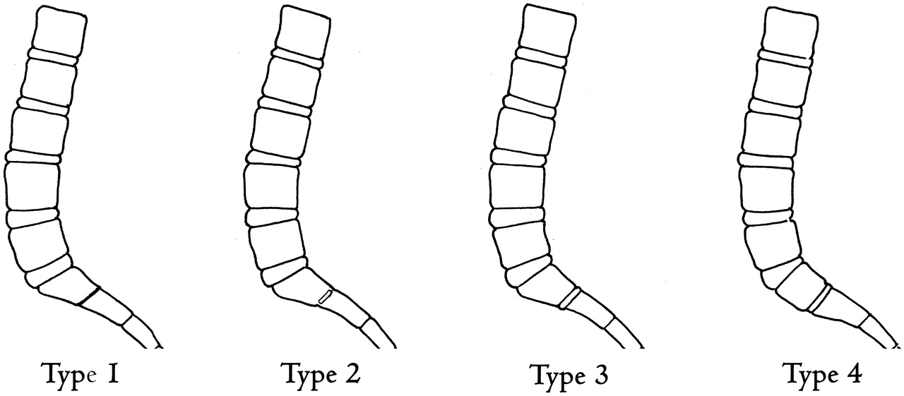
  • Fig 2.
    • Download figure
    • Open in new tab
    • Download powerpoint
    Fig 2.

    Illustration demonstrating the Castellvi classification of LSTVs.

  • Fig 3.
    • Download figure
    • Open in new tab
    • Download powerpoint
    Fig 3.

    Castellvi type Ib LSTV in a 53-year-old woman. AP radiograph demonstrates an LSTV with bilaterally enlarged L5 transverse processes (white arrows). There is no articulation with the sacrum.

  • Fig 4.
    • Download figure
    • Open in new tab
    • Download powerpoint
    Fig 4.

    Castellvi type IIa and IIb LSTVs. A, AP radiograph demonstrates an LSTV with a unilateral anomalous articulation of the right L5 transverse process with the sacrum (white arrow) in a 42-year-old man. B, T2-weighted coronal MR image demonstrates a unilateral anomalous articulation with the sacrum (white arrow) on the left in a 64-year-old man. C, Coronal reconstructed CT image demonstrates bilateral anomalous articulations of broadened transverse processes with the sacrum and the iliac bone on the left (white arrow) in a 52-year-old woman.

  • Fig 5.
    • Download figure
    • Open in new tab
    • Download powerpoint
    Fig 5.

    Castellvi type IIIa and IIIb LSTVs. A, Axial CT image demonstrates osseous fusion of the left L5 transverse process with the sacrum in a 36-year-old woman. B, Coronal reconstructed CT image demonstrates bilateral osseous fusion of L5 to the sacrum in a 31-year-old man. C, Volume-rendered CT image shows a Castellvi type IIIb LSTV with complete osseous fusion of the transverse processes to the sacrum in the same patient as seen in B.

  • Fig 6.
    • Download figure
    • Open in new tab
    • Download powerpoint
    Fig 6.

    Castellvi type IV LSTV in a 61-year-old woman. Coronal T2-weighted MR image demonstrates osseous fusion of the L5 transverse process to the sacrum on the left with an anomalous articulation on the right (white arrow).

  • Fig 7.
    • Download figure
    • Open in new tab
    • Download powerpoint
    Fig 7.

    Transitional S1 vertebral body in a 52-year-old woman. Sagittal CT image demonstrates “squaring” of a lumbarized S1 vertebral body (black arrow). Additionally, there is a fully-sized lumbar type disk between S1 and S2 (white arrow), compared with the characteristic vestigial disk typically seen at this level.
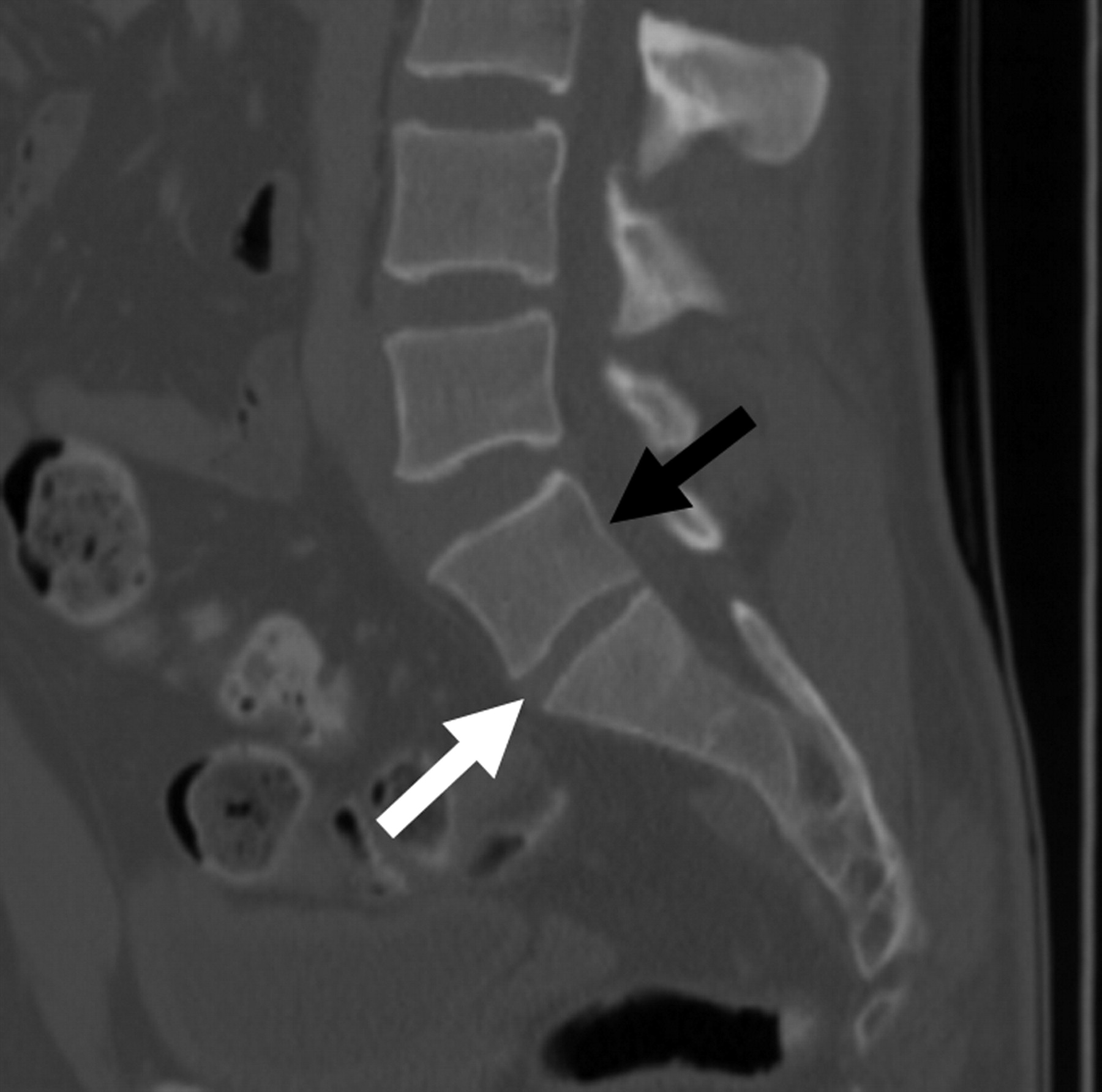
  • Fig 8.
    • Download figure
    • Open in new tab
    • Download powerpoint
    Fig 8.

    Transitional L5 vertebral body in a 52-year-old man. Sagittal CT image demonstrates “wedging” of a sacralized L5 transitional vertebral body (white arrow). A, Note the decreased height between the sacralized L5 vertebral body and S1 (black arrow) compared with the normal height typically seen at this level. B, Coronal CT image shows bilateral osseous fusion of the transverse processes of L5 with the sacrum (Castellvi type IIIb).

  • Fig 9.
    • Download figure
    • Open in new tab
    • Download powerpoint
    Fig 9.

    Illustration depicting the O'Driscoll classification system of S1–2 disk morphology.

  • Fig 10.
    • Download figure
    • Open in new tab
    • Download powerpoint
    Fig 10.

    Axial CT scan in a 52-year-old-woman with a Castellvi type IIa LSTV demonstrates fusion of the ipsilateral facet joint as well as a hypoplastic contralateral facet joint (white arrow) at L5-SI.

  • Fig 11.
    • Download figure
    • Open in new tab
    • Download powerpoint
    Fig 11.

    Axial T2-weighted MR image demonstrates rudimentary facet joints bilaterally (white arrows) at the S1-S2 level in this 79-year-old man with a lumbarized S1.
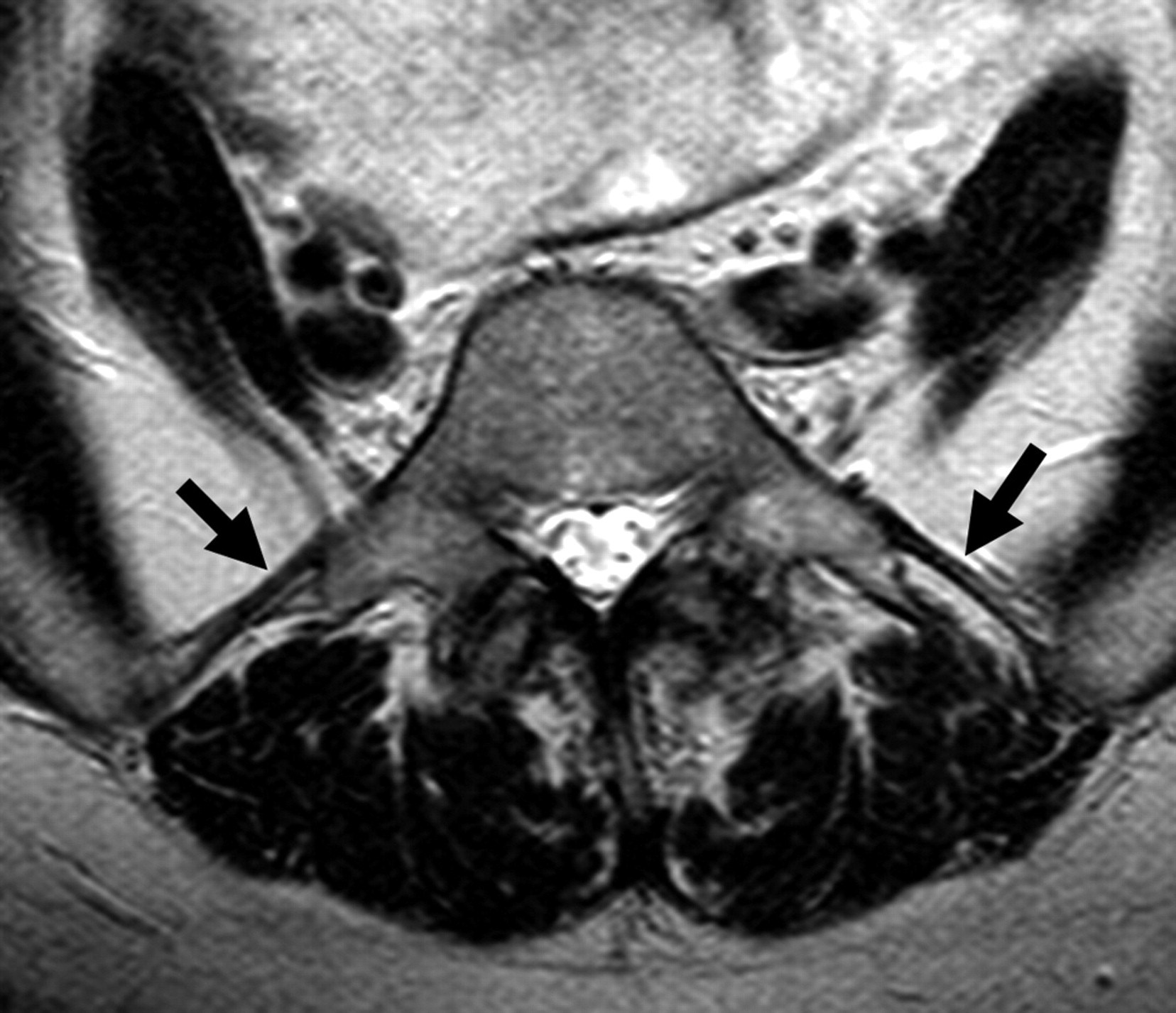
  • Fig 12.
    • Download figure
    • Open in new tab
    • Download powerpoint
    Fig 12.

    Iliolumbar ligaments in a 64-year-old woman. Axial T2-weighted MR image shows low-signal-intensity iliolumbar ligaments extending from the L5 transverse processes to the posteromedial iliac crests (arrows).

  • Fig 13.
    • Download figure
    • Open in new tab
    • Download powerpoint
    Fig 13.

    Adjacent-level disease. Sagittal T2-weighted MR image (A) demonstrates disk desiccation with a small central protrusion and associated annular tear (black arrow) in a 25-year-old female patient with a Castellvi type IIa LSTV as seen on this AP radiograph (B).

  • Fig 14.
    • Download figure
    • Open in new tab
    • Download powerpoint
    Fig 14.

    Adjacent-level disease in a 24-year-old woman. Sagittal T2-weighted MR image in a patient with a Castellvi type IIa LSTV demonstrates grade 1 anterolisthesis, disk desiccation, and disk bulging at the level above the transitional level.

  • Fig 15.
    • Download figure
    • Open in new tab
    • Download powerpoint
    Fig 15.

    Contralateral facet arthrosis in a 45-year-old man. Coronal T1-weighted MR image in a patient with a Castellvi type IIa LSTV demonstrates severe facet arthropathy (white arrow) contralateral to the anomalous articulation.
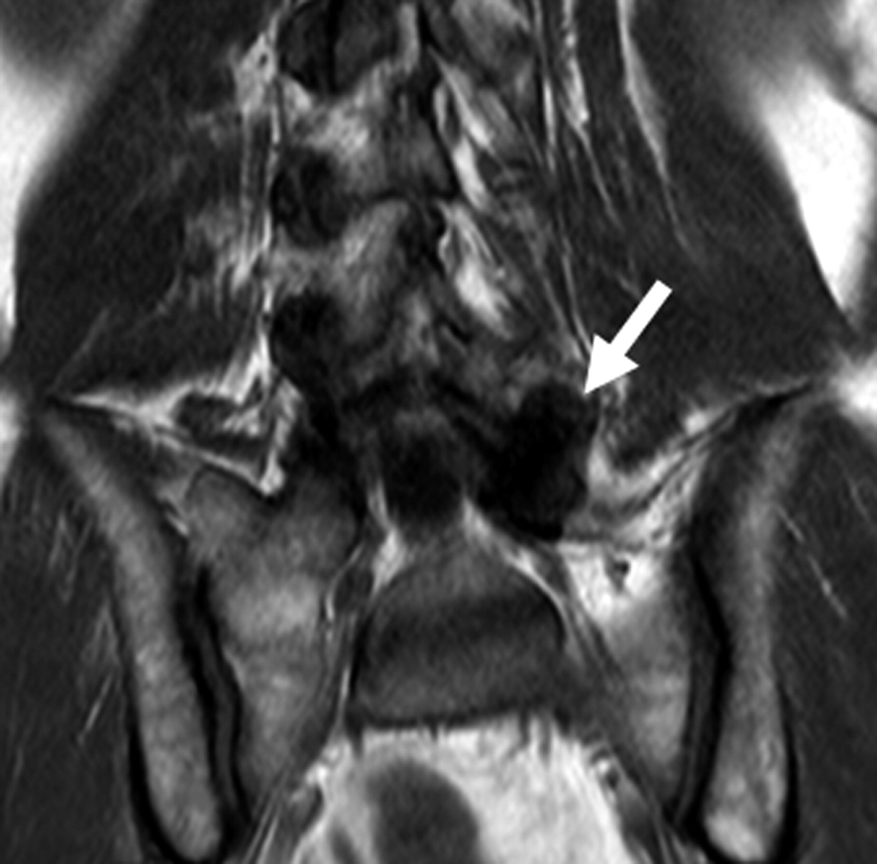
  • Fig 16.
    • Download figure
    • Open in new tab
    • Download powerpoint
    Fig 16.

    Degeneration of an anomalous articulation in an 84-year-old woman. Axial T1-weighted image demonstrates marked degeneration of the anomalous articulation on the right (white arrow) in a patient with a Castellvi type IIb LSTV.
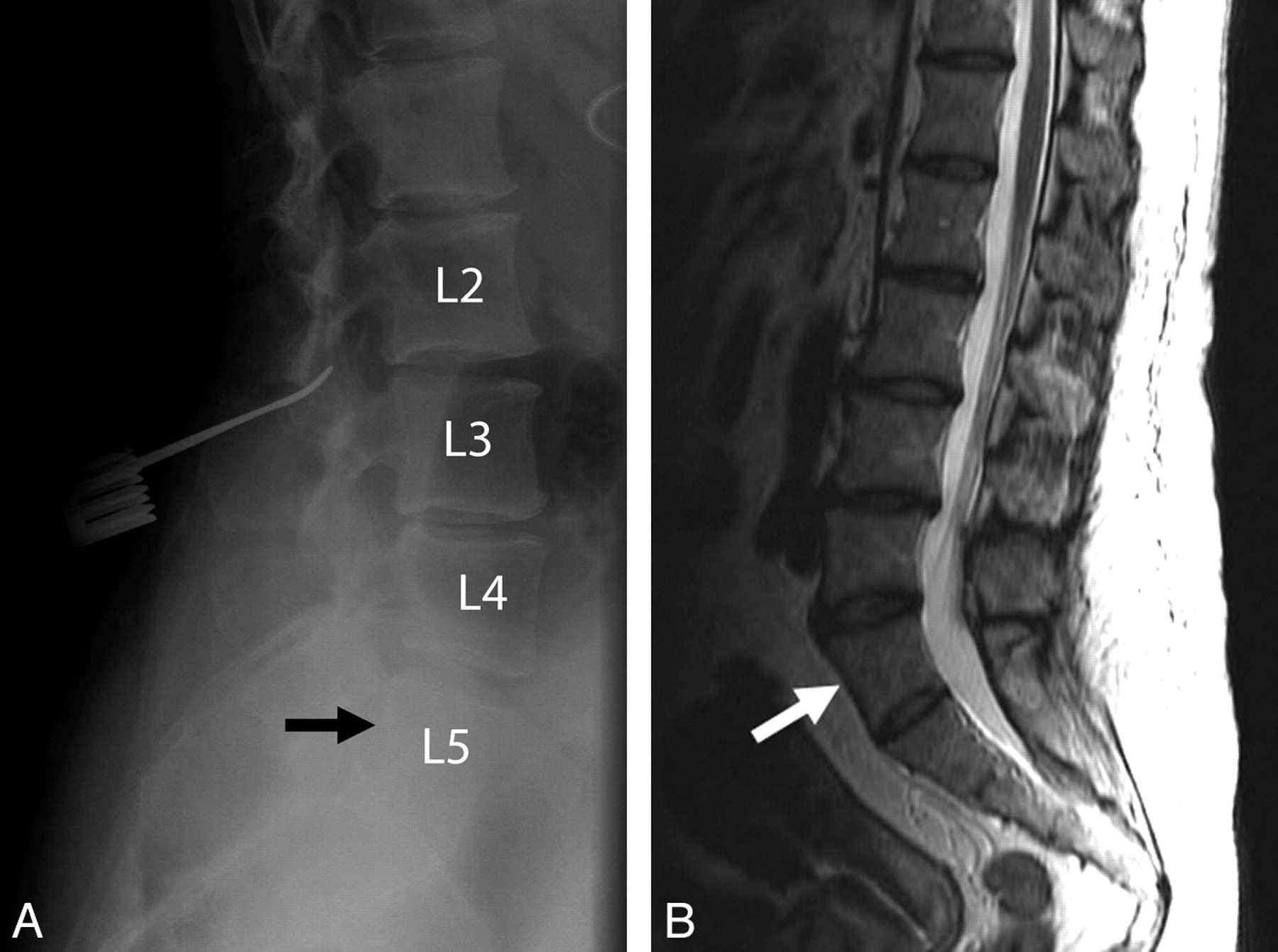
  • Fig 17.
    • Download figure
    • Open in new tab
    • Download powerpoint
    Fig 17.

    Wrong-level spine surgery in a 55-year-old woman. A, Intraoperative radiograph demonstrates a localization device at what was believed to be the L3-L4 level based on misinterpretation of a sacralized L5 vertebral body (black arrow) as S1. B, Correlation with preoperative sagittal T2-weighted MR image better reveals the presence of the LSTV (white arrow) and, if used in combination with the intraoperative films, would have helped to avoid this surgical complication.

  • Fig 18.
    • Download figure
    • Open in new tab
    • Download powerpoint
    Fig 18.

    Injection of an anomalous articulation in a 48-year-old woman with right-sided low back pain and a Castellvi type IIa LSTV. A and B, Fluoroscopic spot images demonstrate degeneration of the anomalous articulation (arrow, A) and needle placement within the anomalous articulation for injection of anesthetic and corticosteroid (B).

PreviousNext
Back to top

In this issue

American Journal of Neuroradiology: 31 (10)
American Journal of Neuroradiology
Vol. 31, Issue 10
1 Nov 2010
  • Table of Contents
  • Index by author
Advertisement
Print
Download PDF
Email Article

Thank you for your interest in spreading the word on American Journal of Neuroradiology.

NOTE: We only request your email address so that the person you are recommending the page to knows that you wanted them to see it, and that it is not junk mail. We do not capture any email address.

Enter multiple addresses on separate lines or separate them with commas.
Lumbosacral Transitional Vertebrae: Classification, Imaging Findings, and Clinical Relevance
(Your Name) has sent you a message from American Journal of Neuroradiology
(Your Name) thought you would like to see the American Journal of Neuroradiology web site.
CAPTCHA
This question is for testing whether or not you are a human visitor and to prevent automated spam submissions.
Cite this article
G.P. Konin, D.M. Walz
Lumbosacral Transitional Vertebrae: Classification, Imaging Findings, and Clinical Relevance
American Journal of Neuroradiology Nov 2010, 31 (10) 1778-1786; DOI: 10.3174/ajnr.A2036

Citation Manager Formats

  • BibTeX
  • Bookends
  • EasyBib
  • EndNote (tagged)
  • EndNote 8 (xml)
  • Medlars
  • Mendeley
  • Papers
  • RefWorks Tagged
  • Ref Manager
  • RIS
  • Zotero
0 Responses
Respond to this article
Share
Bookmark this article
Lumbosacral Transitional Vertebrae: Classification, Imaging Findings, and Clinical Relevance
G.P. Konin, D.M. Walz
American Journal of Neuroradiology Nov 2010, 31 (10) 1778-1786; DOI: 10.3174/ajnr.A2036
del.icio.us logo Twitter logo Facebook logo Mendeley logo
  • Tweet Widget
  • Facebook Like
  • Google Plus One
Purchase

Jump to section

  • Article
    • Abstract
    • Abbreviations
    • Identification and Morphology
    • Numbering Technique
    • Clinical Significance
    • Treatment
    • Conclusions
    • Acknowledgments
    • References
  • Figures & Data
  • Info & Metrics
  • Responses
  • References
  • PDF

Related Articles

  • No related articles found.
  • PubMed
  • Google Scholar

Cited By...

  • Identification of novel vertebral development factors through UK Biobank driven genetic and body imaging analysis reveals markers for back pain
  • Definition of normal vertebral morphology using NHANES-II radiographs
  • Diagnostic treatment-level discrepancies in patients with lumbosacral radicular pain and lumbar spine anomalies
  • Sacral Dysmorphism and Lumbosacral Transitional Vertebrae (LSTV) Review
  • Ultrasound-guided L5 dorsal ramus block: validation of a novel technique
  • Localizing the L5 Vertebra Using Nerve Morphology on MRI: An Accurate and Reliable Technique
  • Determination of lumbosacral transitional vertebrae in kidney urinary bladder x-ray films in the Saudi population
  • Lumbosacral Transitional Vertebra Diagnosed on 99mTc-Methylene Diphosphonate SPECT/CT
  • A Review of Symptomatic Lumbosacral Transitional Vertebrae: Bertolotti's Syndrome
  • Frequency of Discordance between Facet Joint Activity on Technetium Tc99m Methylene Diphosphonate SPECT/CT and Selection for Percutaneous Treatment at a Large Multispecialty Institution
  • A reliable measurement for identifying a lumbosacral transitional vertebra with a solid bony bridge on a single-slice midsagittal MRI or plain lateral radiograph
  • Crossref
  • Google Scholar

This article has not yet been cited by articles in journals that are participating in Crossref Cited-by Linking.

More in this TOC Section

  • An Atlas of Neonatal Neurovascular Imaging Anatomy as Depicted with Microvascular Imaging: The Intracranial Arteries
  • An Atlas of Neonatal Neurovascular Imaging Anatomy as Depicted with Microvascular Imaging: The Intracranial Veins
  • Clinical Translation of Hyperpolarized 13C Metabolic Probes for Glioma Imaging
Show more Review Articles

Similar Articles

Advertisement

Indexed Content

  • Current Issue
  • Accepted Manuscripts
  • Article Preview
  • Past Issues
  • Editorials
  • Editor's Choice
  • Fellows' Journal Club
  • Letters to the Editor
  • Video Articles

Cases

  • Case Collection
  • Archive - Case of the Week
  • Archive - Case of the Month
  • Archive - Classic Case

More from AJNR

  • Trainee Corner
  • Imaging Protocols
  • MRI Safety Corner
  • Book Reviews

Multimedia

  • AJNR Podcasts
  • AJNR Scantastics

Resources

  • Turnaround Time
  • Submit a Manuscript
  • Submit a Video Article
  • Submit an eLetter to the Editor/Response
  • Manuscript Submission Guidelines
  • Statistical Tips
  • Fast Publishing of Accepted Manuscripts
  • Graphical Abstract Preparation
  • Imaging Protocol Submission
  • Evidence-Based Medicine Level Guide
  • Publishing Checklists
  • Author Policies
  • Become a Reviewer/Academy of Reviewers
  • News and Updates

About Us

  • About AJNR
  • Editorial Board
  • Editorial Board Alumni
  • Alerts
  • Permissions
  • Not an AJNR Subscriber? Join Now
  • Advertise with Us
  • Librarian Resources
  • Feedback
  • Terms and Conditions
  • AJNR Editorial Board Alumni

American Society of Neuroradiology

  • Not an ASNR Member? Join Now

© 2025 by the American Society of Neuroradiology All rights, including for text and data mining, AI training, and similar technologies, are reserved.
Print ISSN: 0195-6108 Online ISSN: 1936-959X

Powered by HighWire