Advertisement

Main menu

  • Home
  • Content
    • Current Issue
    • Accepted Manuscripts
    • Article Preview
    • Past Issue Archive
    • AJNR Case Collection
    • Case of the Week Archive
    • Classic Case Archive
    • Case of the Month Archive
  • Special Collections
    • Spinal CSF Leak Articles (Jan 2020-June 2024)
    • 2024 AJNR Journal Awards
    • Most Impactful AJNR Articles
  • Multimedia
    • AJNR Podcast
    • AJNR Scantastics
    • Video Articles
  • For Authors
    • Submit a Manuscript
    • Author Policies
    • Fast publishing of Accepted Manuscripts
    • Graphical Abstract Preparation
    • Manuscript Submission Guidelines
    • Imaging Protocol Submission
    • Submit a Case for the Case Collection
  • About Us
    • About AJNR
    • Editorial Board
  • More
    • Become a Reviewer/Academy of Reviewers
    • Subscribers
    • Permissions
    • Alerts
    • Feedback
    • Advertisers
    • ASNR Home
  • Other Publications
    • ajnr

User menu

  • Alerts
  • Log in

Search

  • Advanced search
American Journal of Neuroradiology

American Journal of Neuroradiology

ASHNR American Society of Functional Neuroradiology ASHNR American Society of Pediatric Neuroradiology ASSR
  • Alerts
  • Log in

Advanced Search

  • Home
  • Content
    • Current Issue
    • Accepted Manuscripts
    • Article Preview
    • Past Issue Archive
    • AJNR Case Collection
    • Case of the Week Archive
    • Classic Case Archive
    • Case of the Month Archive
  • Special Collections
    • Spinal CSF Leak Articles (Jan 2020-June 2024)
    • 2024 AJNR Journal Awards
    • Most Impactful AJNR Articles
  • Multimedia
    • AJNR Podcast
    • AJNR Scantastics
    • Video Articles
  • For Authors
    • Submit a Manuscript
    • Author Policies
    • Fast publishing of Accepted Manuscripts
    • Graphical Abstract Preparation
    • Manuscript Submission Guidelines
    • Imaging Protocol Submission
    • Submit a Case for the Case Collection
  • About Us
    • About AJNR
    • Editorial Board
  • More
    • Become a Reviewer/Academy of Reviewers
    • Subscribers
    • Permissions
    • Alerts
    • Feedback
    • Advertisers
    • ASNR Home
  • Follow AJNR on Twitter
  • Visit AJNR on Facebook
  • Follow AJNR on Instagram
  • Join AJNR on LinkedIn
  • RSS Feeds

Welcome to the new AJNR, Updated Hall of Fame, and more. Read the full announcements.


AJNR is seeking candidates for the position of Associate Section Editor, AJNR Case Collection. Read the full announcement.


Research ArticleInterventional
Open Access

Experience with Microaneurysm Formation at the Basilar Terminus in the Rabbit Elastase Aneurysm Model

D. Dai, Y.H. Ding, R. Kadirvel, D.A. Lewis and D.F. Kallmes
American Journal of Neuroradiology February 2010, 31 (2) 300-303; DOI: https://doi.org/10.3174/ajnr.A1823
D. Dai
  • Find this author on Google Scholar
  • Find this author on PubMed
  • Search for this author on this site
Y.H. Ding
  • Find this author on Google Scholar
  • Find this author on PubMed
  • Search for this author on this site
R. Kadirvel
  • Find this author on Google Scholar
  • Find this author on PubMed
  • Search for this author on this site
D.A. Lewis
  • Find this author on Google Scholar
  • Find this author on PubMed
  • Search for this author on this site
D.F. Kallmes
  • Find this author on Google Scholar
  • Find this author on PubMed
  • Search for this author on this site
  • Article
  • Figures & Data
  • Info & Metrics
  • Responses
  • References
  • PDF
Loading

Article Figures & Data

Figures

  • Fig 1.
    • Download figure
    • Open in new tab
    • Download powerpoint
    Fig 1.

    A, DSA of the right vertebral artery shows the basilar artery (short arrow) and bilateral PcomAs (long arrows). The right PcomA is tortuous and dilated and nearly as large as the basilar artery, compared with the left PcomA (thin long arrow). B, Lateral DSA from the same rabbit subject shows marked enlargement of the right PcomA, which fills, in retrograde fashion, the ECA branches.
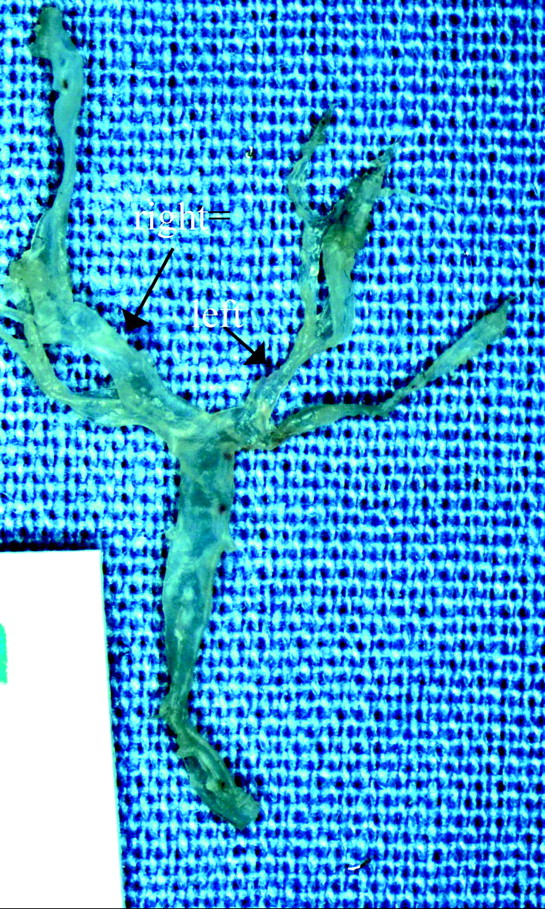
  • Fig 2.
    • Download figure
    • Open in new tab
    • Download powerpoint
    Fig 2.

    A photograph of the basilar artery and its branches shows that both PcomAs are present and that the right PcomA (long arrow) is larger than the left one (short arrow).

  • Fig 3.
    • Download figure
    • Open in new tab
    • Download powerpoint
    Fig 3.

    Typical photomicrographs of the BT show that the IEL is intact and continuous at the BT. A, The basilar artery and its bifurcation (HE, original magnification ×40). B and C, The IEL (black line) at the BT is intact and continuous (B, VVG, original magnification ×100) (C, VVG, original magnification ×200). The medial layer, immediately underneath the IEL and stained light brown, is also intact and continuous. No bulge-like localized dilation or nascent aneurysm formation is observed.

  • Fig 4.
    • Download figure
    • Open in new tab
    • Download powerpoint
    Fig 4.

    Photomicrographs of the BT from a 30-week subject. A, The basilar artery, BT, and 2 concave structures along the P1 segments labeled as 1 and 2 (arrows) for correlation with higher magnification views in B and C (VVG, original magnification ×40). B, The concave structure 1 from A, with an intact IEL, is the black line along the luminal side of the wall, contiguous with the adjacent vessel wall and with the intact but thinned media (VVG, original magnification ×200). C, Concave structure 2 from A, with an intact IEL (VVG, original magnification ×200). D, The serial (adjacent) section of A shows that the concave structures in A−C are branch vessels (arrows) (HE, original magnification ×40). E, Intact and continued IEL of the BT (VVG, original magnification ×200).

PreviousNext
Back to top

In this issue

American Journal of Neuroradiology: 31 (2)
American Journal of Neuroradiology
Vol. 31, Issue 2
1 Feb 2010
  • Table of Contents
  • Index by author
Advertisement
Print
Download PDF
Email Article

Thank you for your interest in spreading the word on American Journal of Neuroradiology.

NOTE: We only request your email address so that the person you are recommending the page to knows that you wanted them to see it, and that it is not junk mail. We do not capture any email address.

Enter multiple addresses on separate lines or separate them with commas.
Experience with Microaneurysm Formation at the Basilar Terminus in the Rabbit Elastase Aneurysm Model
(Your Name) has sent you a message from American Journal of Neuroradiology
(Your Name) thought you would like to see the American Journal of Neuroradiology web site.
CAPTCHA
This question is for testing whether or not you are a human visitor and to prevent automated spam submissions.
Cite this article
D. Dai, Y.H. Ding, R. Kadirvel, D.A. Lewis, D.F. Kallmes
Experience with Microaneurysm Formation at the Basilar Terminus in the Rabbit Elastase Aneurysm Model
American Journal of Neuroradiology Feb 2010, 31 (2) 300-303; DOI: 10.3174/ajnr.A1823

Citation Manager Formats

  • BibTeX
  • Bookends
  • EasyBib
  • EndNote (tagged)
  • EndNote 8 (xml)
  • Medlars
  • Mendeley
  • Papers
  • RefWorks Tagged
  • Ref Manager
  • RIS
  • Zotero
0 Responses
Respond to this article
Share
Bookmark this article
Experience with Microaneurysm Formation at the Basilar Terminus in the Rabbit Elastase Aneurysm Model
D. Dai, Y.H. Ding, R. Kadirvel, D.A. Lewis, D.F. Kallmes
American Journal of Neuroradiology Feb 2010, 31 (2) 300-303; DOI: 10.3174/ajnr.A1823
del.icio.us logo Twitter logo Facebook logo Mendeley logo
  • Tweet Widget
  • Facebook Like
  • Google Plus One
Purchase

Jump to section

  • Article
    • Abstract
    • Abbreviations
    • Materials and Methods
    • Results
    • Discussion
    • Conclusions
    • Footnotes
    • References
  • Figures & Data
  • Info & Metrics
  • Responses
  • References
  • PDF

Related Articles

  • No related articles found.
  • PubMed
  • Google Scholar

Cited By...

  • Aneurysmal Changes at the Basilar Terminus in the Rabbit Elastase Aneurysm Model
  • Reply:
  • Crossref (9)
  • Google Scholar

This article has been cited by the following articles in journals that are participating in Crossref Cited-by Linking.

  • Preclinical Intracranial Aneurysm Models: A Systematic Review
    Fabio Strange, Basil E Grüter, Javier Fandino, Serge Marbacher
    Brain Sciences 2020 10 3
  • Endogenous animal models of intracranial aneurysm development: a review
    Vincent M. Tutino, Hamidreza Rajabzadeh-Oghaz, Sricharan S. Veeturi, Kerry E. Poppenberg, Muhammad Waqas, Max Mandelbaum, Nicholas Liaw, Adnan H. Siddiqui, Hui Meng, John Kolega
    Neurosurgical Review 2021 44 5
  • Comparison of Aneurysm Patency and Mural Inflammation in an Arterial Rabbit Sidewall and Bifurcation Aneurysm Model under Consideration of Different Wall Conditions
    Basil Erwin Grüter, Stefan Wanderer, Fabio Strange, Sivani Sivanrupan, Michael von Gunten, Hans Rudolf Widmer, Daniel Coluccia, Lukas Andereggen, Javier Fandino, Serge Marbacher
    Brain Sciences 2020 10 4
  • Lack of aneurysm formation after carotid artery ligation in rabbits: a polymer MICROFIL® study
    Daying Dai, Yong Hong Ding, Ramanathan Kadirvel, Arash Ehteshami Rad, Debra A. Lewis, David F. Kallmes
    Neuroradiology 2013 55 1
  • Aneurysmal Changes at the Basilar Terminus in the Rabbit Elastase Aneurysm Model
    H. Meng, S.K. Natarajan, L. Gao, C. Ionita, J. Kolega, A.H. Siddiqui, J. Mocco
    American Journal of Neuroradiology 2010 31 3
  • Modelo experimental estável de aneurisma sacular em artéria carótida de suínos utilizando veia jugular interna
    Severino Lourenço da Silva Júnior, Guilherme Benjamin Brandão Pitta, Adamastor Humberto Pereira, Aldemar de Araújo Castro, Maria Helena Tavares de Matos, Fábio Duque Silveira, Leonardo Torres Magalhães, José Adolfo Hurt Almeida de Moraes, Emmylena Karina Cordeiro Machado, Carlos Wagner de Souza Wanderley, Camila Meirelles de Souza Silva, Luciana da Paz dos Santos, João Nicolle Tupiná Nogueira
    Revista do Colégio Brasileiro de Cirurgiões 2013 40 2
  • Scanning electron microscopy analysis of incidence and growth pattern of experimentally induced intracranial aneurysms in rat model
    Lucia Schwyzer, Serge Marbacher, Javier Fandino, Aaron S. Dumont
    Brain Hemorrhages 2021 2 1
  • Monitoring of the heparinization in the rabbit animal model during endovascular interventions
    Anne J. Schmitt, Anna K. Wallner, Shahrzad Afazel, Monika Killer-Oberpfalzer
    Neuroradiology 2013 55 7
  • Reply:: Fig 1.
    D. Dai, D.F. Kallmes
    American Journal of Neuroradiology 2010 31 3

More in this TOC Section

  • SAVE vs. Solumbra Techniques for Thrombectomy
  • CT Perfusion&Reperfusion in Acute Ischemic Stroke
  • Delayed Reperfusion Post-Thrombectomy&Thrombolysis
Show more Interventional

Similar Articles

Advertisement

Indexed Content

  • Current Issue
  • Accepted Manuscripts
  • Article Preview
  • Past Issues
  • Editorials
  • Editors Choice
  • Fellow Journal Club
  • Letters to the Editor

Cases

  • Case Collection
  • Archive - Case of the Week
  • Archive - Case of the Month
  • Archive - Classic Case

Special Collections

  • Special Collections

Resources

  • News and Updates
  • Turn around Times
  • Submit a Manuscript
  • Author Policies
  • Manuscript Submission Guidelines
  • Evidence-Based Medicine Level Guide
  • Publishing Checklists
  • Graphical Abstract Preparation
  • Imaging Protocol Submission
  • Submit a Case
  • Become a Reviewer/Academy of Reviewers
  • Get Peer Review Credit from Publons

Multimedia

  • AJNR Podcast
  • AJNR SCANtastic
  • Video Articles

About Us

  • About AJNR
  • Editorial Board
  • Not an AJNR Subscriber? Join Now
  • Alerts
  • Feedback
  • Advertise with us
  • Librarian Resources
  • Permissions
  • Terms and Conditions

American Society of Neuroradiology

  • Not an ASNR Member? Join Now

© 2025 by the American Society of Neuroradiology All rights, including for text and data mining, AI training, and similar technologies, are reserved.
Print ISSN: 0195-6108 Online ISSN: 1936-959X

Powered by HighWire