
Article Figures & Data
Figures
- Fig 1.
Images from a patient with a benign lesion (parotid pleomorphic adenoma) demonstrate overlay of ADC (A) and cluster maps (B and D) on low b-value images and histograms assuming 2 (C) and 3 (E) clusters. In the 2-cluster model, low ADC is color-coded blue; and high ADC, green. In the 3-cluster model, low ADC is color-coded blue; intermediate ADC, green; and high ADC, red.
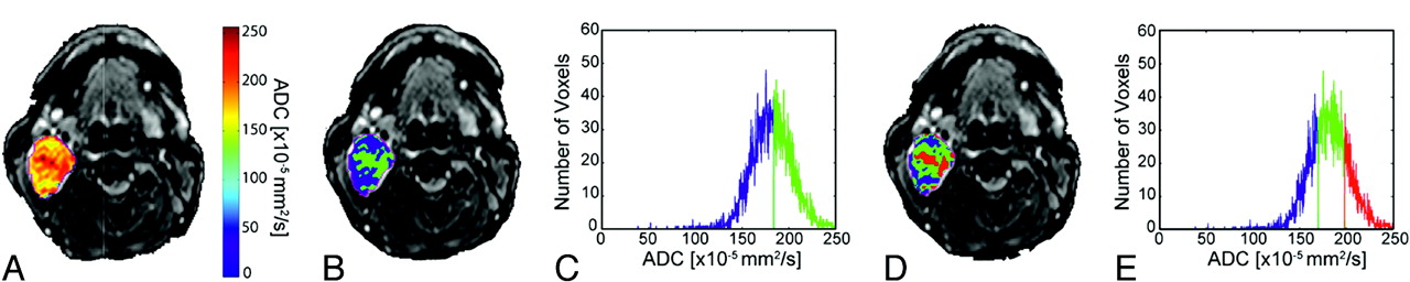
- Fig 2.
Images from a patient with a malignant lesion (sinonasal undifferentiated carcinoma) demonstrate overlay of ADC (A) and cluster maps (B and D) on low b-value images and histograms assuming 2 (C) and 3 (E) clusters. In the 2-cluster model, low ADC is color-coded blue; and high ADC, green. In the 3-cluster model, low ADC is color-coded blue; intermediate ADC, green; and high ADC, red.
- Fig 3.
Bar graphs of whole-tumor ADC, ADCH(k2), ADCL(k2), ADCH(k3), ADCI(k3), and ADCL(k3) obtained from the observations of reader 1 for benign and malignant pathologies are depicted. Significant difference in means are only identified between the ADCL(k3) in benign and malignant lesions (P = .03). For reader 2, significant differences are identified in both the ADCL(k2) and ADCL(k3) clusters (P = .04 and 0.022, respectively) (k2–2-cluster model; k3–3-cluster model).
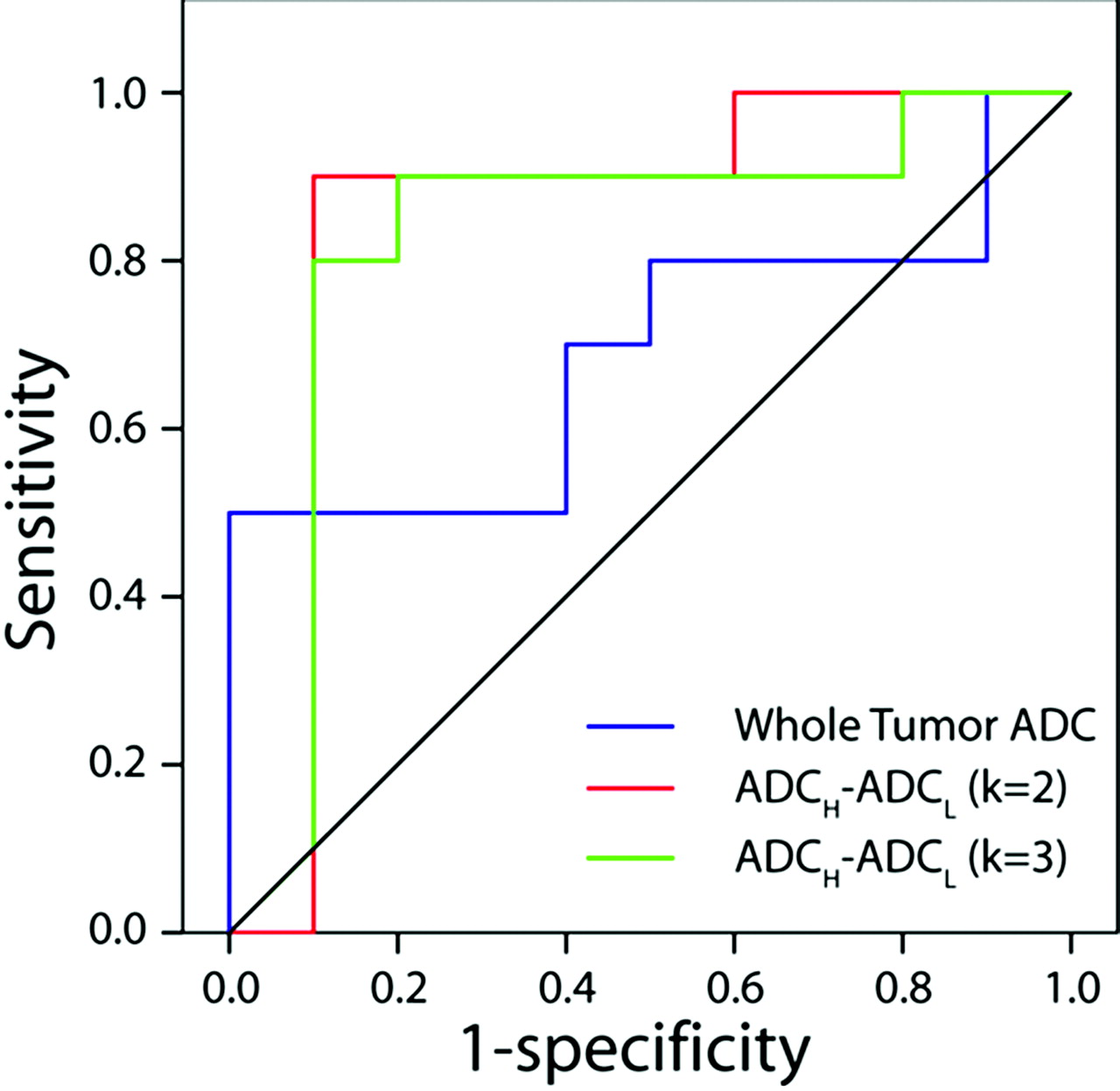
- Fig 4.
ROC curves for whole-lesion mean ADC (blue line) and difference in mean ADCH and ADCL as determined from K-means analysis assuming a cluster number of 2 (red line) and 3 (green line). There was no predictive value of whole-lesion mean ADC for malignancy (P = .2, area under curve = 0.310). However, the quantitative difference in the means of high ADC and low ADC clusters in both the 2- and 3-cluster models was found to be predictive of malignancy (2 clusters: P = .008, area under curve = 0.850; 3 clusters: P = .01, area under curve = 0.825).


Tables
Pathology No. Patient No. Age (yr)/Sex Diagnosis Reader 1, Mean ADC (10−5 mm2/s) Reader 2, Mean ADC (10−5 mm2/s) 1 1 23/F Masseteric hemangioma 176 181 2 2 26/F Sympathetic schwannoma 127 128 3 2 26/F Vestibular nerve schwannoma 180 181 4 3 56/M Vestibular nerve schwannoma 182 182 5 4 41/F Trigeminal schwannoma 128 126 6 5 33/F Neurosarcoidosis 180 174 7 6 61/F Fungal sinusitis 110 110 8 7 60/F Vagal schwannoma 85 85 9 8 63/F Pleomorphic adenoma parotid 202 204 10 9 76/F Stable parapharyngeal mass for 3 years 92 92 Pathology No./ Patient No. Age (yr)/Sex Diagnosis Reader 1, Mean ADC (10−5 mm2/s) Reader 2, Mean ADC (10−5 mm2/s) 1 41/F Adenosquamous cell carcinoma, skull base 99 116 2 37/M SNUC 104 100 3 58/M SCC hypopharynx 82 76 4 61/M SCC, palatine tonsil 154 121 5 62/M SCC, palatine tonsil 113 114 6 60/M SCC, tongue base 134 114 7 66/M SNUC 167 167 8 82/M SCC, supraglottis 96 89 9 43/M SCC, oropharynx 161 148 10 18/F Spindle cell sarcoma, parotid gland 98 98
In this issue


Jump to section
Related Articles
Cited By...
- No citing articles found.