
Article Figures & Data
Figures
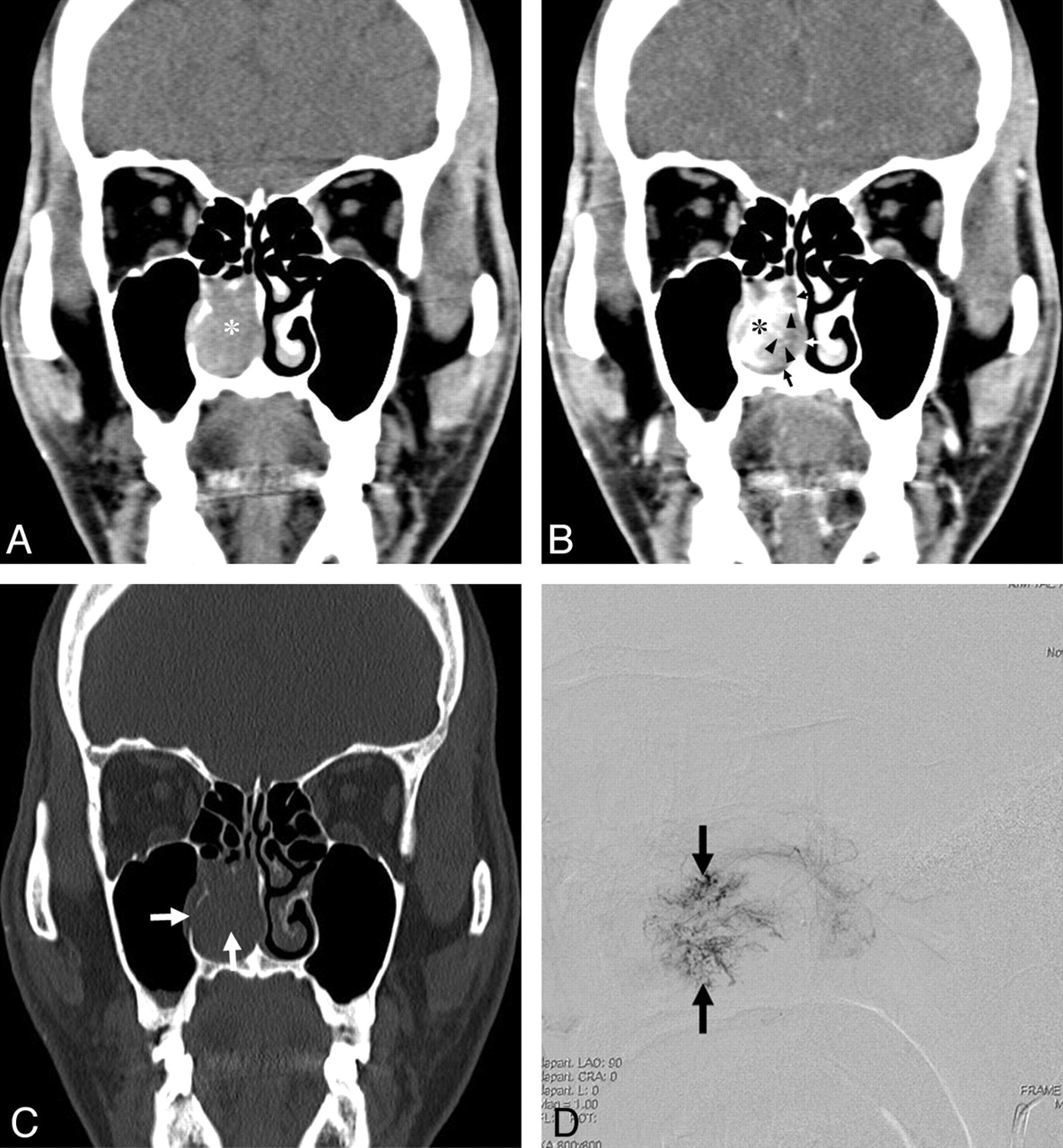
- Fig 1.
A 56-year-old woman with a lobular capillary hemangioma arising from the nasal septum. A, An axial NECT image shows a homogeneous hypoattenuating mass (asterisk) in the right anterior nasal cavity. The nasal septum is eroded (arrow), and the right middle turbinate is displaced laterally (small arrows). B, An axial CECT image reveals a lobular intensely enhancing mass (asterisk) arising from the nasal septum and a hypoattenuating cap (arrows). C, A Gd-enhanced SE T1WI (TR/TE, 700/19 ms) demonstrates only a lobular intensely enhancing mass (arrow), resulting from fill-in of Gd in the hypoattenuating cap of the CECT image. Note the tubular high-velocity SI voids (arrowheads) within the mass. D, A lateral view of the delayed phase of a carotid angiogram demonstrates a lobular capillary blush of the mass (arrows).
- Fig 2.
A 53-year-old man with a lobular capillary hemangioma arising from the right inferior turbinate. A, A coronal NECT image shows a homogeneous isoattenuating mass (asterisk) in the right nasal cavity. B, A coronal CECT image reveals a lobular intensely enhancing mass (asterisk) arising from the right inferior turbinate and an isoattenuating cap (arrows) containing linear and spotty enhancing foci (arrowheads). C, A coronal NECT image of a bone algorithm demonstrates erosion of the medial wall of the right maxillary sinus and bony destruction of the right inferior turbinate (arrows). D, A lateral view of the delayed phase of a microcatheter angiogram of the right sphenopalatine artery demonstrates a lobular capillary blush of the mass (arrows).
- Fig 3.
A 56-year-old man with a lobular capillary hemangioma arising from the left inferior turbinate. A, An axial NECT image shows a homogeneous isoattenuating mass arising from the destroyed left inferior turbinate (asterisk) with lateral displacement and erosion of the medial wall of the left maxillary sinus (arrow). B, An axial CECT image reveals a lobular intensely enhancing mass (asterisk) and a hypoattenuating area containing spotty enhancing foci (arrowheads). C, A SE T1WI (TR/TE, 600/11 ms) shows a hypointense LCHNC lesion (asterisk) compared with the masticator muscle. D, A dynamic Gd-enhanced SE T1WI (TR/TE, 350/11 ms) obtained at 1 minute after intravenous administration of Gd demonstrates a lobular intensely enhancing mass (asterisk) and a less intensely enhancing area containing tubular high-velocity SI voids (arrowheads). E, A dynamic Gd-enhanced SE T1WI (TR/TE, 350/11 ms) obtained at 4 minutes after intravenous administration of Gd reveals more homogeneous enhancement of the entire LCHNC lesion.
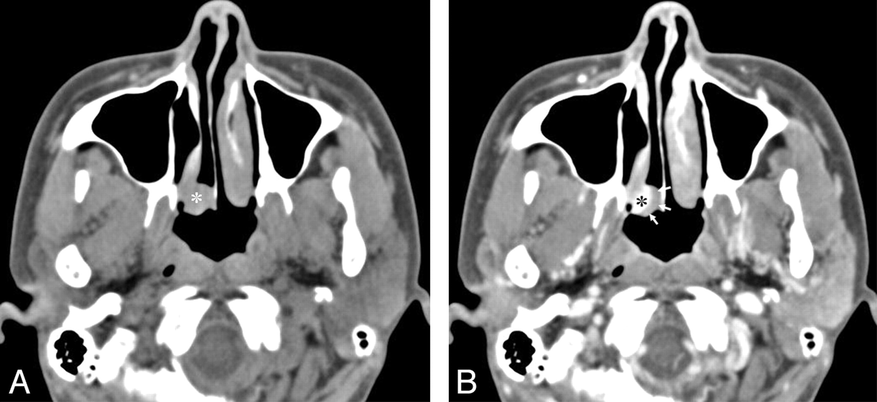
- Fig 4.
A 35-year-old woman with a lobular capillary hemangioma arising from the posterior tip of the right inferior turbinate. A, An axial NECT image shows a homogeneous isoattenuating mass (asterisk) arising from the posterior tip of the right inferior turbinate near the posterior choana. B, An axial CECT image demonstrates an intensely enhancing mass (asterisk) and an isoattenuating cap (arrows).


Tables
Patient No./Age (yr)/Sex Size (mm) Location NECT CECT Bony Changes Intensely Enhancing Mass Iso or Hypo Cap Erosion Displacement 1/56/F 22 NS Hypo + + NS MT 2/30/F 25 IT Hypo + + – – 3/65/F 19 IT Iso + + – – 4/53/M 28 IT Iso + + IT, MS – 5/56/M 45 IT Iso + – IT, MS MS 6/35/F 13 IT Iso + + – – -
a + indicates present; –, absent.
-
Authors No. of Cases CT Features MR Imaging Features Bony Changes CT MRI Erosion Displacement Puxeddu et al4 6 4 Soft-tissue mass with intense CE (6/6) High SI on T2WI, low SI on T1WI with intense CE (4/4) NM NM Katori and Tsukuda14 1 1 Soft-tissue mass Isointense to muscle on T1WI, hyperintense with numerous small signal voids on T2WI NM NM Iwata et al23 3 3 A well-circumscribed mass (3/3) High SI on T2WI with CE (3/3) – NM Miller et al1 1 0 Soft-tissue mass – – NM Ozcan et al3 1 0 Soft-tissue mass – – NM Kurtaran et al13 1 0 Soft-tissue mass – – NM -
–, indicates not present.
-
In this issue


Jump to section
Related Articles
Cited By...
- Middle turbinate angiofibroma in an adolescent boy
- Pyogenic granuloma gravidarum: a case in the nasal cavity and the use of MRI as a preoperative surgical aide
- Intranasal lobular capillary haemangioma
- Routine and Dynamic MR Imaging Study of Lobular Capillary Hemangioma of the Nasal Cavity with Comparison to Inverting Papilloma
- MR Evaluation of Sinonasal Angiomatous Polyp
- CT findings in two cases of lobular capillary haemangioma of the nasal cavity: focusing on the enhancement pattern