
Article Figures & Data
Figures
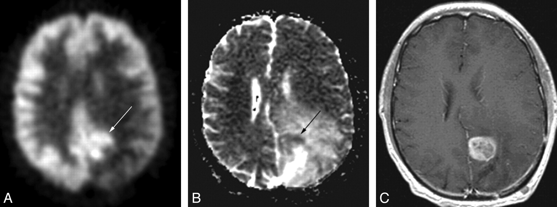
- Fig 1.
A 49-year-old man with an anaplastic astrocytoma. The PET scan (A) demonstrates a C-shaped area of increased radiopharmaceutical uptake (arrow), which exquisitely matches the area of restricted diffusion on the ADC map (arrow, B). C, The correspondence between the FDG-PET scan and the ADC map is better than that in the gadolinium-enhanced MR image (C).
- Fig 2.
A 20-year-old woman with an anaplastic astrocytoma. The area of FDG-PET (arrow, A) uptake closely matches the area of restricted diffusion on the ADC map (arrow, B). This area only shows subtle enhancement (C). The area of bright enhancement does not correlate with either increased FDG-PET uptake or restricted diffusion.
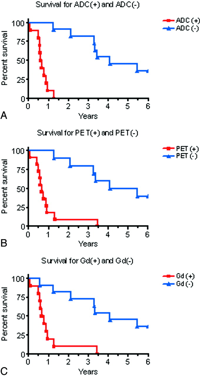
- Fig 3.
Top, Survival curves for the 10 patients with restricted diffusion on ADC maps and for the 11 patients without restricted diffusion on ADC maps, P < .0001. Middle, Survival curves for the 11 patients with increased FDG uptake on PET and the 10 patients with no increased FDG uptake on PET, P = .0004. Bottom, Survival curves for the 10 patients with gadolinium enhancement and the 11 patients without gadolinium enhancement.


Tables
Patient Tumor Age (yr) Sex Survival from Date of Diagnosis Imaging 1 GBM 31 M 10 m 26 d 11 m 5 d 2 GBM 36 F 11 m 13 d 10 m 19 d 3 GBM 49 M 6 m 12 d 7 m 2 d 4 GBM 56 F 12 m 8 d 6 m 20 d 5 GBM 56 M 29 m 19 d 15 m 19 d 6 AA 20 F 35 m 6 d 25 d 7 AA 50 F 17 m 3 d 5 m 22 d 8 AA 49 M 10 m 30 d 7 m 25 d 9 AA 45 M 7 m 15 d 9 m 7 d 10 AA 51 F 21 m 6 m 18 d 11 AODG 43 M 81 m 14 d 65 m 11 d 12 AODG 41 M 63 m 6 d 41 m 10 d 13 LGA 51 F >76 m >75 m 14 LGA 35 F 53 m 17 d 39 m 16 d 15 LGA 49 M >157 m >86 m 16 LGA 54 M >127 m >78 m 17 LGA 79 F >105 m >74 m 18 LGODG 42 F 50 m 14 d 49 m 1 d 19 LGODG 34 F 125 m 5 d 25 m 11 d 20 LGODG 47 M 21 m 15 d 15 m 4 d 21 LGA 45 M 48 m 14 d 40 m 9 d Patient PET ADC Gd 1 2 3 4 5 6 R Value A ∩ F / A A ∩ F / F A ∩ F / AF G ∩ F / G G ∩ F / F G ∩ F / GF 1 + + + 58.1 81 51.2 15.5 69.5 14.5 0.369 2 + + + 58.8 34.5 48.7 41.8 80 37.9 −0.629 3 + + + 63.6 56.2 42.5 30.7 66.7 26.6 −0.66 4 + + + 85 71.8 64 82.3 60.3 53.4 −0.686 5 + + – 55.8 71.4 45.7 – – – −0.125 6 + + + 72.6 76.3 59.4 46.7 83.8 42.9 −0.722 7 + + + 88 73 66.8 74.1 78.7 61.8 −0.451 8 + + + 84 81.5 70.8 87 75.1 67.5 −0.207 9 + + + 46.8 40.6 27.7 44.1 72.7 37.8 −0.28 10 + + + 54.6 74.8 46.1 31.6 48.7 23.8 −0.38 11 – – – – – – – – – −0.222 12 + – + – – – 38 44.9 25.9 −0.044 13 – – – – – – – – – 0.59 14 – – – – – – – – – 0.015 15 – – – – – – – – – −0.569 16 – – – – – – – – – −0.0569 17 – – – – – – – – – −0.611 18 – – – – – – – – – 0.201 19 – – – – – – – – – −0.319 20 – – – – – – – – – −0.006 21 – – – – – – – – – −0.469 -
a + indicates subjective evaluation of positive findings on imaging; –, no findings on imaging. See ″Materials and Methods″ for further explanation of Table values and their derivation.
-
In this issue


Jump to section
Related Articles
Cited By...
- Non-Contrast-Enhancing Tumor: A New Frontier in Glioblastoma Research
- Biological tumor volume in 18FET-PET before radiochemotherapy correlates with survival in GBM
- Focal Changes in Diffusivity on Apparent Diffusion Coefficient MR Imaging and Amino Acid Uptake on PET Do Not Colocalize in Nonenhancing Low-Grade Gliomas
- Longitudinal Restriction Spectrum Imaging Is Resistant to Pseudoresponse in Patients with High-Grade Gliomas Treated with Bevacizumab
- Exploratory Evaluation of MR Permeability with 18F-FDG PET Mapping in Pediatric Brain Tumors: A Report from the Pediatric Brain Tumor Consortium
- Correlation of MRI-Derived Apparent Diffusion Coefficients in Newly Diagnosed Gliomas with [18F]-Fluoro-L-Dopa PET: What Are We Really Measuring with Minimum ADC?
- Single-Shot Turbo Spin-Echo Diffusion-Weighted Imaging for Retinoblastoma: Initial Experience
- Evaluation of 18F-FDG PET and MRI Associations in Pediatric Diffuse Intrinsic Brain Stem Glioma: A Report from the Pediatric Brain Tumor Consortium