
Article Figures & Data
Figures
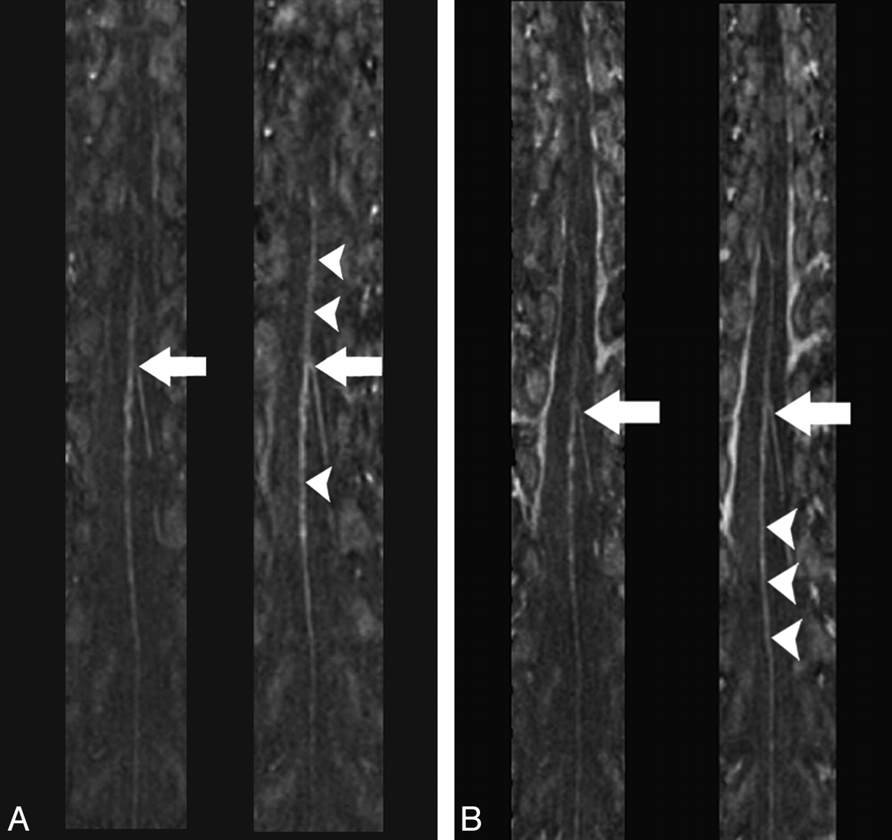
- Fig 1.
Multiplanar reformation images of the spinal MRA obtained during the predominantly arterial (A) and predominantly venous (B) phases in the same healthy individual by using gadodiamide (left) and gadobenate dimeglumine (right) (TR/TE, 5.9/1.9 ms; flip angle, 30°; 0.2 mmol/kg). The hairpin configuration of the connection between the ASA and AKA is seen with both contrast agents (arrows); however, intravascular enhancement of the ASA appears greater in the gadobenate dimeglumine study (arrowheads).
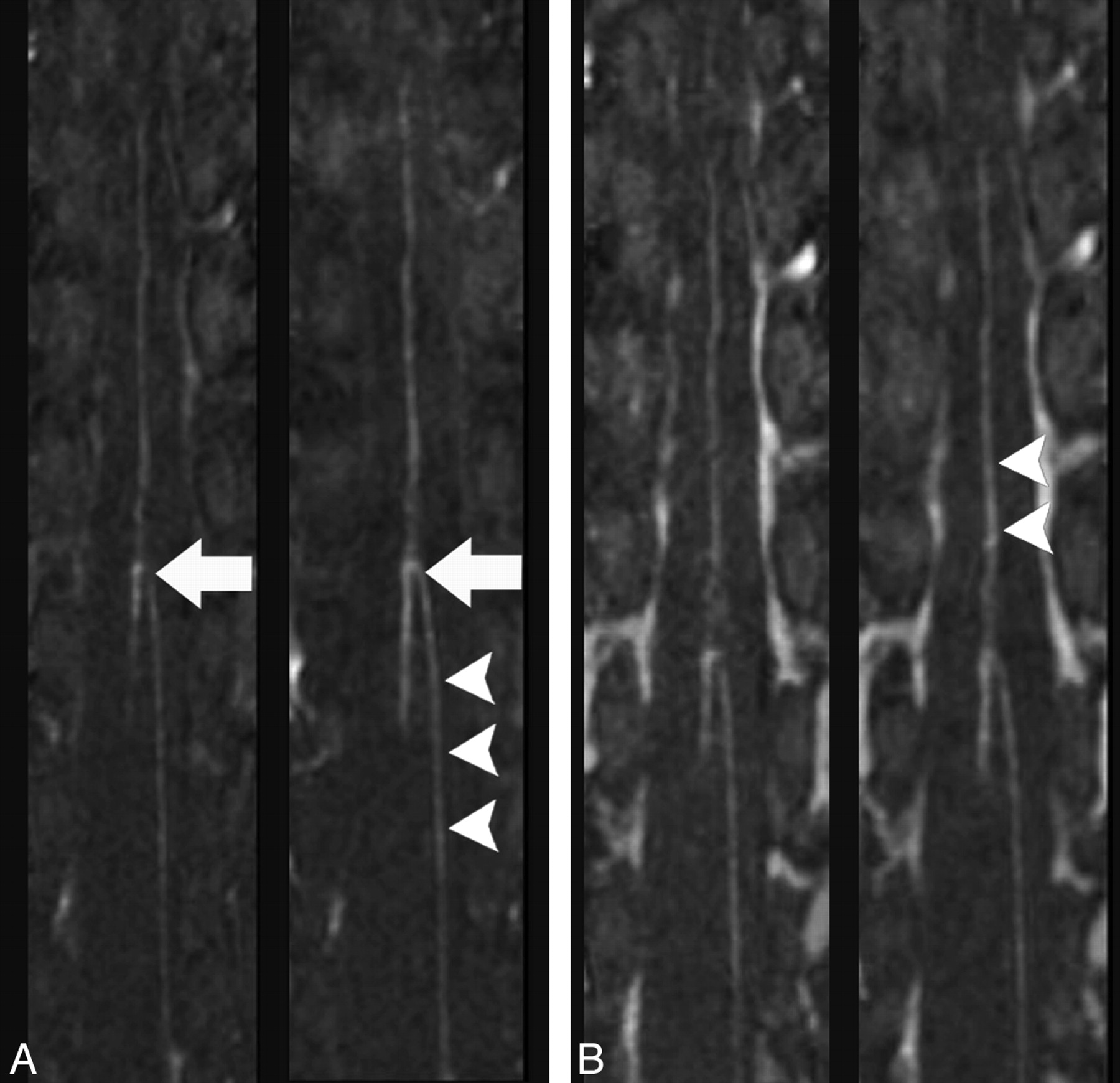
- Fig 2.
Multiplanar reformation images of the spinal MRA obtained during the predominantly arterial (A) and predominantly venous (B) phases in the same healthy individual by using gadodiamide (left) and gadobenate dimeglumine (right) (TR/TE, 5.9/1.9 ms; flip angle, 30°; 0.2 mmol/kg). The hairpin configuration of the connection between the ASA and AKA is better visualized by using gadobenate dimeglumine (arrows); intravascular enhancement appears greater in the gadobenate dimeglumine study (arrowheads).


Tables
Comparison of gadodiamide- and gadobenate dimeglumine−enhanced MRAa
Observer A Observer B GD G GD G Background homogeneity 3.6 (0.7) 3.5 (0.7) 3.6 (0.9) 3.45 (0.8) Sharpness 3.3 (1) 2.9 (1) 3.2 (0.8) 2.9 (0.8) Continuity 3.3 (0.9) 2.6 (1.1) 3.5 (0.9) 2.4 (0.7) Contrast 3.3 (1) 2.7 (1) 3.6 (0.8) 3.0 (0.8) -
a A 5-point scale (1 = very poor; 2 = poor; 3 = moderate; 4 = good; 5 = excellent) was used. The number represents the average rating (SD).
-


