Advertisement

Main menu

  • Home
  • Content
    • Current Issue
    • Accepted Manuscripts
    • Article Preview
    • Past Issue Archive
    • AJNR Case Collection
    • Case of the Week Archive
    • Classic Case Archive
    • Case of the Month Archive
  • Special Collections
    • Spinal CSF Leak Articles (Jan 2020-June 2024)
    • 2024 AJNR Journal Awards
    • Most Impactful AJNR Articles
  • Multimedia
    • AJNR Podcast
    • AJNR Scantastics
    • Video Articles
  • For Authors
    • Submit a Manuscript
    • Author Policies
    • Fast publishing of Accepted Manuscripts
    • Graphical Abstract Preparation
    • Manuscript Submission Guidelines
    • Imaging Protocol Submission
    • Submit a Case for the Case Collection
  • About Us
    • About AJNR
    • Editorial Board
  • More
    • Become a Reviewer/Academy of Reviewers
    • Subscribers
    • Permissions
    • Alerts
    • Feedback
    • Advertisers
    • ASNR Home
  • Other Publications
    • ajnr

User menu

  • Alerts
  • Log in

Search

  • Advanced search
American Journal of Neuroradiology

American Journal of Neuroradiology

ASHNR American Society of Functional Neuroradiology ASHNR American Society of Pediatric Neuroradiology ASSR
  • Alerts
  • Log in

Advanced Search

  • Home
  • Content
    • Current Issue
    • Accepted Manuscripts
    • Article Preview
    • Past Issue Archive
    • AJNR Case Collection
    • Case of the Week Archive
    • Classic Case Archive
    • Case of the Month Archive
  • Special Collections
    • Spinal CSF Leak Articles (Jan 2020-June 2024)
    • 2024 AJNR Journal Awards
    • Most Impactful AJNR Articles
  • Multimedia
    • AJNR Podcast
    • AJNR Scantastics
    • Video Articles
  • For Authors
    • Submit a Manuscript
    • Author Policies
    • Fast publishing of Accepted Manuscripts
    • Graphical Abstract Preparation
    • Manuscript Submission Guidelines
    • Imaging Protocol Submission
    • Submit a Case for the Case Collection
  • About Us
    • About AJNR
    • Editorial Board
  • More
    • Become a Reviewer/Academy of Reviewers
    • Subscribers
    • Permissions
    • Alerts
    • Feedback
    • Advertisers
    • ASNR Home
  • Follow AJNR on Twitter
  • Visit AJNR on Facebook
  • Follow AJNR on Instagram
  • Join AJNR on LinkedIn
  • RSS Feeds

Welcome to the new AJNR, Updated Hall of Fame, and more. Read the full announcements.


AJNR is seeking candidates for the position of Associate Section Editor, AJNR Case Collection. Read the full announcement.


LetterLetter

MR Imaging Findings in Wernicke Encephalopathy: Nonalcoholics May Be Similar to Alcoholics

L.C. Hygino da Cruz, R.C. Domingues, I. Vilanova and E.L. Gasparetto
American Journal of Neuroradiology June 2010, 31 (6) E54; DOI: https://doi.org/10.3174/ajnr.A2113
L.C. Hygino da Cruz Jr
  • Find this author on Google Scholar
  • Find this author on PubMed
  • Search for this author on this site
R.C. Domingues
  • Find this author on Google Scholar
  • Find this author on PubMed
  • Search for this author on this site
I. Vilanova
  • Find this author on Google Scholar
  • Find this author on PubMed
  • Search for this author on this site
E.L. Gasparetto
  • Find this author on Google Scholar
  • Find this author on PubMed
  • Search for this author on this site
  • Article
  • Figures & Data
  • Info & Metrics
  • Responses
  • References
  • PDF
Loading

We read with special interest the article of Zuccoli et al1 entitled “MR Imaging Findings in 56 Patients with Wernicke Encephalopathy: Nonalcoholics May Differ From Alcoholics.” The authors reported the typical and atypical imaging findings of Wernicke encephalopathy (WE). They suggested that alcoholic and nonalcoholic patients present different MR imaging patterns due to differences in the pathophysiologic process. Typical imaging findings, which were more common in alcoholic patients, consist of symmetric signal-intensity abnormalities in the thalami, mamillary bodies, tectal plate, and periaqueductal area, as well as contrast enhancement in the mamillary bodies and thalami, whereas signal-intensity alterations in the cerebellum, cerebral vermis, cranial nerve nuclei, red nuclei, dentate nuclei, caudate nuclei, splenium, and cerebral cortex represent atypical MR imaging findings and are more frequent in nonalcoholic patients. The authors speculated that alcohol might have a protective effect on the brain areas that show atypical lesions in WE, which could explain why atypical findings are seen only in nonalcoholic patients.

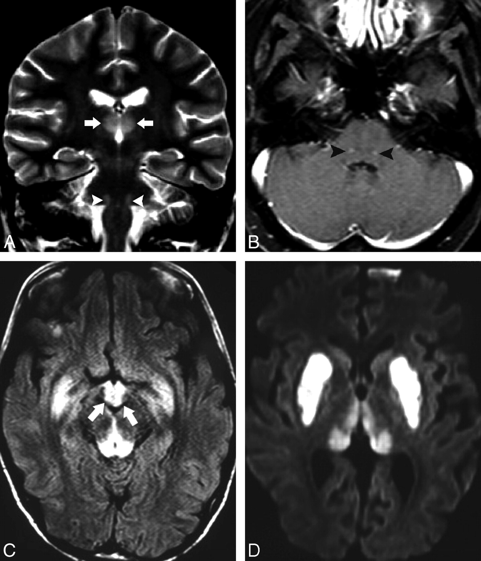
We report 2 cases of nonalcoholic WE: 1 presenting with typical and another with atypical MR imaging findings. The first case is a 21-year-old woman with non-Hodgkin lymphoma, having undergone chemotherapy and bone marrow transplant (BMT). Thirty-one days after BMT and receiving parenteral nutrition, she presented neurologic and visual disturbances first attributed to cyclosporine. However, MR imaging findings revealed, on T2-weighted images, high signal intensity symmetrically and bilaterally in the medial vestibular nuclei, facial nuclei, medial and pulvinar thalami (Fig 1A), and periventricular region of the third ventricle, as well as in the mamillary bodies. After gadolinium administration, there was enhancement in the mamillary bodies and in the facial nuclei (Fig 1B).

Fig 1.
  • Download figure
  • Open in new tab
  • Download powerpoint
Fig 1.

Case 1. Coronal T2-weighted image (A) shows high signal intensity in the thalami (arrows) and facial nuclei (arrowheads), which demonstrates contrast enhancement (arrowheads) in the T1-weighted image (B) after gadolinium administration. Case 2. FLAIR images (C and D) show bilateral and symmetric areas of high signal intensity on the mamillary bodies (arrows), periaqueductal gray matter, thalami, and putamina.

The additional case was a 13-year-old boy with suppurative appendicitis, who underwent laparotomy complicated with mesenteric infarction, adhesion, and hematoma of the rectum. Thus, he underwent a colostomy, and after a month of parenteral nutrition, he developed a sixth-nerve palsy, seizures, and coma. The MR imaging demonstrated well-defined symmetric and bilateral lesions hyperintense on T2-weighted and fluid-attenuated inversion recovery (FLAIR) images in the tectum of the midbrain, the periaqueductal gray matter, mamillary bodies, thalami, putamina, and periventricular region of the third ventricle, as well as in the frontal cortex. The lesions also presented restricted diffusion and no contrast enhancement. In both cases, the diagnosis of WE was suggested and appropriate therapy was initiated. However, the patients did not respond and died in the following days.

We report these cases of nonalcoholic WE demonstrating that both typical and atypical MR imaging findings may coexist. Although Zuccoli et al1 suggested that contrast enhancement of the mamillary bodies and thalami indicate alcoholic WE findings, these findings were already demonstrated in nonalcoholic patients,2 corroborating our cases. Our findings indicate that the WE physiopathology might not differ between alcoholic and nonalcoholic patients. Alcohol may not play as important a role as thiamine deficiency does in the whole process. Many clinical conditions can directly or indirectly cause thiamine deficiency, which will lead to WE.

Finally, as suggested by Zuccoli et al,1,3 the correct diagnosis is the main challenge in the clinical approach to these patients. The initiation of the required treatment can avoid or delay the appearance of severe neurologic deficits and even death, no matter the cause of thiamine deficiency.

References

  1. 1.↵
    1. Zuccoli G,
    2. Santa Cruz D,
    3. Bertolini M,
    4. et al
    . MR imaging findings in 56 patients with Wernicke encephalopathy: nonalcoholics may differ from alcoholics. AJNR Am J Neuroradiol 2009;30:171–76. Epub 2008 Oct 22
    Abstract/FREE Full Text
  2. 2.↵
    1. Fei GQ,
    2. Zhong C,
    3. Jin L,
    4. et al
    . Clinical characteristics and MR imaging features of nonalcoholic Wernicke encephalopathy. AJNR Am J Neuroradiol 2008;29:164–69
    Abstract/FREE Full Text
  3. 3.↵
    1. Zuccoli G,
    2. Gallucci M,
    3. Capellades J,
    4. et al
    . Wernicke encephalopathy: MR findings in twenty-six alcoholic and non-alcoholic patients. AJNR Am J Neuroradiol 2007;28:1328–31
    Abstract/FREE Full Text
  • Copyright © American Society of Neuroradiology
PreviousNext
Back to top

In this issue

American Journal of Neuroradiology: 31 (6)
American Journal of Neuroradiology
Vol. 31, Issue 6
1 Jun 2010
  • Table of Contents
  • Index by author
Advertisement
Print
Download PDF
Email Article

Thank you for your interest in spreading the word on American Journal of Neuroradiology.

NOTE: We only request your email address so that the person you are recommending the page to knows that you wanted them to see it, and that it is not junk mail. We do not capture any email address.

Enter multiple addresses on separate lines or separate them with commas.
MR Imaging Findings in Wernicke Encephalopathy: Nonalcoholics May Be Similar to Alcoholics
(Your Name) has sent you a message from American Journal of Neuroradiology
(Your Name) thought you would like to see the American Journal of Neuroradiology web site.
CAPTCHA
This question is for testing whether or not you are a human visitor and to prevent automated spam submissions.
Cite this article
L.C. Hygino da Cruz, R.C. Domingues, I. Vilanova, E.L. Gasparetto
MR Imaging Findings in Wernicke Encephalopathy: Nonalcoholics May Be Similar to Alcoholics
American Journal of Neuroradiology Jun 2010, 31 (6) E54; DOI: 10.3174/ajnr.A2113

Citation Manager Formats

  • BibTeX
  • Bookends
  • EasyBib
  • EndNote (tagged)
  • EndNote 8 (xml)
  • Medlars
  • Mendeley
  • Papers
  • RefWorks Tagged
  • Ref Manager
  • RIS
  • Zotero
0 Responses
Respond to this article
Share
Bookmark this article
MR Imaging Findings in Wernicke Encephalopathy: Nonalcoholics May Be Similar to Alcoholics
L.C. Hygino da Cruz, R.C. Domingues, I. Vilanova, E.L. Gasparetto
American Journal of Neuroradiology Jun 2010, 31 (6) E54; DOI: 10.3174/ajnr.A2113
del.icio.us logo Twitter logo Facebook logo Mendeley logo
  • Tweet Widget
  • Facebook Like
  • Google Plus One
Purchase

Jump to section

  • Article
    • References
  • Figures & Data
  • Info & Metrics
  • Responses
  • References
  • PDF

Related Articles

  • No related articles found.
  • PubMed
  • Google Scholar

Cited By...

  • Spectrum of MR Imaging Findings in Wernicke Encephalopathy: Are Atypical Areas of Involvement Only Present in Nonalcoholic Patients?
  • Reply:
  • MR Imaging: An Increasingly Important Tool in the Early Diagnosis of Wernicke Encephalopathy
  • Nonalcoholic Wernicke Encephalopathy with Extensive Cortical Involvement: Cortical Laminar Necrosis and Hemorrhage Demonstrated with Susceptibility-Weighted MR Phase Images
  • Crossref (7)
  • Google Scholar

This article has been cited by the following articles in journals that are participating in Crossref Cited-by Linking.

  • MR Imaging Findings in Alcoholic and Nonalcoholic Acute Wernicke’s Encephalopathy: A Review
    Gaetana Manzo, Angela De Gennaro, Attilio Cozzolino, Antonietta Serino, Giacomo Fenza, Andrea Manto
    BioMed Research International 2014 2014
  • Differences Between Alcoholic and Nonalcoholic Patients With Wernicke Encephalopathy: A Multicenter Observational Study
    Antonio J. Chamorro, Beatriz Rosón-Hernández, José-A. Medina-García, Roberto Muga-Bustamante, Joaquín Fernández-Solá, M.-Candelaria Martín-González, Elena Seco-Hernández, Ignacio Novo-Veleiro, Carlos Suárez-Cuervo, Ana M. Mateos-Díaz, Rafael Monte-Secades, Begoña Machado-Prieto, Rubén Puerta-Louro, Cristina Prada-González, Álvaro Fernández-Rial, Patricia Sabio-Repiso, Rocío Vázquez-Vigo, Ana-C. Antolí-Royo, Aina Gomila-Grange, Nieves-C. Felipe-Pérez, Arantza Sanvisens-Bergé, Emilia Antúnez-Jorge, Camino-M. Fernández-Rodríguez, Lucía Alvela-Suárez, Alba Fidalgo-Navarro, Miguel Marcos, Joaquín Castro, María-A. Polvorosa Gómez, Mario Del Valle-Sánchez, José Lopez-Castro, Arturo González-Quintela, F.-Javier Laso
    Mayo Clinic Proceedings 2017 92 6
  • Spectrum of MR Imaging Findings in Wernicke Encephalopathy: Are Atypical Areas of Involvement Only Present in Nonalcoholic Patients?
    N.D. Ha, Y.C. Weon, J.C. Jang, B.S. Kang, S.H. Choi
    American Journal of Neuroradiology 2012 33 7
  • Nonalcoholic Wernicke Encephalopathy with Extensive Cortical Involvement: Cortical Laminar Necrosis and Hemorrhage Demonstrated with Susceptibility-Weighted MR Phase Images: Fig 1.
    D.B. Pereira, M.L. Pereira, E.L. Gasparetto
    American Journal of Neuroradiology 2011 32 2
  • MR Imaging: An Increasingly Important Tool in the Early Diagnosis of Wernicke Encephalopathy
    G. Zuccoli, N. Pipitone
    American Journal of Neuroradiology 2012 33 6
  • Reply:
    N. Du Ha, Y. Cheol Weon
    American Journal of Neuroradiology 2012 33 6
  • Wernicke encephalopathy with lesions in the bilateral abducens nuclei: a case report
    Daisuke Kuzume, Yuko Morimoto, Masahiro Yamasaki, Naohisa Hosomi
    Rinsho Shinkeigaku 2022 62 11

More in this TOC Section

  • Brain AVM’s Nidus: What if We Hadn’t Understood Anything?
  • Letter to the Editor regarding “Automated Volumetric Software in Dementia: Help or Hindrance to the Neuroradiologist?”
  • Reply:
Show more Letters

Similar Articles

Advertisement

Indexed Content

  • Current Issue
  • Accepted Manuscripts
  • Article Preview
  • Past Issues
  • Editorials
  • Editors Choice
  • Fellow Journal Club
  • Letters to the Editor

Cases

  • Case Collection
  • Archive - Case of the Week
  • Archive - Case of the Month
  • Archive - Classic Case

Special Collections

  • Special Collections

Resources

  • News and Updates
  • Turn around Times
  • Submit a Manuscript
  • Author Policies
  • Manuscript Submission Guidelines
  • Evidence-Based Medicine Level Guide
  • Publishing Checklists
  • Graphical Abstract Preparation
  • Imaging Protocol Submission
  • Submit a Case
  • Become a Reviewer/Academy of Reviewers
  • Get Peer Review Credit from Publons

Multimedia

  • AJNR Podcast
  • AJNR SCANtastic
  • Video Articles

About Us

  • About AJNR
  • Editorial Board
  • Not an AJNR Subscriber? Join Now
  • Alerts
  • Feedback
  • Advertise with us
  • Librarian Resources
  • Permissions
  • Terms and Conditions

American Society of Neuroradiology

  • Not an ASNR Member? Join Now

© 2025 by the American Society of Neuroradiology All rights, including for text and data mining, AI training, and similar technologies, are reserved.
Print ISSN: 0195-6108 Online ISSN: 1936-959X

Powered by HighWire