Advertisement

Main menu

  • Home
  • Content
    • Current Issue
    • Accepted Manuscripts
    • Article Preview
    • Past Issue Archive
    • Video Articles
    • AJNR Case Collection
    • Case of the Week Archive
    • Case of the Month Archive
    • Classic Case Archive
  • Special Collections
    • AJNR Awards
    • Low-Field MRI
    • Alzheimer Disease
    • ASNR Foundation Special Collection
    • Photon-Counting CT
    • View All
  • Multimedia
    • AJNR Podcasts
    • AJNR SCANtastic
    • Trainee Corner
    • MRI Safety Corner
    • Imaging Protocols
  • For Authors
    • Submit a Manuscript
    • Submit a Video Article
    • Submit an eLetter to the Editor/Response
    • Manuscript Submission Guidelines
    • Statistical Tips
    • Fast Publishing of Accepted Manuscripts
    • Graphical Abstract Preparation
    • Imaging Protocol Submission
    • Author Policies
  • About Us
    • About AJNR
    • Editorial Board
    • Editorial Board Alumni
  • More
    • Become a Reviewer/Academy of Reviewers
    • Subscribers
    • Permissions
    • Alerts
    • Feedback
    • Advertisers
    • ASNR Home

User menu

  • Alerts
  • Log in

Search

  • Advanced search
American Journal of Neuroradiology

American Journal of Neuroradiology

ASHNR American Society of Functional Neuroradiology ASHNR American Society of Pediatric Neuroradiology ASSR
  • Alerts
  • Log in

Advanced Search

  • Home
  • Content
    • Current Issue
    • Accepted Manuscripts
    • Article Preview
    • Past Issue Archive
    • Video Articles
    • AJNR Case Collection
    • Case of the Week Archive
    • Case of the Month Archive
    • Classic Case Archive
  • Special Collections
    • AJNR Awards
    • Low-Field MRI
    • Alzheimer Disease
    • ASNR Foundation Special Collection
    • Photon-Counting CT
    • View All
  • Multimedia
    • AJNR Podcasts
    • AJNR SCANtastic
    • Trainee Corner
    • MRI Safety Corner
    • Imaging Protocols
  • For Authors
    • Submit a Manuscript
    • Submit a Video Article
    • Submit an eLetter to the Editor/Response
    • Manuscript Submission Guidelines
    • Statistical Tips
    • Fast Publishing of Accepted Manuscripts
    • Graphical Abstract Preparation
    • Imaging Protocol Submission
    • Author Policies
  • About Us
    • About AJNR
    • Editorial Board
    • Editorial Board Alumni
  • More
    • Become a Reviewer/Academy of Reviewers
    • Subscribers
    • Permissions
    • Alerts
    • Feedback
    • Advertisers
    • ASNR Home
  • Follow AJNR on Twitter
  • Visit AJNR on Facebook
  • Follow AJNR on Instagram
  • Join AJNR on LinkedIn
  • RSS Feeds

AJNR Awards, New Junior Editors, and more. Read the latest AJNR updates

Getting new auth cookie, if you see this message a lot, tell someone!
Research ArticleNeurointerventionE

Feasibility of Angiographic CT in Peri-Interventional Diagnostic Imaging: A Comparative Study with Multidetector CT

M.-N. Psychogios, J.-H. Buhk, P. Schramm, A. Xyda, A. Mohr and M. Knauth
American Journal of Neuroradiology August 2010, 31 (7) 1226-1231; DOI: https://doi.org/10.3174/ajnr.A2086
M.-N. Psychogios
  • Find this author on Google Scholar
  • Find this author on PubMed
  • Search for this author on this site
J.-H. Buhk
  • Find this author on Google Scholar
  • Find this author on PubMed
  • Search for this author on this site
P. Schramm
  • Find this author on Google Scholar
  • Find this author on PubMed
  • Search for this author on this site
A. Xyda
  • Find this author on Google Scholar
  • Find this author on PubMed
  • Search for this author on this site
A. Mohr
  • Find this author on Google Scholar
  • Find this author on PubMed
  • Search for this author on this site
M. Knauth
  • Find this author on Google Scholar
  • Find this author on PubMed
  • Search for this author on this site
  • Article
  • Figures & Data
  • Supplemental
  • Info & Metrics
  • Responses
  • References
  • PDF
Loading

Article Figures & Data

Figures

  • Tables
  • Fig 1.
    • Download figure
    • Open in new tab
    • Download powerpoint
    Fig 1.

    A, A 51-year-old man with a symptomatic middle cerebral artery stenosis. B, After percutaneous transluminal angioplasty and application of a Wingspan stent (Boston Scientific, Natick, Massachusetts), DSA depicts the successful vessel reconstruction. C, Stent conformity and deployment are displayed on 3D reconstructions after postprocessing the same volume dataset that provided us with the ACT. The postinterventional ACT (D) shows an SAH and contrast-agent accumulation primarily in the right lateral sulcus. MDCT (E) verifies this finding. The supratentorial ventricular system can be sufficiently evaluated in both examinations. The old right anterior cerebral artery infarction can be diagnosed on MDCT (E) but is undetectable on the ACT examination (D). F, Follow-up MDCT after 3 days shows complete resolution of SAH.

  • Fig 2.
    • Download figure
    • Open in new tab
    • Download powerpoint
    Fig 2.

    A 65-year-old man. A, Lateral occipital artery angiogram shows a tentorial dural fistula and a venous aneurysm. B–E, The ACT examination (B ) after EVD application and diagnostic angiography depicts a large IVH and a small IPH of the right parietal lobe. Small amounts of SAH in a frontal sulcus on the left side can also be detected on the ACT examination (D, arrow ). These findings can be confirmed on MDCT images (C and E ). The exact position of EVD catheter tip can be accurately depicted on both examinations.
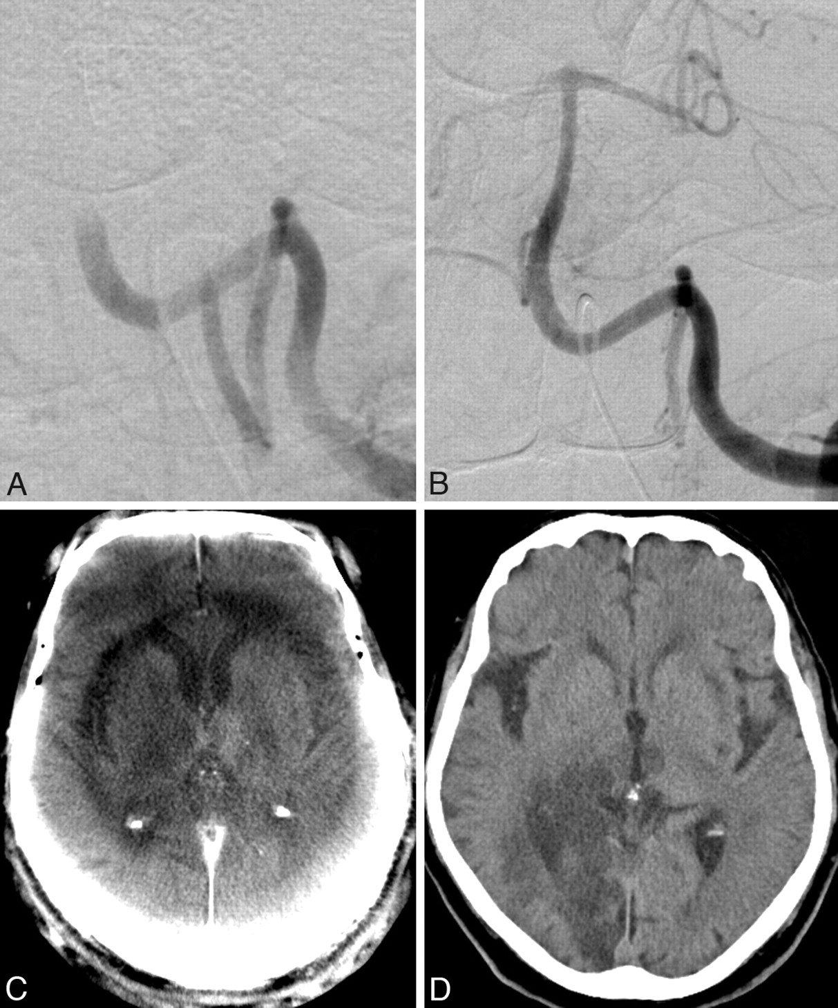
  • Fig 3.
    • Download figure
    • Open in new tab
    • Download powerpoint
    Fig 3.

    A, Ruptured distal carotid artery aneurysm of a 72-year-old man with acute SAH, Hunt and Hess 4. B, After endovascular treatment with Guglielmi detachable coils (Boston Scientific). C–F, ACT images (C and E) show the SAH and IVH as well as dilation of the supratentorial ventricular system. In contrast to MDCT (F), the fourth ventricle cannot be evaluated in the ACT examination (E) due to beam hardening artifacts. After implantation of a lumbar drain, follow-up MDCT scans (D and F) show a slight decrease of the lateral ventricle size.

  • Fig 4.
    • Download figure
    • Open in new tab
    • Download powerpoint
    Fig 4.

    A, A 63-year-old woman with acute basilar artery thrombosis. B and C, After recanalization of the artery with a Penumbra System (Penumbra, Alameda, California) (B), an acute ischemic lesion of the right thalamus can be seen on the ACT images (C). There is no complication in the form of an IPH or SAH. Typical ring artifacts can be seen on ACT and should not be confused with areas of cerebral edema. D, The follow-up MDCT after 2 hours confirms the thalamic infarction but also depicts a right occipital lobe infarction, which was undetectable on the ACT examination.

Tables

  • Figures
  • Interobserver agreement

    MDCTACT
    Observer 1 vs 20.910.82
    Observer 1 vs 30.870.72
    Observer 2 vs 30.860.69
    Mean κw0.880.74
PreviousNext
Back to top

In this issue

American Journal of Neuroradiology: 31 (7)
American Journal of Neuroradiology
Vol. 31, Issue 7
1 Aug 2010
  • Table of Contents
  • Index by author
Advertisement
Print
Download PDF
Email Article

Thank you for your interest in spreading the word on American Journal of Neuroradiology.

NOTE: We only request your email address so that the person you are recommending the page to knows that you wanted them to see it, and that it is not junk mail. We do not capture any email address.

Enter multiple addresses on separate lines or separate them with commas.
Feasibility of Angiographic CT in Peri-Interventional Diagnostic Imaging: A Comparative Study with Multidetector CT
(Your Name) has sent you a message from American Journal of Neuroradiology
(Your Name) thought you would like to see the American Journal of Neuroradiology web site.
CAPTCHA
This question is for testing whether or not you are a human visitor and to prevent automated spam submissions.
Cite this article
M.-N. Psychogios, J.-H. Buhk, P. Schramm, A. Xyda, A. Mohr, M. Knauth
Feasibility of Angiographic CT in Peri-Interventional Diagnostic Imaging: A Comparative Study with Multidetector CT
American Journal of Neuroradiology Aug 2010, 31 (7) 1226-1231; DOI: 10.3174/ajnr.A2086

Citation Manager Formats

  • BibTeX
  • Bookends
  • EasyBib
  • EndNote (tagged)
  • EndNote 8 (xml)
  • Medlars
  • Mendeley
  • Papers
  • RefWorks Tagged
  • Ref Manager
  • RIS
  • Zotero
0 Responses
Respond to this article
Share
Bookmark this article
Feasibility of Angiographic CT in Peri-Interventional Diagnostic Imaging: A Comparative Study with Multidetector CT
M.-N. Psychogios, J.-H. Buhk, P. Schramm, A. Xyda, A. Mohr, M. Knauth
American Journal of Neuroradiology Aug 2010, 31 (7) 1226-1231; DOI: 10.3174/ajnr.A2086
del.icio.us logo Twitter logo Facebook logo Mendeley logo
  • Tweet Widget
  • Facebook Like
  • Google Plus One
Purchase

Jump to section

  • Article
    • Abstract
    • Abbreviations
    • Materials and Methods
    • Results
    • Discussion
    • Conclusions
    • Footnotes
    • References
  • Figures & Data
  • Supplemental
  • Info & Metrics
  • Responses
  • References
  • PDF

Related Articles

  • PubMed
  • Google Scholar

Cited By...

  • Incidence, Risk Factors, and Clinical Implications of Subarachnoid Hyperdensities on Flat-Panel Detector CT following Mechanical Thrombectomy in Patients with Anterior Circulation Acute Ischemic Stroke
  • Evaluation of Sine Spin flat detector CT imaging compared with multidetector CT
  • The impact of software-based metal artifact reduction on the liquid embolic agent Onyx in cone-beam CT: a systematic in vitro and in vivo study
  • Diagnosing Early Ischemic Changes with the Latest-Generation Flat Detector CT: A Comparative Study with Multidetector CT
  • Latest generation of flat detector CT as a peri-interventional diagnostic tool: a comparative study with multidetector CT
  • One-Stop Management of Acute Stroke Patients: Minimizing Door-to-Reperfusion Times
  • Flat Detector Angio-CT following Intra-Arterial Therapy of Acute Ischemic Stroke: Identification of Hemorrhage and Distinction from Contrast Accumulation due to Blood-Brain Barrier Disruption
  • Angiographic CT for Intraprocedural Monitoring of Complex Neuroendovascular Procedures
  • Feasibility of Intravenous Flat Panel Detector CT Angiography for Intracranial Arterial Stenosis
  • Recanalization of Large Intracranial Vessels Using the Penumbra System: A Single-Center Experience
  • Feasibility of Cerebral Blood Volume Mapping by Flat Panel Detector CT in the Angiography Suite: First Experience in Patients with Acute Middle Cerebral Artery Occlusions
  • Feasibility of Flat Panel Angiographic CT after Intravenous Contrast Agent Application in the Postoperative Evaluation of Patients with Clipped Aneurysms
  • Angiographic CT after Intravenous Contrast Agent Application: A Noninvasive Follow-Up Tool after Intracranial Angioplasty and Stenting
  • Crossref
  • Google Scholar

This article has not yet been cited by articles in journals that are participating in Crossref Cited-by Linking.

More in this TOC Section

  • A Retrospective Study in Tentorial DAVFs
  • Proximal Protection Devices for Carotid Stenting
  • Guided vs Conventional Angiography Systems
Show more NEUROINTERVENTION

Similar Articles

Advertisement

Indexed Content

  • Current Issue
  • Accepted Manuscripts
  • Article Preview
  • Past Issues
  • Editorials
  • Editor's Choice
  • Fellows' Journal Club
  • Letters to the Editor
  • Video Articles

Cases

  • Case Collection
  • Archive - Case of the Week
  • Archive - Case of the Month
  • Archive - Classic Case

More from AJNR

  • Trainee Corner
  • Imaging Protocols
  • MRI Safety Corner
  • Book Reviews

Multimedia

  • AJNR Podcasts
  • AJNR Scantastics

Resources

  • Turnaround Time
  • Submit a Manuscript
  • Submit a Video Article
  • Submit an eLetter to the Editor/Response
  • Manuscript Submission Guidelines
  • Statistical Tips
  • Fast Publishing of Accepted Manuscripts
  • Graphical Abstract Preparation
  • Imaging Protocol Submission
  • Evidence-Based Medicine Level Guide
  • Publishing Checklists
  • Author Policies
  • Become a Reviewer/Academy of Reviewers
  • News and Updates

About Us

  • About AJNR
  • Editorial Board
  • Editorial Board Alumni
  • Alerts
  • Permissions
  • Not an AJNR Subscriber? Join Now
  • Advertise with Us
  • Librarian Resources
  • Feedback
  • Terms and Conditions
  • AJNR Editorial Board Alumni

American Society of Neuroradiology

  • Not an ASNR Member? Join Now

© 2025 by the American Society of Neuroradiology All rights, including for text and data mining, AI training, and similar technologies, are reserved.
Print ISSN: 0195-6108 Online ISSN: 1936-959X

Powered by HighWire