Advertisement

Main menu

  • Home
  • Content
    • Current Issue
    • Accepted Manuscripts
    • Article Preview
    • Past Issue Archive
    • Video Articles
    • AJNR Case Collection
    • Case of the Week Archive
    • Case of the Month Archive
    • Classic Case Archive
  • Special Collections
    • AJNR Awards
    • Low-Field MRI
    • Alzheimer Disease
    • ASNR Foundation Special Collection
    • Photon-Counting CT
    • View All
  • Multimedia
    • AJNR Podcasts
    • AJNR SCANtastic
    • Trainee Corner
    • MRI Safety Corner
    • Imaging Protocols
  • For Authors
    • Submit a Manuscript
    • Submit a Video Article
    • Submit an eLetter to the Editor/Response
    • Manuscript Submission Guidelines
    • Statistical Tips
    • Fast Publishing of Accepted Manuscripts
    • Graphical Abstract Preparation
    • Imaging Protocol Submission
    • Author Policies
  • About Us
    • About AJNR
    • Editorial Board
    • Editorial Board Alumni
  • More
    • Become a Reviewer/Academy of Reviewers
    • Subscribers
    • Permissions
    • Alerts
    • Feedback
    • Advertisers
    • ASNR Home

User menu

  • Alerts
  • Log in

Search

  • Advanced search
American Journal of Neuroradiology

American Journal of Neuroradiology

ASHNR American Society of Functional Neuroradiology ASHNR American Society of Pediatric Neuroradiology ASSR
  • Alerts
  • Log in

Advanced Search

  • Home
  • Content
    • Current Issue
    • Accepted Manuscripts
    • Article Preview
    • Past Issue Archive
    • Video Articles
    • AJNR Case Collection
    • Case of the Week Archive
    • Case of the Month Archive
    • Classic Case Archive
  • Special Collections
    • AJNR Awards
    • Low-Field MRI
    • Alzheimer Disease
    • ASNR Foundation Special Collection
    • Photon-Counting CT
    • View All
  • Multimedia
    • AJNR Podcasts
    • AJNR SCANtastic
    • Trainee Corner
    • MRI Safety Corner
    • Imaging Protocols
  • For Authors
    • Submit a Manuscript
    • Submit a Video Article
    • Submit an eLetter to the Editor/Response
    • Manuscript Submission Guidelines
    • Statistical Tips
    • Fast Publishing of Accepted Manuscripts
    • Graphical Abstract Preparation
    • Imaging Protocol Submission
    • Author Policies
  • About Us
    • About AJNR
    • Editorial Board
    • Editorial Board Alumni
  • More
    • Become a Reviewer/Academy of Reviewers
    • Subscribers
    • Permissions
    • Alerts
    • Feedback
    • Advertisers
    • ASNR Home
  • Follow AJNR on Twitter
  • Visit AJNR on Facebook
  • Follow AJNR on Instagram
  • Join AJNR on LinkedIn
  • RSS Feeds

AJNR Awards, New Junior Editors, and more. Read the latest AJNR updates

Research ArticleSpine

T2 Relaxation Times Correlated with Stage of Lumbar Intervertebral Disk Degeneration and Patient Age

N.L. Marinelli, V.M. Haughton and P.A. Anderson
American Journal of Neuroradiology August 2010, 31 (7) 1278-1282; DOI: https://doi.org/10.3174/ajnr.A2080
N.L. Marinelli
  • Find this author on Google Scholar
  • Find this author on PubMed
  • Search for this author on this site
V.M. Haughton
  • Find this author on Google Scholar
  • Find this author on PubMed
  • Search for this author on this site
P.A. Anderson
  • Find this author on Google Scholar
  • Find this author on PubMed
  • Search for this author on this site
  • Article
  • Figures & Data
  • Info & Metrics
  • Responses
  • References
  • PDF
Loading

Article Figures & Data

Figures

  • Tables
  • Fig 1.
    • Download figure
    • Open in new tab
    • Download powerpoint
    Fig 1.

    Color image of a T2 relaxation time map. The color scale ranges from 25 to 146 ms. The image shows 5 lumbar disks. A grade II disk is illustrated at L1–2; grade III, at L3–4 and L4–5; grade IV, at L2–3; and grade V, at L5–S1. Areas with T2 relaxation times that are off the scale are black, for example, as shown by the CSF in the central spinal canal. The color scale shows relaxation times from 25 ms (red end of scale) to 146 ms (blue end of scale).
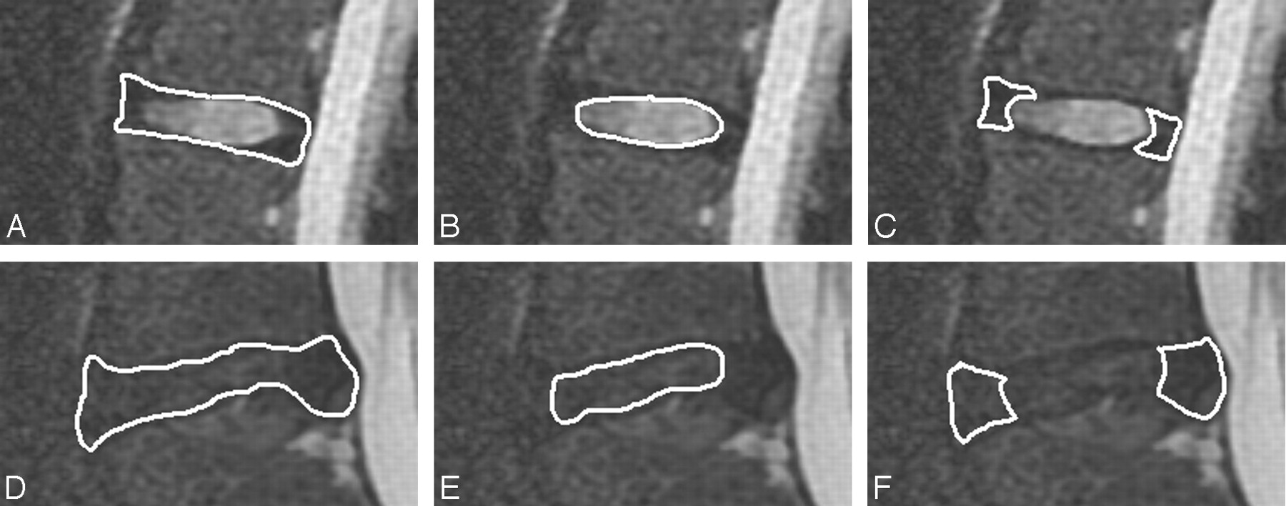
  • Fig 2.
    • Download figure
    • Open in new tab
    • Download powerpoint
    Fig 2.

    Region-of-interest placement for the whole disk (A and D), nucleus pulposus region (B and E), and annulus fibrosus (C and F) for a Pfirrmann grade II disk (A–C) and a Pfirrmann grade IV disk (D–F).

  • Fig 3.
    • Download figure
    • Open in new tab
    • Download powerpoint
    Fig 3.

    T2-weighted images illustrating 4 Pfirrmann disk grades. Midsagittal images show a grade II disk (A) with high signal intensity in the central region and lower signal intensity in the peripheral region, clearly distinguishing the nucleus pulposus region from the annulus fibrosus; a grade III disk (B) with lower signal intensity in the central region and less clear distinction between the nucleus pulposus region and annulus; a grade IV disk (C) with homogeneously low signal intensity and with no distinction of the nucleus pulposus region and annulus; and a grade V disk (D), which is collapsed.

  • Fig 4.
    • Download figure
    • Open in new tab
    • Download powerpoint
    Fig 4.

    T2-weighted image of a disk initially interpreted by 1 reader as Pfirrmann grade III and by the other reader as grade IV. By consensus, it was classified as grade IV.

  • Fig 5.
    • Download figure
    • Open in new tab
    • Download powerpoint
    Fig 5.

    Mean T2 relaxation times versus Pfirrmann grade for whole disks (A) and the nucleus pulposus region (B). The means (±SDs) for all disks of each grade are illustrated. The vertical bars represent the SD from the mean.

  • Fig 6.
    • Download figure
    • Open in new tab
    • Download powerpoint
    Fig 6.

    Mean T2 relaxation times for the nucleus pulposus region of Pfirrmann grade II disks for each decade of life. Mean T2 relaxation times are calculated as the averages for those in their 20s, 30s, 40s, 50s, and 60s. The vertical bars represent the SD from the mean.

Tables

  • Figures
  • Demographic, clinical, and L1–2 through L5–S1 imaging data for the 20 patients

    Patient No.Age (yr)SexClinical Indication for ImagingImaging Findings
    154MLow back pain, leg painL4–5 stenosis
    269FDegenerative spondylolisthesis L4/L5
    364FSevere leg painL5–S1 herniated disk
    445FLow back painL3–4 disk collapse
    551FLow back painL5–S1 spondylolisthesis and disk bulge
    634MBack painL5–S1 herniated disk, sacral metastasis from male breast cancer
    760MLow back pain3-Level disk degeneration
    821FSciaticaNormal MRI findings
    930FLow back pain2-Level disk degeneration
    1036MLow back painPostop L4–5 laminectomy, small L5–S1 protrusion
    1144FLow back painFacet joint disease 2 levels
    1235MLow back pain, saddle paresthesiasL3–4 herniated disk, L4–5 microdiskectomy, L5–S1 high-intensity zone
    1349FRight buttock painPostop L3–4 and L4–5 laminectomy
    1427FLow back painL5–S1 disk degeneration
    1569MLow back painHematoma at L2, bulging disks at L2–3 and L3–4
    1665FLow back painPostop L4–5 laminectomy, bulging annulus, 5 levels
    17a36FLow back painNormal MRI findings
    1840FLow back painBreast metastasis S1
    1965MLow back painFacet degeneration, stenosis, and spondylolisthesis at L4–5, disk degeneration at L5–S1
    2062MParesthesiasMildly degenerated disks L3–4, L4–5
    • a Patient No. 17 was excluded due to motion artifacts.

PreviousNext
Back to top

In this issue

American Journal of Neuroradiology: 31 (7)
American Journal of Neuroradiology
Vol. 31, Issue 7
1 Aug 2010
  • Table of Contents
  • Index by author
Advertisement
Print
Download PDF
Email Article

Thank you for your interest in spreading the word on American Journal of Neuroradiology.

NOTE: We only request your email address so that the person you are recommending the page to knows that you wanted them to see it, and that it is not junk mail. We do not capture any email address.

Enter multiple addresses on separate lines or separate them with commas.
T2 Relaxation Times Correlated with Stage of Lumbar Intervertebral Disk Degeneration and Patient Age
(Your Name) has sent you a message from American Journal of Neuroradiology
(Your Name) thought you would like to see the American Journal of Neuroradiology web site.
CAPTCHA
This question is for testing whether or not you are a human visitor and to prevent automated spam submissions.
Cite this article
N.L. Marinelli, V.M. Haughton, P.A. Anderson
T2 Relaxation Times Correlated with Stage of Lumbar Intervertebral Disk Degeneration and Patient Age
American Journal of Neuroradiology Aug 2010, 31 (7) 1278-1282; DOI: 10.3174/ajnr.A2080

Citation Manager Formats

  • BibTeX
  • Bookends
  • EasyBib
  • EndNote (tagged)
  • EndNote 8 (xml)
  • Medlars
  • Mendeley
  • Papers
  • RefWorks Tagged
  • Ref Manager
  • RIS
  • Zotero
0 Responses
Respond to this article
Share
Bookmark this article
T2 Relaxation Times Correlated with Stage of Lumbar Intervertebral Disk Degeneration and Patient Age
N.L. Marinelli, V.M. Haughton, P.A. Anderson
American Journal of Neuroradiology Aug 2010, 31 (7) 1278-1282; DOI: 10.3174/ajnr.A2080
del.icio.us logo Twitter logo Facebook logo Mendeley logo
  • Tweet Widget
  • Facebook Like
  • Google Plus One
Purchase

Jump to section

  • Article
    • Abstract
    • Abbreviations
    • Materials and Methods
    • Results
    • Discussion
    • Conclusions
    • Footnotes
    • References
  • Figures & Data
  • Info & Metrics
  • Responses
  • References
  • PDF

Related Articles

  • No related articles found.
  • PubMed
  • Google Scholar

Cited By...

  • Running acceleration correlates with T2 magnetic resonance imaging values of the lumber intervertebral disc
  • Intervertebral Disc Elastography to Relate Shear Modulus and Relaxometry in Compression and Bending
  • Variability of T2-Relaxation Times of Healthy Lumbar Intervertebral Discs is More Homogeneous within an Individual Than across Healthy Individuals
  • We Need to Talk about Lumbar Total Disc Replacement
  • T1{rho} and T2 Mapping of the Intervertebral Disk: Comparison of Different Methods of Segmentation
  • Comparison of the T2 Relaxation Time of the Temporomandibular Joint Articular Disk between Patients with Temporomandibular Disorders and Asymptomatic Volunteers
  • MR Imaging Assessment of Lumbar Intervertebral Disk Degeneration and Age-Related Changes: Apparent Diffusion Coefficient versus T2 Quantitation
  • Crossref
  • Google Scholar

This article has not yet been cited by articles in journals that are participating in Crossref Cited-by Linking.

More in this TOC Section

  • CT-Guided C2 Dorsal Root Ganglion Radiofrequency Ablation for the Treatment of Cervicogenic Headache: Case Series and Clinical Outcomes
  • The Evaluation and Prediction of Laminoplasty Surgery Outcome in Patients with Degenerative Cervical Myelopathy Using Diffusion Tensor MRI
  • Subject-Specific Studies of CSF Bulk Flow Patterns in the Spinal Canal: Implications for the Dispersion of Solute Particles in Intrathecal Drug Delivery
Show more Spine

Similar Articles

Advertisement

Indexed Content

  • Current Issue
  • Accepted Manuscripts
  • Article Preview
  • Past Issues
  • Editorials
  • Editor's Choice
  • Fellows' Journal Club
  • Letters to the Editor
  • Video Articles

Cases

  • Case Collection
  • Archive - Case of the Week
  • Archive - Case of the Month
  • Archive - Classic Case

More from AJNR

  • Trainee Corner
  • Imaging Protocols
  • MRI Safety Corner
  • Book Reviews

Multimedia

  • AJNR Podcasts
  • AJNR Scantastics

Resources

  • Turnaround Time
  • Submit a Manuscript
  • Submit a Video Article
  • Submit an eLetter to the Editor/Response
  • Manuscript Submission Guidelines
  • Statistical Tips
  • Fast Publishing of Accepted Manuscripts
  • Graphical Abstract Preparation
  • Imaging Protocol Submission
  • Evidence-Based Medicine Level Guide
  • Publishing Checklists
  • Author Policies
  • Become a Reviewer/Academy of Reviewers
  • News and Updates

About Us

  • About AJNR
  • Editorial Board
  • Editorial Board Alumni
  • Alerts
  • Permissions
  • Not an AJNR Subscriber? Join Now
  • Advertise with Us
  • Librarian Resources
  • Feedback
  • Terms and Conditions
  • AJNR Editorial Board Alumni

American Society of Neuroradiology

  • Not an ASNR Member? Join Now

© 2025 by the American Society of Neuroradiology All rights, including for text and data mining, AI training, and similar technologies, are reserved.
Print ISSN: 0195-6108 Online ISSN: 1936-959X

Powered by HighWire