Advertisement

Main menu

  • Home
  • Content
    • Current Issue
    • Accepted Manuscripts
    • Article Preview
    • Past Issue Archive
    • Video Articles
    • AJNR Case Collection
    • Case of the Week Archive
    • Case of the Month Archive
    • Classic Case Archive
  • Special Collections
    • AJNR Awards
    • Low-Field MRI
    • Alzheimer Disease
    • ASNR Foundation Special Collection
    • Photon-Counting CT
    • View All
  • Multimedia
    • AJNR Podcasts
    • AJNR SCANtastic
    • Trainee Corner
    • MRI Safety Corner
    • Imaging Protocols
  • For Authors
    • Submit a Manuscript
    • Submit a Video Article
    • Submit an eLetter to the Editor/Response
    • Manuscript Submission Guidelines
    • Statistical Tips
    • Fast Publishing of Accepted Manuscripts
    • Graphical Abstract Preparation
    • Imaging Protocol Submission
    • Author Policies
  • About Us
    • About AJNR
    • Editorial Board
    • Editorial Board Alumni
  • More
    • Become a Reviewer/Academy of Reviewers
    • Subscribers
    • Permissions
    • Alerts
    • Feedback
    • Advertisers
    • ASNR Home

User menu

  • Alerts
  • Log in

Search

  • Advanced search
American Journal of Neuroradiology

American Journal of Neuroradiology

ASHNR American Society of Functional Neuroradiology ASHNR American Society of Pediatric Neuroradiology ASSR
  • Alerts
  • Log in

Advanced Search

  • Home
  • Content
    • Current Issue
    • Accepted Manuscripts
    • Article Preview
    • Past Issue Archive
    • Video Articles
    • AJNR Case Collection
    • Case of the Week Archive
    • Case of the Month Archive
    • Classic Case Archive
  • Special Collections
    • AJNR Awards
    • Low-Field MRI
    • Alzheimer Disease
    • ASNR Foundation Special Collection
    • Photon-Counting CT
    • View All
  • Multimedia
    • AJNR Podcasts
    • AJNR SCANtastic
    • Trainee Corner
    • MRI Safety Corner
    • Imaging Protocols
  • For Authors
    • Submit a Manuscript
    • Submit a Video Article
    • Submit an eLetter to the Editor/Response
    • Manuscript Submission Guidelines
    • Statistical Tips
    • Fast Publishing of Accepted Manuscripts
    • Graphical Abstract Preparation
    • Imaging Protocol Submission
    • Author Policies
  • About Us
    • About AJNR
    • Editorial Board
    • Editorial Board Alumni
  • More
    • Become a Reviewer/Academy of Reviewers
    • Subscribers
    • Permissions
    • Alerts
    • Feedback
    • Advertisers
    • ASNR Home
  • Follow AJNR on Twitter
  • Visit AJNR on Facebook
  • Follow AJNR on Instagram
  • Join AJNR on LinkedIn
  • RSS Feeds

AJNR Awards, New Junior Editors, and more. Read the latest AJNR updates

Research ArticleBrain
Open Access

The Triple Rule-Out for Acute Ischemic Stroke: Imaging the Brain, Carotid Arteries, Aorta, and Heart

A.D. Furtado, D.D. Adraktas, N. Brasic, S.-C. Cheng, K. Ordovas, W.S. Smith, M.R. Lewin, K. Chun, J.D. Chien, S. Schaeffer and M. Wintermark
American Journal of Neuroradiology August 2010, 31 (7) 1290-1296; DOI: https://doi.org/10.3174/ajnr.A2075
A.D. Furtado
  • Find this author on Google Scholar
  • Find this author on PubMed
  • Search for this author on this site
D.D. Adraktas
  • Find this author on Google Scholar
  • Find this author on PubMed
  • Search for this author on this site
N. Brasic
  • Find this author on Google Scholar
  • Find this author on PubMed
  • Search for this author on this site
S.-C. Cheng
  • Find this author on Google Scholar
  • Find this author on PubMed
  • Search for this author on this site
K. Ordovas
  • Find this author on Google Scholar
  • Find this author on PubMed
  • Search for this author on this site
W.S. Smith
  • Find this author on Google Scholar
  • Find this author on PubMed
  • Search for this author on this site
M.R. Lewin
  • Find this author on Google Scholar
  • Find this author on PubMed
  • Search for this author on this site
K. Chun
  • Find this author on Google Scholar
  • Find this author on PubMed
  • Search for this author on this site
J.D. Chien
  • Find this author on Google Scholar
  • Find this author on PubMed
  • Search for this author on this site
S. Schaeffer
  • Find this author on Google Scholar
  • Find this author on PubMed
  • Search for this author on this site
M. Wintermark
  • Find this author on Google Scholar
  • Find this author on PubMed
  • Search for this author on this site
  • Article
  • Figures & Data
  • Supplemental
  • Info & Metrics
  • Responses
  • References
  • PDF
Loading

Article Figures & Data

Figures

  • Fig 1.
    • Download figure
    • Open in new tab
    • Download powerpoint
    Fig 1.

    Stroke CT protocol design. A bolus of 30 cc of contrast was injected into the right or left cubital vein, followed by a 15 cc saline flush, both at an injection rate of 5 cc per second. The first acquisition (1) (not ECG gated) ascends from the aortic arch origin to the vertex of the head, taking place after a delay determined from perfusion CT used as a bolus test, typically 15 seconds. A second bolus (2) of 60 cc of contrast was injected 3 seconds later and followed by a 60 cc saline flush, again at an injection rate of 5 cc per second. The second acquisition descends from the aortic arch origin to the diaphragm and is ECG gated.
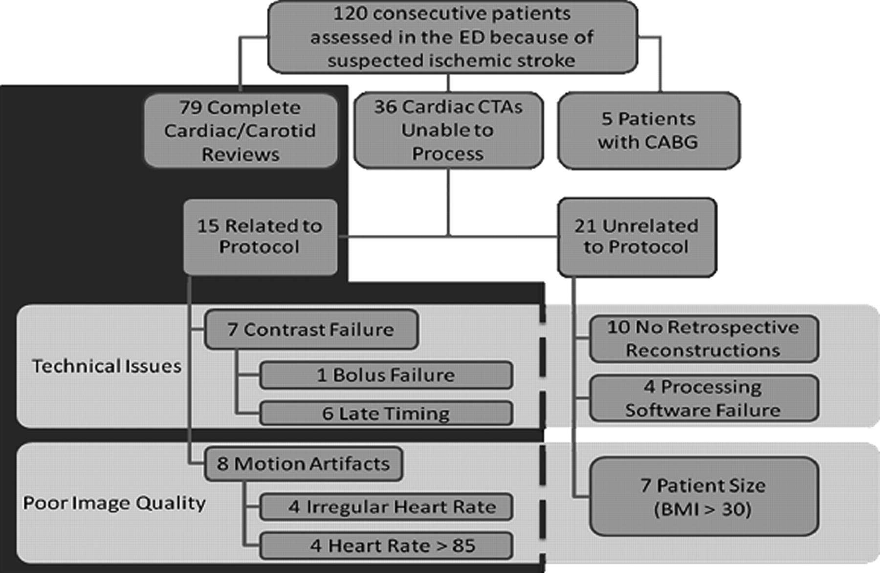
  • Fig 2.
    • Download figure
    • Open in new tab
    • Download powerpoint
    Fig 2.

    Study participants. One hundred twenty consecutive patients receiving standard-of-care stroke CT evaluation were prospectively enrolled in our study. Five patients were excluded because of CABGs. In 79 patients, both the carotid and cardiac portions of the CTA succeeded. In 36 patients, the carotid portion succeeded, but the cardiac portion failed. Fifteen of these 36 failures were directly related to our protocol, 7 were related to patient size, and 14, to technical issues. The 21 cases in which failure was unrelated to our protocol were excluded, and our image-quality analysis was performed on the remaining 94 patients.

  • Fig 3.
    • Download figure
    • Open in new tab
    • Download powerpoint
    Fig 3.

    A, Segment-level analysis of coronary artery image quality. Each coronary artery segment was given an image-quality score (0 = excellent, 1 = good, and 2 = nondiagnostic). The nondiagnostic segments were separated into 3 categories: motion artifacts, poor contrast, and streak artifacts. Those segments that were nondiagnostic due to tiny vessel size, heavy calcification, or pacemaker artifacts were excluded because they were nondiagnostic for reasons not directly related to our protocol. B, Vessel-level analysis of coronary artery image quality. A coronary artery was considered excellent if the entire artery was diagnostic, good if 1 or 2 segments were diagnostic, and nondiagnostic if no segments were diagnostic. Those segments that were nondiagnostic for reasons unrelated to our protocol (ie, tiny vessel size, heavy calcification, or pacemaker artifacts) were omitted from this analysis. C, Patient-level analysis of coronary artery image quality. Cardiac examinations that were 75%–100% diagnostic were considered excellent, cardiac examinations that were 50%–74% diagnostic were considered good, and cardiac examinations that were 0%–49% diagnostic were considered failed. Most patients (65%, n = 61) had cardiac examinations that were 75%–100% diagnostic. Nondiagnostic segments in the failed cases were primarily due to motion artifacts.

PreviousNext
Back to top

In this issue

American Journal of Neuroradiology: 31 (7)
American Journal of Neuroradiology
Vol. 31, Issue 7
1 Aug 2010
  • Table of Contents
  • Index by author
Advertisement
Print
Download PDF
Email Article

Thank you for your interest in spreading the word on American Journal of Neuroradiology.

NOTE: We only request your email address so that the person you are recommending the page to knows that you wanted them to see it, and that it is not junk mail. We do not capture any email address.

Enter multiple addresses on separate lines or separate them with commas.
The Triple Rule-Out for Acute Ischemic Stroke: Imaging the Brain, Carotid Arteries, Aorta, and Heart
(Your Name) has sent you a message from American Journal of Neuroradiology
(Your Name) thought you would like to see the American Journal of Neuroradiology web site.
CAPTCHA
This question is for testing whether or not you are a human visitor and to prevent automated spam submissions.
Cite this article
A.D. Furtado, D.D. Adraktas, N. Brasic, S.-C. Cheng, K. Ordovas, W.S. Smith, M.R. Lewin, K. Chun, J.D. Chien, S. Schaeffer, M. Wintermark
The Triple Rule-Out for Acute Ischemic Stroke: Imaging the Brain, Carotid Arteries, Aorta, and Heart
American Journal of Neuroradiology Aug 2010, 31 (7) 1290-1296; DOI: 10.3174/ajnr.A2075

Citation Manager Formats

  • BibTeX
  • Bookends
  • EasyBib
  • EndNote (tagged)
  • EndNote 8 (xml)
  • Medlars
  • Mendeley
  • Papers
  • RefWorks Tagged
  • Ref Manager
  • RIS
  • Zotero
0 Responses
Respond to this article
Share
Bookmark this article
The Triple Rule-Out for Acute Ischemic Stroke: Imaging the Brain, Carotid Arteries, Aorta, and Heart
A.D. Furtado, D.D. Adraktas, N. Brasic, S.-C. Cheng, K. Ordovas, W.S. Smith, M.R. Lewin, K. Chun, J.D. Chien, S. Schaeffer, M. Wintermark
American Journal of Neuroradiology Aug 2010, 31 (7) 1290-1296; DOI: 10.3174/ajnr.A2075
del.icio.us logo Twitter logo Facebook logo Mendeley logo
  • Tweet Widget
  • Facebook Like
  • Google Plus One
Purchase

Jump to section

  • Article
    • Abstract
    • Abbreviations
    • Materials and Methods
    • Results
    • Discussion
    • Conclusions
    • Footnotes
    • References
  • Figures & Data
  • Supplemental
  • Info & Metrics
  • Responses
  • References
  • PDF

Related Articles

  • PubMed
  • Google Scholar

Cited By...

  • Diagnostic Yield of ECG-Gated Cardiac CT in theAcute Phase of Ischemic Stroke vsTransthoracic Echocardiography
  • High prevalence of intracranial aneurysms in patients with aortic dissection or aneurysm: feasibility of extended aorta CT angiography with involvement of intracranial arteries
  • Cerebral CTA with Low Tube Voltage and Low Contrast Material Volume for Detection of Intracranial Aneurysms
  • CTA for Screening of Complicated Atherosclerotic Carotid Plaque--American Heart Association Type VI Lesions as Defined by MRI
  • Crossref
  • Google Scholar

This article has not yet been cited by articles in journals that are participating in Crossref Cited-by Linking.

More in this TOC Section

  • Statin Therapy Does Not Affect the Radiographic and Clinical Profile of Patients with TIA and Minor Stroke
  • Usefulness of Quantitative Susceptibility Mapping for the Diagnosis of Parkinson Disease
  • White Matter Alterations in the Brains of Patients with Active, Remitted, and Cured Cushing Syndrome: A DTI Study
Show more Brain

Similar Articles

Advertisement

Indexed Content

  • Current Issue
  • Accepted Manuscripts
  • Article Preview
  • Past Issues
  • Editorials
  • Editor's Choice
  • Fellows' Journal Club
  • Letters to the Editor
  • Video Articles

Cases

  • Case Collection
  • Archive - Case of the Week
  • Archive - Case of the Month
  • Archive - Classic Case

More from AJNR

  • Trainee Corner
  • Imaging Protocols
  • MRI Safety Corner
  • Book Reviews

Multimedia

  • AJNR Podcasts
  • AJNR Scantastics

Resources

  • Turnaround Time
  • Submit a Manuscript
  • Submit a Video Article
  • Submit an eLetter to the Editor/Response
  • Manuscript Submission Guidelines
  • Statistical Tips
  • Fast Publishing of Accepted Manuscripts
  • Graphical Abstract Preparation
  • Imaging Protocol Submission
  • Evidence-Based Medicine Level Guide
  • Publishing Checklists
  • Author Policies
  • Become a Reviewer/Academy of Reviewers
  • News and Updates

About Us

  • About AJNR
  • Editorial Board
  • Editorial Board Alumni
  • Alerts
  • Permissions
  • Not an AJNR Subscriber? Join Now
  • Advertise with Us
  • Librarian Resources
  • Feedback
  • Terms and Conditions
  • AJNR Editorial Board Alumni

American Society of Neuroradiology

  • Not an ASNR Member? Join Now

© 2025 by the American Society of Neuroradiology All rights, including for text and data mining, AI training, and similar technologies, are reserved.
Print ISSN: 0195-6108 Online ISSN: 1936-959X

Powered by HighWire