Advertisement

Main menu

  • Home
  • Content
    • Current Issue
    • Accepted Manuscripts
    • Article Preview
    • Past Issue Archive
    • Video Articles
    • AJNR Case Collection
    • Case of the Week Archive
    • Case of the Month Archive
    • Classic Case Archive
  • Special Collections
    • AJNR Awards
    • Low-Field MRI
    • Alzheimer Disease
    • ASNR Foundation Special Collection
    • Photon-Counting CT
    • View All
  • Multimedia
    • AJNR Podcasts
    • AJNR SCANtastic
    • Trainee Corner
    • MRI Safety Corner
    • Imaging Protocols
  • For Authors
    • Submit a Manuscript
    • Submit a Video Article
    • Submit an eLetter to the Editor/Response
    • Manuscript Submission Guidelines
    • Statistical Tips
    • Fast Publishing of Accepted Manuscripts
    • Graphical Abstract Preparation
    • Imaging Protocol Submission
    • Author Policies
  • About Us
    • About AJNR
    • Editorial Board
    • Editorial Board Alumni
  • More
    • Become a Reviewer/Academy of Reviewers
    • Subscribers
    • Permissions
    • Alerts
    • Feedback
    • Advertisers
    • ASNR Home

User menu

  • Alerts
  • Log in

Search

  • Advanced search
American Journal of Neuroradiology

American Journal of Neuroradiology

ASHNR American Society of Functional Neuroradiology ASHNR American Society of Pediatric Neuroradiology ASSR
  • Alerts
  • Log in

Advanced Search

  • Home
  • Content
    • Current Issue
    • Accepted Manuscripts
    • Article Preview
    • Past Issue Archive
    • Video Articles
    • AJNR Case Collection
    • Case of the Week Archive
    • Case of the Month Archive
    • Classic Case Archive
  • Special Collections
    • AJNR Awards
    • Low-Field MRI
    • Alzheimer Disease
    • ASNR Foundation Special Collection
    • Photon-Counting CT
    • View All
  • Multimedia
    • AJNR Podcasts
    • AJNR SCANtastic
    • Trainee Corner
    • MRI Safety Corner
    • Imaging Protocols
  • For Authors
    • Submit a Manuscript
    • Submit a Video Article
    • Submit an eLetter to the Editor/Response
    • Manuscript Submission Guidelines
    • Statistical Tips
    • Fast Publishing of Accepted Manuscripts
    • Graphical Abstract Preparation
    • Imaging Protocol Submission
    • Author Policies
  • About Us
    • About AJNR
    • Editorial Board
    • Editorial Board Alumni
  • More
    • Become a Reviewer/Academy of Reviewers
    • Subscribers
    • Permissions
    • Alerts
    • Feedback
    • Advertisers
    • ASNR Home
  • Follow AJNR on Twitter
  • Visit AJNR on Facebook
  • Follow AJNR on Instagram
  • Join AJNR on LinkedIn
  • RSS Feeds

AJNR Awards, New Junior Editors, and more. Read the latest AJNR updates

Research ArticleBrain
Open Access

Correlation of Automated Volumetric Analysis of Brain MR Imaging with Cognitive Impairment in a Natural History Study of Mucopolysaccharidosis II

Z. Fan, M. Styner, J. Muenzer, M. Poe and M. Escolar
American Journal of Neuroradiology August 2010, 31 (7) 1319-1323; DOI: https://doi.org/10.3174/ajnr.A2032
Z. Fan
  • Find this author on Google Scholar
  • Find this author on PubMed
  • Search for this author on this site
M. Styner
  • Find this author on Google Scholar
  • Find this author on PubMed
  • Search for this author on this site
J. Muenzer
  • Find this author on Google Scholar
  • Find this author on PubMed
  • Search for this author on this site
M. Poe
  • Find this author on Google Scholar
  • Find this author on PubMed
  • Search for this author on this site
M. Escolar
  • Find this author on Google Scholar
  • Find this author on PubMed
  • Search for this author on this site
  • Article
  • Figures & Data
  • Info & Metrics
  • Responses
  • References
  • PDF
Loading

Article Figures & Data

Figures

  • Tables
  • Fig 1.
    • Download figure
    • Open in new tab
    • Download powerpoint
    Fig 1.

    Correlation of brain tissue volume (A) and brain tissue/ICV ratio (B) with cognitive status. The black vertical crossbars indicate the timeline when VP shunt was placed.
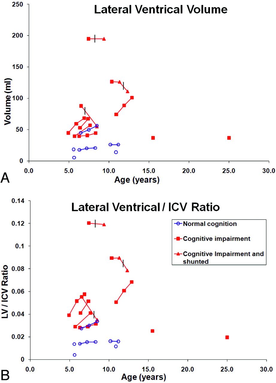
  • Fig 2.
    • Download figure
    • Open in new tab
    • Download powerpoint
    Fig 2.

    Correlation of LV volume (A) and LV/ICV ratio (B) with cognitive status. The vertical crossbars indicate the timeline when VP shunt was placed.

  • Fig 3.
    • Download figure
    • Open in new tab
    • Download powerpoint
    Fig 3.

    Examples of the automated segmentation of brain and semi-automated segmentation of lateral ventricles. A, Original axial T1 image; B, aligned and registered T1 image; C, segmented image, where green voxels represent gray matter, pink voxels represent white matter, and dark blue voxels represent CSF; D, registered T1 axial image; E, 2D view of the segmented lateral ventricles in red; F, 3D view of the lateral ventricles (enlarged).

Tables

  • Figures
  • Volumetric analysis of brain MR Imaging in MPS II Patients

    VariablesAll Patients MR Imaging Studies (N = 33), Mean (SD)Patients with Cognitive Impairment MR Imaging Studies (N = 22), Mean (SD)Patients with Normal Cognition MR Imaging Studies (N = 11), Mean (SD)P Valuea
    Age (years)9.0 (5.0)9.9 (6.2)7.5 (2.37).296
    ICV (mL)1443 (189)1464 (201)1407 (176).96
    Brain tissue (mL)1242 (173)1234 (195)1254 (143).25
    LV (mL)54 (48)73 (52)21 (13).046b
    Brain tissue/ICV0.86 (0.012)0.84 (0.03)0.89 (0.01)<.001b
    LV/ICV0.04 (0.015)0.05 (0.03)0.01 (0.01).029b
    • a P values were comparisons between patients with cognitive impairment and those with normal cognition.

    • b P values reached statistical significance.

PreviousNext
Back to top

In this issue

American Journal of Neuroradiology: 31 (7)
American Journal of Neuroradiology
Vol. 31, Issue 7
1 Aug 2010
  • Table of Contents
  • Index by author
Advertisement
Print
Download PDF
Email Article

Thank you for your interest in spreading the word on American Journal of Neuroradiology.

NOTE: We only request your email address so that the person you are recommending the page to knows that you wanted them to see it, and that it is not junk mail. We do not capture any email address.

Enter multiple addresses on separate lines or separate them with commas.
Correlation of Automated Volumetric Analysis of Brain MR Imaging with Cognitive Impairment in a Natural History Study of Mucopolysaccharidosis II
(Your Name) has sent you a message from American Journal of Neuroradiology
(Your Name) thought you would like to see the American Journal of Neuroradiology web site.
CAPTCHA
This question is for testing whether or not you are a human visitor and to prevent automated spam submissions.
Cite this article
Z. Fan, M. Styner, J. Muenzer, M. Poe, M. Escolar
Correlation of Automated Volumetric Analysis of Brain MR Imaging with Cognitive Impairment in a Natural History Study of Mucopolysaccharidosis II
American Journal of Neuroradiology Aug 2010, 31 (7) 1319-1323; DOI: 10.3174/ajnr.A2032

Citation Manager Formats

  • BibTeX
  • Bookends
  • EasyBib
  • EndNote (tagged)
  • EndNote 8 (xml)
  • Medlars
  • Mendeley
  • Papers
  • RefWorks Tagged
  • Ref Manager
  • RIS
  • Zotero
0 Responses
Respond to this article
Share
Bookmark this article
Correlation of Automated Volumetric Analysis of Brain MR Imaging with Cognitive Impairment in a Natural History Study of Mucopolysaccharidosis II
Z. Fan, M. Styner, J. Muenzer, M. Poe, M. Escolar
American Journal of Neuroradiology Aug 2010, 31 (7) 1319-1323; DOI: 10.3174/ajnr.A2032
del.icio.us logo Twitter logo Facebook logo Mendeley logo
  • Tweet Widget
  • Facebook Like
  • Google Plus One
Purchase

Jump to section

  • Article
    • Abstract
    • Abbreviations
    • Materials and Methods
    • Results
    • Discussion
    • Conclusions
    • Footnotes
    • References
  • Figures & Data
  • Info & Metrics
  • Responses
  • References
  • PDF

Related Articles

  • No related articles found.
  • PubMed
  • Google Scholar

Cited By...

  • Brain and Spinal MR Imaging Findings in Mucopolysaccharidoses: A Review
  • Crossref
  • Google Scholar

This article has not yet been cited by articles in journals that are participating in Crossref Cited-by Linking.

More in this TOC Section

  • Evaluating the Effects of White Matter Multiple Sclerosis Lesions on the Volume Estimation of 6 Brain Tissue Segmentation Methods
  • Quiet PROPELLER MRI Techniques Match the Quality of Conventional PROPELLER Brain Imaging Techniques
  • Predictors of Reperfusion in Patients with Acute Ischemic Stroke
Show more Brain

Similar Articles

Advertisement

Indexed Content

  • Current Issue
  • Accepted Manuscripts
  • Article Preview
  • Past Issues
  • Editorials
  • Editor's Choice
  • Fellows' Journal Club
  • Letters to the Editor
  • Video Articles

Cases

  • Case Collection
  • Archive - Case of the Week
  • Archive - Case of the Month
  • Archive - Classic Case

More from AJNR

  • Trainee Corner
  • Imaging Protocols
  • MRI Safety Corner
  • Book Reviews

Multimedia

  • AJNR Podcasts
  • AJNR Scantastics

Resources

  • Turnaround Time
  • Submit a Manuscript
  • Submit a Video Article
  • Submit an eLetter to the Editor/Response
  • Manuscript Submission Guidelines
  • Statistical Tips
  • Fast Publishing of Accepted Manuscripts
  • Graphical Abstract Preparation
  • Imaging Protocol Submission
  • Evidence-Based Medicine Level Guide
  • Publishing Checklists
  • Author Policies
  • Become a Reviewer/Academy of Reviewers
  • News and Updates

About Us

  • About AJNR
  • Editorial Board
  • Editorial Board Alumni
  • Alerts
  • Permissions
  • Not an AJNR Subscriber? Join Now
  • Advertise with Us
  • Librarian Resources
  • Feedback
  • Terms and Conditions
  • AJNR Editorial Board Alumni

American Society of Neuroradiology

  • Not an ASNR Member? Join Now

© 2025 by the American Society of Neuroradiology All rights, including for text and data mining, AI training, and similar technologies, are reserved.
Print ISSN: 0195-6108 Online ISSN: 1936-959X

Powered by HighWire