Advertisement

Main menu

  • Home
  • Content
    • Current Issue
    • Accepted Manuscripts
    • Article Preview
    • Past Issue Archive
    • AJNR Case Collection
    • Case of the Week Archive
    • Classic Case Archive
    • Case of the Month Archive
  • Special Collections
    • Spinal CSF Leak Articles (Jan 2020-June 2024)
    • 2024 AJNR Journal Awards
    • Most Impactful AJNR Articles
  • Multimedia
    • AJNR Podcast
    • AJNR Scantastics
    • Video Articles
  • For Authors
    • Submit a Manuscript
    • Author Policies
    • Fast publishing of Accepted Manuscripts
    • Graphical Abstract Preparation
    • Manuscript Submission Guidelines
    • Imaging Protocol Submission
    • Submit a Case for the Case Collection
  • About Us
    • About AJNR
    • Editorial Board
  • More
    • Become a Reviewer/Academy of Reviewers
    • Subscribers
    • Permissions
    • Alerts
    • Feedback
    • Advertisers
    • ASNR Home
  • Other Publications
    • ajnr

User menu

  • Alerts
  • Log in

Search

  • Advanced search
American Journal of Neuroradiology

American Journal of Neuroradiology

ASHNR American Society of Functional Neuroradiology ASHNR American Society of Pediatric Neuroradiology ASSR
  • Alerts
  • Log in

Advanced Search

  • Home
  • Content
    • Current Issue
    • Accepted Manuscripts
    • Article Preview
    • Past Issue Archive
    • AJNR Case Collection
    • Case of the Week Archive
    • Classic Case Archive
    • Case of the Month Archive
  • Special Collections
    • Spinal CSF Leak Articles (Jan 2020-June 2024)
    • 2024 AJNR Journal Awards
    • Most Impactful AJNR Articles
  • Multimedia
    • AJNR Podcast
    • AJNR Scantastics
    • Video Articles
  • For Authors
    • Submit a Manuscript
    • Author Policies
    • Fast publishing of Accepted Manuscripts
    • Graphical Abstract Preparation
    • Manuscript Submission Guidelines
    • Imaging Protocol Submission
    • Submit a Case for the Case Collection
  • About Us
    • About AJNR
    • Editorial Board
  • More
    • Become a Reviewer/Academy of Reviewers
    • Subscribers
    • Permissions
    • Alerts
    • Feedback
    • Advertisers
    • ASNR Home
  • Follow AJNR on Twitter
  • Visit AJNR on Facebook
  • Follow AJNR on Instagram
  • Join AJNR on LinkedIn
  • RSS Feeds

Welcome to the new AJNR, Updated Hall of Fame, and more. Read the full announcements.


AJNR is seeking candidates for the position of Associate Section Editor, AJNR Case Collection. Read the full announcement.


Research ArticleBrain
Open Access

Predicting Language Improvement in Acute Stroke Patients Presenting with Aphasia: A Multivariate Logistic Model Using Location-Weighted Atlas-Based Analysis of Admission CT Perfusion Scans

S. Payabvash, S. Kamalian, S. Fung, Y. Wang, J. Passanese, S. Kamalian, L.C.S. Souza, A. Kemmling, G.J. Harris, E.F. Halpern, R.G. González, K.L. Furie and M.H. Lev
American Journal of Neuroradiology October 2010, 31 (9) 1661-1668; DOI: https://doi.org/10.3174/ajnr.A2125
S. Payabvash
  • Find this author on Google Scholar
  • Find this author on PubMed
  • Search for this author on this site
S. Kamalian
  • Find this author on Google Scholar
  • Find this author on PubMed
  • Search for this author on this site
S. Fung
  • Find this author on Google Scholar
  • Find this author on PubMed
  • Search for this author on this site
Y. Wang
  • Find this author on Google Scholar
  • Find this author on PubMed
  • Search for this author on this site
J. Passanese
  • Find this author on Google Scholar
  • Find this author on PubMed
  • Search for this author on this site
S. Kamalian
  • Find this author on Google Scholar
  • Find this author on PubMed
  • Search for this author on this site
L.C.S. Souza
  • Find this author on Google Scholar
  • Find this author on PubMed
  • Search for this author on this site
A. Kemmling
  • Find this author on Google Scholar
  • Find this author on PubMed
  • Search for this author on this site
G.J. Harris
  • Find this author on Google Scholar
  • Find this author on PubMed
  • Search for this author on this site
E.F. Halpern
  • Find this author on Google Scholar
  • Find this author on PubMed
  • Search for this author on this site
R.G. González
  • Find this author on Google Scholar
  • Find this author on PubMed
  • Search for this author on this site
K.L. Furie
  • Find this author on Google Scholar
  • Find this author on PubMed
  • Search for this author on this site
M.H. Lev
  • Find this author on Google Scholar
  • Find this author on PubMed
  • Search for this author on this site
  • Article
  • Figures & Data
  • Info & Metrics
  • Responses
  • References
  • PDF
Loading

Article Figures & Data

Figures

  • Tables
  • Fig 1.
    • Download figure
    • Open in new tab
    • Download powerpoint
    Fig 1.

    Of 119 patients with acute ischemic stroke who underwent CTP at our hospital between December 2006 and April 2008, 58 were included in this study.

  • Fig 2.
    • Download figure
    • Open in new tab
    • Download powerpoint
    Fig 2.

    Upper row: orthogonal sections of the lower third of the sublobar insular ribbon. Middle row: orthogonal sections passing through the angular gyrus GM of BA 39. The mean relative rCBF values of these 2 regions were the only CTP parameters that were independent predictors of early aphasia improvement. Lower row: a 3D standard MNI-152 brain space reflecting the main cortical BAs directly involved in language function (labeled by corresponding number), as well as the lower third subinsular ribbon and angular gyrus GM, as noted above.
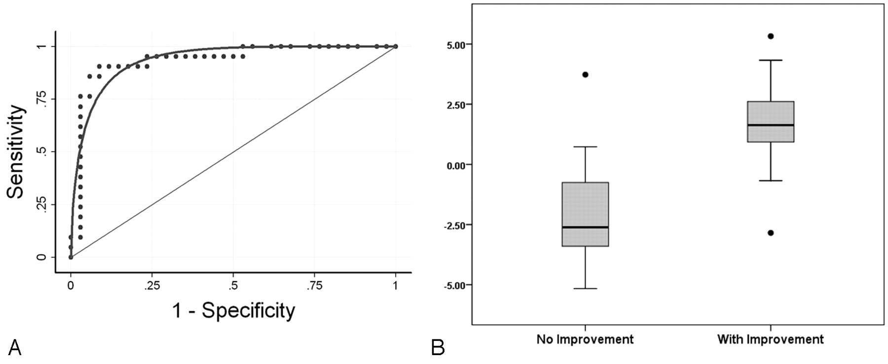
  • Fig 3.
    • Download figure
    • Open in new tab
    • Download powerpoint
    Fig 3.

    A, The ROC curve for the forced-entry multiple logistic regression model derived from the admission CTP/CTA imaging and the clinically independent predictors of language improvement (Table 3). B, Boxplot graph shows how this model can distinguish between those patients with improvement of language function and those without; the y-axis represents the values of the regression equation (Table 3). Patients with aphasic with positive regression scores (>0) have a >50% probability of language improvement by discharge.

Tables

  • Figures
    • View popup
    Table 1:

    Comparison of clinical/CTA characteristics between patients with and without improvement of aphasia (univariate analysis)

    Aphasia Improvement (n = 21)No Improvement (n = 37)P Value
    Age69.5 ± 3.876.8 ± 1.9.10
    Female11 (52%)21 (57%).78
    Admission NIHSS score8 (2–23)19 (5–28).001a
    Admission aphasia NIHSS score2 (1–3)3 (1–3).002a
    Major proximal arterial occlusion7 (33%)30 (81%).001a
    Thrombolytic therapy (IV)5 (24%)11 (29%).76
    IA thrombectomyb1 (5%)3 (8%)1.00
    Ictus to IV treatment interval (hour)2.11 ± 0.251.93 ± 0.25.67
    IV treatment to CTP interval (hour)c1.13 ± 0.491.51 ± 0.52.68
    Ictus to CTP interval (hour)4.46 ± 0.574.78 ± 0.48.68
    • a Significant.

    • b Of the 4 patients who received thrombectomy, only 1, who had complete recanalization, subsequently showed language improvement. All patients who received thrombectomy had first received IV thrombolysis.

    • c Only 1 patient with and 4 without language improvement received IV thrombolysis following CTP; negative treatment-to-CTP-time values were calculated for these patients.

    • View popup
    Table 2:

    Details of the forced-entry multiple logistic regression model derived only from the admission CTP/CTA independent predictors of language improvement

    BP ValueEXP(B)95% CI for EXP(B)
    rCBF of sublobar insular ribbon (lower third)2.89.0116.761.87-149.80
    rCBF of angular gyrus GM BA 391.76.275.820.24-139.82
    Presence of proximal cerebral artery occlusion−1.46.08.230.04-1.21
    Constant−3.87.050.21
    • View popup
    Table 3:

    Details of the forced-entry multiple logistic regression model derived from both the admission CTP/CTA imaging and the clinically independent predictors of language improvement

    BP ValueEXP(B)95% CI for EXP(B)
    Admission aphasia score (NIHSS, section 9)−1.21.030.290.09-0.90
    rCBF of sublobar insular ribbon (lower third)2.23.049.341.09-80.02
    Presence of proximal cerebral artery occlusion−1.94.040.140.02-0.93
    rCBF of angular gyrus GM BA 391.64.375.160.14-191.01
    Constant−0.47.840.62
    • View popup
    Table 4:

    Calculation of the 8-point aphasia improvement scorea

    VariablesPoints
    Aphasia score on admission NIHSS examination1–3
    Proximal cerebral artery occlusion Absent0
        on admission CTAPresent2
    rCBF of the sublobar insular ribbon >1.5−2
        (lower third)0.66–1.50
    0.34–0.661
    <0.342
    rCBF of angular gyrus GM (BA 39)>0.660
    ≤0.661
    • a The aphasia outcome score is the sum of all points based on the admission language component of the NIHSS score, CTP (2 regions), and the presence or absence of CTA primal intracranial occlusion. Note that an rCBF > 1.5 corresponds to a region of marked hyperemia compared with the baseline normal cerebral blood flow in the rCBF of that region.

    • View popup
    Table 5:

    Predictive value of the aphasia improvement score

    Degree of Language ImprovementAphasia Improvement ScoreaNo. of PatientsProbability of ImprovementbNo. of Patients with This Score Showing Improvement
    Excellent1 or 21365%–99% (1 FP)12 (93%)
    Fair3 or 42018%–96% (2 FP, 2 FN)9 (45%)
    Poor5 or 6123%–32% (1 FN)1 (12%)
    Dismal7 or 8131%–6%0 (0%)
    • a The aphasia improvement score is calculated as described in Table 4.

    • b The probability of improvement is estimated on the basis of the logistic regression equation of Table 3; FP and FN cases are defined on the basis of the results of a multivariate model.

PreviousNext
Back to top

In this issue

American Journal of Neuroradiology: 31 (9)
American Journal of Neuroradiology
Vol. 31, Issue 9
1 Oct 2010
  • Table of Contents
  • Index by author
Advertisement
Print
Download PDF
Email Article

Thank you for your interest in spreading the word on American Journal of Neuroradiology.

NOTE: We only request your email address so that the person you are recommending the page to knows that you wanted them to see it, and that it is not junk mail. We do not capture any email address.

Enter multiple addresses on separate lines or separate them with commas.
Predicting Language Improvement in Acute Stroke Patients Presenting with Aphasia: A Multivariate Logistic Model Using Location-Weighted Atlas-Based Analysis of Admission CT Perfusion Scans
(Your Name) has sent you a message from American Journal of Neuroradiology
(Your Name) thought you would like to see the American Journal of Neuroradiology web site.
CAPTCHA
This question is for testing whether or not you are a human visitor and to prevent automated spam submissions.
Cite this article
S. Payabvash, S. Kamalian, S. Fung, Y. Wang, J. Passanese, S. Kamalian, L.C.S. Souza, A. Kemmling, G.J. Harris, E.F. Halpern, R.G. González, K.L. Furie, M.H. Lev
Predicting Language Improvement in Acute Stroke Patients Presenting with Aphasia: A Multivariate Logistic Model Using Location-Weighted Atlas-Based Analysis of Admission CT Perfusion Scans
American Journal of Neuroradiology Oct 2010, 31 (9) 1661-1668; DOI: 10.3174/ajnr.A2125

Citation Manager Formats

  • BibTeX
  • Bookends
  • EasyBib
  • EndNote (tagged)
  • EndNote 8 (xml)
  • Medlars
  • Mendeley
  • Papers
  • RefWorks Tagged
  • Ref Manager
  • RIS
  • Zotero
0 Responses
Respond to this article
Share
Bookmark this article
Predicting Language Improvement in Acute Stroke Patients Presenting with Aphasia: A Multivariate Logistic Model Using Location-Weighted Atlas-Based Analysis of Admission CT Perfusion Scans
S. Payabvash, S. Kamalian, S. Fung, Y. Wang, J. Passanese, S. Kamalian, L.C.S. Souza, A. Kemmling, G.J. Harris, E.F. Halpern, R.G. González, K.L. Furie, M.H. Lev
American Journal of Neuroradiology Oct 2010, 31 (9) 1661-1668; DOI: 10.3174/ajnr.A2125
del.icio.us logo Twitter logo Facebook logo Mendeley logo
  • Tweet Widget
  • Facebook Like
  • Google Plus One
Purchase

Jump to section

  • Article
    • Abstract
    • Abbreviations
    • Methods and Materials
    • Results
    • Discussion
    • Conclusions
    • Footnotes
    • References
  • Figures & Data
  • Info & Metrics
  • Responses
  • References
  • PDF

Related Articles

  • No related articles found.
  • PubMed
  • Google Scholar

Cited By...

  • Differential Contribution of ASPECTS Regions to Clinical Outcome after Thrombectomy for Acute Ischemic Stroke
  • Location-Specific ASPECTS Paradigm in Acute Ischemic Stroke: A Systematic Review and Meta-Analysis
  • Acute Ischemic Stroke Infarct Topology: Association with Lesion Volume and Severity of Symptoms at Admission and Discharge
  • Imaging for Prediction of Functional Outcome and Assessment of Recovery in Ischemic Stroke
  • Prediction of Aphasia Outcome Using Diffusion Tensor Tractography for Arcuate Fasciculus in Stroke
  • Cerebral perfusion in acute stroke prognostication: Go with the flow, or know with the quo?
  • Location-weighted CTP analysis predicts early motor improvement in stroke: A preliminary study
  • Crossref (41)
  • Google Scholar

This article has been cited by the following articles in journals that are participating in Crossref Cited-by Linking.

  • Neuroplasticity of Language Networks in Aphasia: Advances, Updates, and Future Challenges
    Swathi Kiran, Cynthia K. Thompson
    Frontiers in Neurology 2019 10
  • Regional Ischemic Vulnerability of the Brain to Hypoperfusion
    Seyedmehdi Payabvash, Leticia C.S. Souza, Yifei Wang, Pamela W. Schaefer, Karen L. Furie, Elkan F. Halpern, R. Gilberto Gonzalez, Michael H. Lev
    Stroke 2011 42 5
  • Predictors of Functional Outcome Following Stroke
    Richard L. Harvey
    Physical Medicine and Rehabilitation Clinics of North America 2015 26 4
  • Decoding post-stroke motor function from structural brain imaging
    Jane M. Rondina, Maurizio Filippone, Mark Girolami, Nick S. Ward
    NeuroImage: Clinical 2016 12
  • Predicting language outcomes after stroke: Is structural disconnection a useful predictor?
    Thomas M.H. Hope, Alex P. Leff, Cathy J. Price
    NeuroImage: Clinical 2018 19
  • Prediction of Aphasia Outcome Using Diffusion Tensor Tractography for Arcuate Fasciculus in Stroke
    S.H. Kim, S.H. Jang
    American Journal of Neuroradiology 2013 34 4
  • Imaging for Prediction of Functional Outcome and Assessment of Recovery in Ischemic Stroke
    Wolf-Dieter Heiss, Chelsea S. Kidwell
    Stroke 2014 45 4
  • Transcranial near-infrared laser therapy applied to promote clinical recovery in acute and chronic neurodegenerative diseases
    Paul A Lapchak
    Expert Review of Medical Devices 2012 9 1
  • The Aphasia Rapid Test: an NIHSS-like aphasia test
    C. Azuar, A. Leger, C. Arbizu, F. Henry-Amar, S. Chomel-Guillaume, Y. Samson
    Journal of Neurology 2013 260 8
  • Acute Ischemic Stroke Infarct Topology: Association with Lesion Volume and Severity of Symptoms at Admission and Discharge
    S. Payabvash, S. Taleb, J.C. Benson, A.M. McKinney
    American Journal of Neuroradiology 2017 38 1

More in this TOC Section

  • Optimal MRI Sequence for Identifying Occlusion Location in Acute Stroke: Which Value of Time-Resolved Contrast-Enhanced MRA?
  • SWI or T2*: Which MRI Sequence to Use in the Detection of Cerebral Microbleeds? The Karolinska Imaging Dementia Study
  • Progression of Microstructural Damage in Spinocerebellar Ataxia Type 2: A Longitudinal DTI Study
Show more Brain

Similar Articles

Advertisement

Indexed Content

  • Current Issue
  • Accepted Manuscripts
  • Article Preview
  • Past Issues
  • Editorials
  • Editors Choice
  • Fellow Journal Club
  • Letters to the Editor

Cases

  • Case Collection
  • Archive - Case of the Week
  • Archive - Case of the Month
  • Archive - Classic Case

Special Collections

  • Special Collections

Resources

  • News and Updates
  • Turn around Times
  • Submit a Manuscript
  • Author Policies
  • Manuscript Submission Guidelines
  • Evidence-Based Medicine Level Guide
  • Publishing Checklists
  • Graphical Abstract Preparation
  • Imaging Protocol Submission
  • Submit a Case
  • Become a Reviewer/Academy of Reviewers
  • Get Peer Review Credit from Publons

Multimedia

  • AJNR Podcast
  • AJNR SCANtastic
  • Video Articles

About Us

  • About AJNR
  • Editorial Board
  • Not an AJNR Subscriber? Join Now
  • Alerts
  • Feedback
  • Advertise with us
  • Librarian Resources
  • Permissions
  • Terms and Conditions

American Society of Neuroradiology

  • Not an ASNR Member? Join Now

© 2025 by the American Society of Neuroradiology All rights, including for text and data mining, AI training, and similar technologies, are reserved.
Print ISSN: 0195-6108 Online ISSN: 1936-959X

Powered by HighWire