
Article Figures & Data
Figures
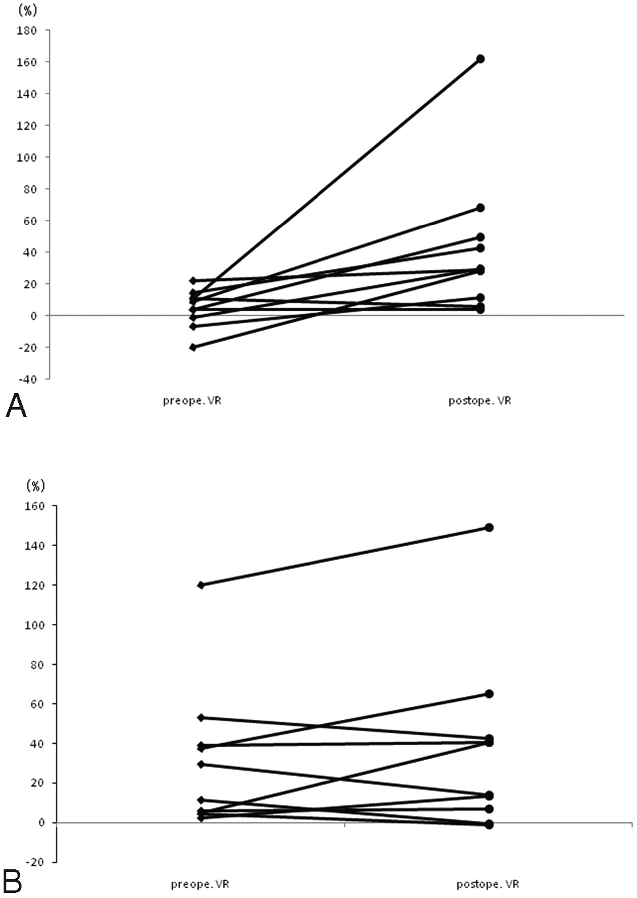
- Fig 1.
Postoperative change in rVR in the bypass-established hemisphere. The increased rVR rate is significantly higher on the bypass-established hemisphere (A) than that on the contralateral hemisphere (B) (P = .0018).
- Fig 2.
Postoperative change in the misery perfusion area in the bypass-established hemisphere. The proportion of stage II area on the bypass-established hemisphere (A) is significantly lower than that on the contralateral hemisphere (B) (P = .0088).
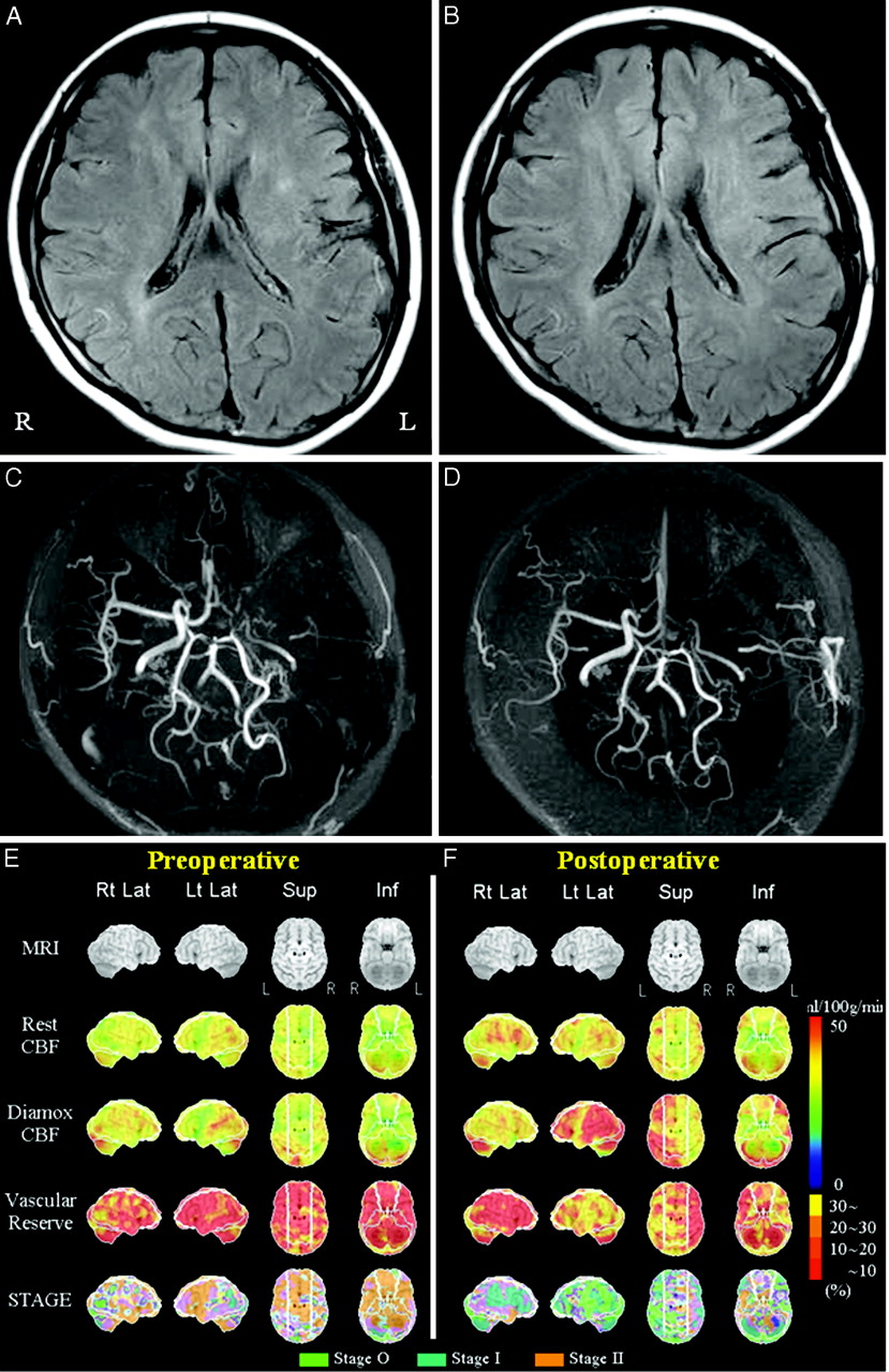
- Fig 3.
Adult Moyamoya disease. Data from a 25-year-old female patient who had a right transient ischemic attack. She underwent direct bypass surgery on the left cerebral hemisphere. MR imaging of the preoperative (A) and postoperative (B) views shows a decrease in the ivy sign in the left MCA territory after bypass surgery. MRA of the preoperative (C) and postoperative (D) views shows increased flow-related enhancement via left superficial temporal arteries to the left MCA region after surgery. Quantitative analysis of preoperative (E) and postoperative (F) basal/ACZ stress brain perfusion SPECT. The lower 4 columns show 3D stereotactic surface projection format view sets of resting CBF, Diamox CBF, VR, and staging by a JET study from the top. VR is impaired preoperatively in most of the left MCA territory (E). Both VR and staging in the left MCA territory have improved postoperatively (F).
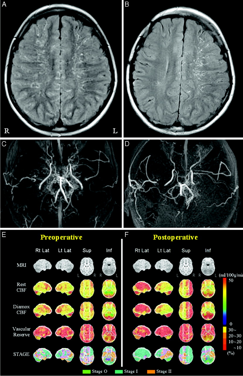
- Fig 4.
A child with Moyamoya disease. Data from an 11-year-old male patient who had left involuntary movement. He underwent indirect bypass surgery on the right cerebral hemisphere. MR imaging of the preoperative (A) and postoperative (B) views shows a decrease in the ivy sign on the right hemisphere after bypass surgery. MRA of the preoperative (C) and postoperative (D) views shows increased flow-related enhancement via the right temporal subcutaneous tissue to the right MCA region after surgery. Quantitative analysis of preoperative (E) and postoperative (F) basal/ACZ stress brain perfusion SPECT. VRs in bilateral MCA territories are impaired preoperatively (E). VR and staging in the right hemisphere are improved postoperatively (F).


In this issue


Jump to section
Related Articles
Cited By...
- The Possible Difference of Underlying Pathophysiologies between "Ivy Sign" on Contrast-Enhanced MRI and FLAIR
- Ivy Sign in Moyamoya Disease: A Comparative Study of the FLAIR Vascular Hyperintensity Sign Against Contrast-Enhanced MRI
- FLAIR vascular hyperintensity resolution in a TIA patient: Clinical-radiologic correlation
- Moyamoya syndrome in sickle cell anaemia: a cause of recurrent stroke
- Elevated Cerebral Blood Volume Contributes to Increased FLAIR Signal in the Cerebral Sulci of Propofol-Sedated Children