Advertisement

Main menu

  • Home
  • Content
    • Current Issue
    • Accepted Manuscripts
    • Article Preview
    • Past Issue Archive
    • AJNR Case Collection
    • Case of the Week Archive
    • Classic Case Archive
    • Case of the Month Archive
  • Special Collections
    • Spinal CSF Leak Articles (Jan 2020-June 2024)
    • 2024 AJNR Journal Awards
    • Most Impactful AJNR Articles
  • Multimedia
    • AJNR Podcast
    • AJNR Scantastics
    • Video Articles
  • For Authors
    • Submit a Manuscript
    • Author Policies
    • Fast publishing of Accepted Manuscripts
    • Graphical Abstract Preparation
    • Manuscript Submission Guidelines
    • Imaging Protocol Submission
    • Submit a Case for the Case Collection
  • About Us
    • About AJNR
    • Editorial Board
  • More
    • Become a Reviewer/Academy of Reviewers
    • Subscribers
    • Permissions
    • Alerts
    • Feedback
    • Advertisers
    • ASNR Home
  • Other Publications
    • ajnr

User menu

  • Alerts
  • Log in

Search

  • Advanced search
American Journal of Neuroradiology

American Journal of Neuroradiology

ASHNR American Society of Functional Neuroradiology ASHNR American Society of Pediatric Neuroradiology ASSR
  • Alerts
  • Log in

Advanced Search

  • Home
  • Content
    • Current Issue
    • Accepted Manuscripts
    • Article Preview
    • Past Issue Archive
    • AJNR Case Collection
    • Case of the Week Archive
    • Classic Case Archive
    • Case of the Month Archive
  • Special Collections
    • Spinal CSF Leak Articles (Jan 2020-June 2024)
    • 2024 AJNR Journal Awards
    • Most Impactful AJNR Articles
  • Multimedia
    • AJNR Podcast
    • AJNR Scantastics
    • Video Articles
  • For Authors
    • Submit a Manuscript
    • Author Policies
    • Fast publishing of Accepted Manuscripts
    • Graphical Abstract Preparation
    • Manuscript Submission Guidelines
    • Imaging Protocol Submission
    • Submit a Case for the Case Collection
  • About Us
    • About AJNR
    • Editorial Board
  • More
    • Become a Reviewer/Academy of Reviewers
    • Subscribers
    • Permissions
    • Alerts
    • Feedback
    • Advertisers
    • ASNR Home
  • Follow AJNR on Twitter
  • Visit AJNR on Facebook
  • Follow AJNR on Instagram
  • Join AJNR on LinkedIn
  • RSS Feeds

Welcome to the new AJNR, Updated Hall of Fame, and more. Read the full announcements.


AJNR is seeking candidates for the position of Associate Section Editor, AJNR Case Collection. Read the full announcement.


Research ArticleBrain

“Giant” Arachnoid Granulations Just Like CSF?: NOT!!

C.R. Trimble, H.R. Harnsberger, M. Castillo, M. Brant-Zawadzki and A.G. Osborn
American Journal of Neuroradiology October 2010, 31 (9) 1724-1728; DOI: https://doi.org/10.3174/ajnr.A2157
C.R. Trimble
  • Find this author on Google Scholar
  • Find this author on PubMed
  • Search for this author on this site
H.R. Harnsberger
  • Find this author on Google Scholar
  • Find this author on PubMed
  • Search for this author on this site
M. Castillo
  • Find this author on Google Scholar
  • Find this author on PubMed
  • Search for this author on this site
M. Brant-Zawadzki
  • Find this author on Google Scholar
  • Find this author on PubMed
  • Search for this author on this site
A.G. Osborn
  • Find this author on Google Scholar
  • Find this author on PubMed
  • Search for this author on this site
  • Article
  • Figures & Data
  • Info & Metrics
  • Responses
  • References
  • PDF
Loading

Article Figures & Data

Figures

  • Fig 1.
    • Download figure
    • Open in new tab
    • Download powerpoint
    Fig 1.

    Transverse sinus giant AG. A, Axial nonenhanced CT with a bone algorithm shows cystic expansion of the diploic space adjacent to the left TS (arrow). B, Axial T2WI shows a large AG in the left TS with 2 internal septations (arrows) and subarachnoid spaces converging at the base of the AG (open arrow). C, Axial FLAIR image demonstrates incomplete suppression of fluid within the margins of the intra-AG septations (arrow) and complete suppression of intra-AG fluid outside the septations. D, Axial fat-saturated postcontrast T1WI shows linear enhancement at the posteromedial margin of the AG, most likely representing an intra-AG vein. The soft-tissue septations themselves do not enhance.

  • Fig 2.
    • Download figure
    • Open in new tab
    • Download powerpoint
    Fig 2.

    Superior sagittal sinus giant AG. A, Sagittal T1WI shows a giant AG in the SSS. Note that fluid in the AG (arrow) is hyperintense to CSF. A distinct linear flow void (open arrow) is seen. B, Sagittal T2WI shows that fluid is mixed iso- and hypointense to CSF. A distinct intra-AG vein is present (open arrow). C, Axial FLAIR image shows that intra-AG fluid (arrow) is not suppressed. Phase dispersion (curved arrow) is present around the linear flow void entering the AG. D, Postcontrast fat-saturated T1WI shows an enhancing vein entering the AG (arrow) and enhancing veins (open arrows) within the AG itself. E, Axial DWI shows that fluid within the AG does not demonstrate restricted diffusion. F, Lateral DSA, venous phase, shows a filling defect in the SSS caused by the giant AG (arrow). Note veins (open arrows) within the AG.

  • Fig 3.
    • Download figure
    • Open in new tab
    • Download powerpoint
    Fig 3.

    TS giant AG with a soft-tissue mass. A, Coronal T2WI shows a 1-cm AG in the right TS. Fluid (arrow) is of CSF signal intensity; soft tissue (curved arrow) projects into the AG lumen through an opening in the dura. Note the flow void from a vein (open arrow) in the AG. B, Coronal inversion recovery image shows that of pedunculated soft tissue (arrow) at the base of the AG is isointense with adjacent gray matter, while the fluid is isointense with CSF. C, Coronal contrast-enhanced scan shows enhancement of a vein (open arrow) within the AG. Soft tissue (curved arrow) does not enhance.
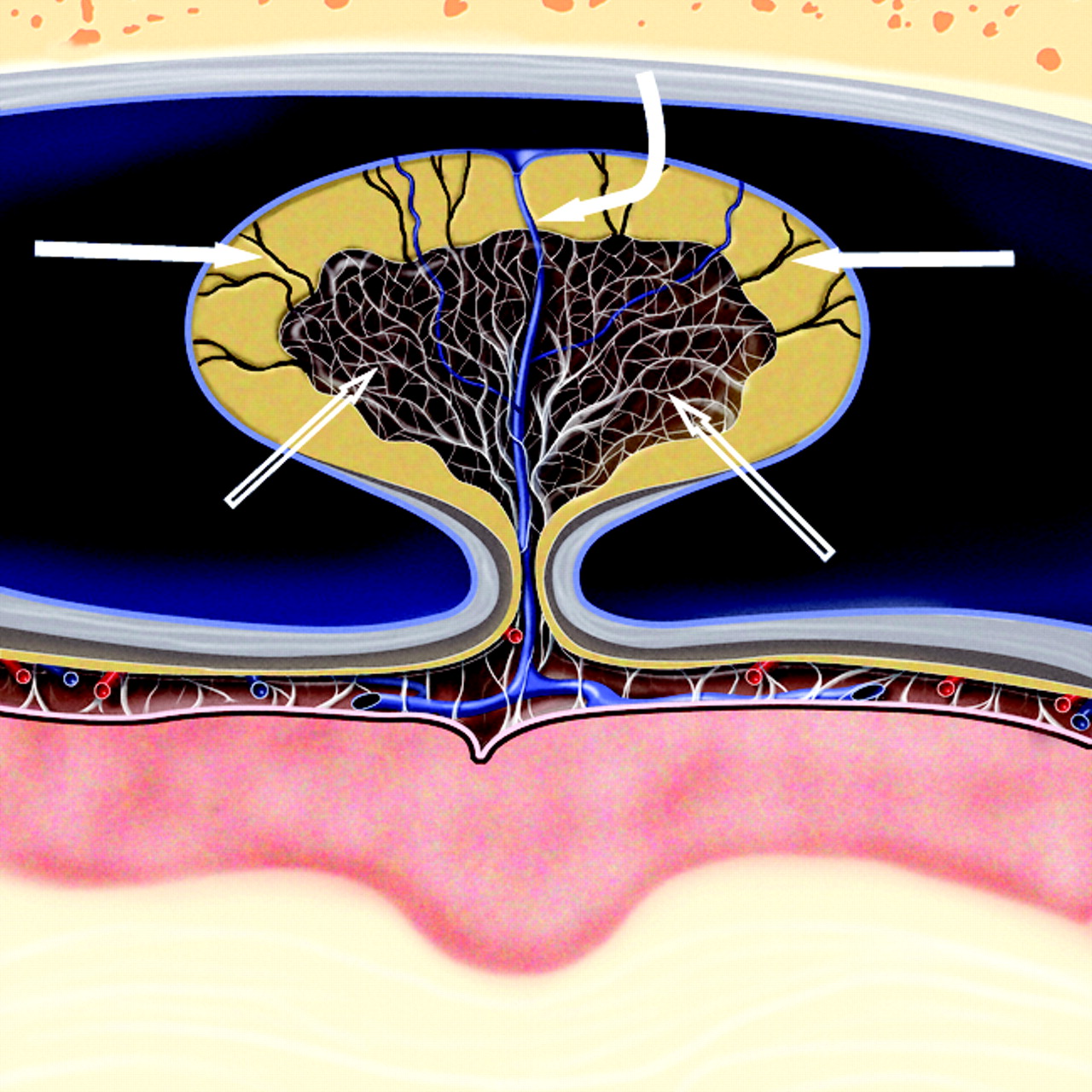
  • Fig 4.
    • Download figure
    • Open in new tab
    • Download powerpoint
    Fig 4.

    Cross-sectional graphic of a giant venous sinus AG projecting into a dural venous sinus. A core of CSF-filled collagenous trabeculation (open arrows) extends from the subarachnoid space into the granulation and is covered by an apical cap of arachnoid cells. CSF channels (arrows) extend through the cap to the sinus endothelium and drain CSF into the venous circulation. A vein (curved arrow) also courses through the body of the AG, penetrates the arachnoid cap layer, and empties into the dural venous sinus. Graphic is used with permission from Amirsys Inc., Salt Lake City, Utah

PreviousNext
Back to top

In this issue

American Journal of Neuroradiology: 31 (9)
American Journal of Neuroradiology
Vol. 31, Issue 9
1 Oct 2010
  • Table of Contents
  • Index by author
Advertisement
Print
Download PDF
Email Article

Thank you for your interest in spreading the word on American Journal of Neuroradiology.

NOTE: We only request your email address so that the person you are recommending the page to knows that you wanted them to see it, and that it is not junk mail. We do not capture any email address.

Enter multiple addresses on separate lines or separate them with commas.
“Giant” Arachnoid Granulations Just Like CSF?: NOT!!
(Your Name) has sent you a message from American Journal of Neuroradiology
(Your Name) thought you would like to see the American Journal of Neuroradiology web site.
CAPTCHA
This question is for testing whether or not you are a human visitor and to prevent automated spam submissions.
Cite this article
C.R. Trimble, H.R. Harnsberger, M. Castillo, M. Brant-Zawadzki, A.G. Osborn
“Giant” Arachnoid Granulations Just Like CSF?: NOT!!
American Journal of Neuroradiology Oct 2010, 31 (9) 1724-1728; DOI: 10.3174/ajnr.A2157

Citation Manager Formats

  • BibTeX
  • Bookends
  • EasyBib
  • EndNote (tagged)
  • EndNote 8 (xml)
  • Medlars
  • Mendeley
  • Papers
  • RefWorks Tagged
  • Ref Manager
  • RIS
  • Zotero
0 Responses
Respond to this article
Share
Bookmark this article
“Giant” Arachnoid Granulations Just Like CSF?: NOT!!
C.R. Trimble, H.R. Harnsberger, M. Castillo, M. Brant-Zawadzki, A.G. Osborn
American Journal of Neuroradiology Oct 2010, 31 (9) 1724-1728; DOI: 10.3174/ajnr.A2157
del.icio.us logo Twitter logo Facebook logo Mendeley logo
  • Tweet Widget
  • Facebook Like
  • Google Plus One
Purchase

Jump to section

  • Article
    • Abstract
    • Abbreviations
    • Materials and Methods
    • Results
    • Discussion
    • Conclusions
    • Footnotes
    • References
  • Figures & Data
  • Info & Metrics
  • Responses
  • References
  • PDF

Related Articles

  • No related articles found.
  • PubMed
  • Google Scholar

Cited By...

  • Author response: Idiopathic intracranial hypertension: The veno glymphatic connections
  • Giant arachnoid granulation with a thrombosed dural arteriovenous fistula
  • Cranial Arachnoid Protrusions and Contiguous Diploic Veins in CSF Drainage
  • Dural sinus filling defect: intrasigmoid encephalocele
  • Crossref (49)
  • Google Scholar

This article has been cited by the following articles in journals that are participating in Crossref Cited-by Linking.

  • Cerebrospinal fluid tracer efflux to parasagittal dura in humans
    Geir Ringstad, Per Kristian Eide
    Nature Communications 2020 11 1
  • Molecular trans-dural efflux to skull bone marrow in humans with CSF disorders
    Geir Ringstad, Per Kristian Eide
    Brain 2022 145 4
  • Spectrum of lytic lesions of the skull: a pictorial essay
    Lorenzo Ugga, Renato Cuocolo, Sirio Cocozza, Andrea Ponsiglione, Arnaldo Stanzione, Vito Chianca, Alessandra D’Amico, Arturo Brunetti, Massimo Imbriaco
    Insights into Imaging 2018 9 5
  • Giant Arachnoid Granulations Mimicking Pathology
    Bart De Keyzer, Sven Bamps, Frank Van Calenbergh, Philippe Demaerel, Guido Wilms
    The Neuroradiology Journal 2014 27 3
  • Cranial Arachnoid Protrusions and Contiguous Diploic Veins in CSF Drainage
    S. Tsutsumi, I. Ogino, M. Miyajima, M. Nakamura, Y. Yasumoto, H. Arai, M. Ito
    American Journal of Neuroradiology 2014 35 9
  • Brain Herniation into Arachnoid Granulations: Clinical and Neuroimaging Features
    Greta B. Liebo, John (Jack) I. Lane, Jamie J. Van Gompel, Laurence J. Eckel, Kara M. Schwartz, Vance T. Lehman
    Journal of Neuroimaging 2016 26 6
  • Normal neuroanatomical variants that may be misinterpreted as disease entities
    S. Ramji, P. Touska, P. Rich, A.D. MacKinnon
    Clinical Radiology 2017 72 10
  • ‘Do not touch’ lesions of the skull base
    Mircea C Dobre, Nancy Fischbein
    Journal of Medical Imaging and Radiation Oncology 2014 58 4
  • Evaluation of giant arachnoid granulations with high-resolution 3D-volumetric MR sequences at 3T
    Hayri Ogul, Fadime Guven, Emine Izgi, Mecit Kantarci
    European Journal of Radiology 2019 121
  • Focal herniation of cerebral parenchyma into transverse sinus
    Ali Kocyigit, Duygu Herek, Yasemin Isik Balci
    Journal of Neuroradiology 2015 42 2

More in this TOC Section

  • Statin Therapy Does Not Affect the Radiographic and Clinical Profile of Patients with TIA and Minor Stroke
  • Optimal MRI Sequence for Identifying Occlusion Location in Acute Stroke: Which Value of Time-Resolved Contrast-Enhanced MRA?
  • SWI or T2*: Which MRI Sequence to Use in the Detection of Cerebral Microbleeds? The Karolinska Imaging Dementia Study
Show more Brain

Similar Articles

Advertisement

Indexed Content

  • Current Issue
  • Accepted Manuscripts
  • Article Preview
  • Past Issues
  • Editorials
  • Editors Choice
  • Fellow Journal Club
  • Letters to the Editor

Cases

  • Case Collection
  • Archive - Case of the Week
  • Archive - Case of the Month
  • Archive - Classic Case

Special Collections

  • Special Collections

Resources

  • News and Updates
  • Turn around Times
  • Submit a Manuscript
  • Author Policies
  • Manuscript Submission Guidelines
  • Evidence-Based Medicine Level Guide
  • Publishing Checklists
  • Graphical Abstract Preparation
  • Imaging Protocol Submission
  • Submit a Case
  • Become a Reviewer/Academy of Reviewers
  • Get Peer Review Credit from Publons

Multimedia

  • AJNR Podcast
  • AJNR SCANtastic
  • Video Articles

About Us

  • About AJNR
  • Editorial Board
  • Not an AJNR Subscriber? Join Now
  • Alerts
  • Feedback
  • Advertise with us
  • Librarian Resources
  • Permissions
  • Terms and Conditions

American Society of Neuroradiology

  • Not an ASNR Member? Join Now

© 2025 by the American Society of Neuroradiology All rights, including for text and data mining, AI training, and similar technologies, are reserved.
Print ISSN: 0195-6108 Online ISSN: 1936-959X

Powered by HighWire