
Article Figures & Data
Figures
- Fig 1.
Laterally projecting left carotid aneurysm with residual filling after prior coiling. A, Subtracted carotid angiography in the frontal-projection working angle demonstrates residual filling of the aneurysm neck extending into the proximal fundus. B, Unsubtracted frontal projection in the working angle demonstrates a PED in position across the aneurysm neck. No additional embolization coils were added. The gap between the coil mass and PED construct (arrow) indicates the region of residual aneurysm filling. C, Follow-up angiogram in the frontal projection 6 months after treatment shows complete occlusion of the aneurysm. D, A native image in the frontal projection shows that the gap between the coil mass and PED construct has resolved (arrow). This finding indicates that the aneurysm has not only undergone complete thrombosis, but that the thrombus mass has been resorbed in the interim with contraction of the intra-aneurysmal thrombus-coil mass complex around the outside of the construct.
- Fig 2.
Dysplastic wide-neck partially thrombosed aneurysm arising from the proximal cavernous segment of the internal carotid artery. A and B, 3D images reconstructed from rotational angiographic source data demonstrate that the neck of the aneurysm incorporates >180° of the vessel circumference over a 10-mm segment of the carotid artery. C, Conventional angiography in the lateral working projection depicts the partially thrombosed wide-neck aneurysm arising from the circumferentially diseased segment. Just proximal to the aneurysmal segment is a mild focal stenosis (arrow). D, Native image in the lateral working-angle projection after treatment demonstrates the Pipeline construct in place across the aneurysm neck, with a loose packing of the aneurysm with embolization coils. E, Follow-up angiography at 180 days in the lateral working projection shows total occlusion of the aneurysm as well as complete anatomic remodeling of the diseased parent artery. The dysplastic aneurysmal vascular segment now has a smooth tubular configuration. The proximal stenosis has also completely remodeled and now is normal in caliber.
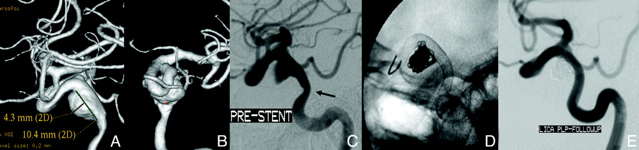
- Fig 3.
Left ICA parophthamalic-segment aneurysm in a patient with progressive ipsilateral vision loss. A and B, 3D reconstructed images from rotational source data show a very large wide-neck aneurysm arising from the parophthalmic segment of the left ICA. C, Initial angiogram in the frontal working projection demonstrates a very large aneurysm arising from the left ICA. D, Following reconstruction with a PED, filling of the aneurysm with contrast is diminished because flow has been redirected along the normal course of the ICA and into the left anterior circulation. The patient emerged from general anesthesia with an improvement in left-eye vision. E, A native image depicts the PED construct in position across the aneurysmal segment. F, Subtracted image from the 6-month follow-up angiogram shows anatomic reconstruction of the parent artery with complete aneurysm occlusion. G, Axial CT image at the level of the optic chiasm before treatment demonstrates the peripherally calcified fundus of the very large left ICA aneurysm projecting into the suprasellar cistern. H, Six-month follow-up axial CT image depicts a PED in place within the parent ICA with complete resolution of the aneurysm-thrombus mass. The suprasellar cistern, which was formerly effaced by the aneurysm, now appears normally filled with CSF. These serial images depict the physiologic progression that is possible in some cases after PED reconstruction—starting with mechanical flow diversion, progressing to physiologic aneurysm thrombosis and complete occlusion, followed by endoluminal parent artery reconstruction and, ultimately, anatomical restoration with resolution of the aneurysm-thrombus mass and dissipation of the regional mass effect.


Tables
Characteristic No. (%) or Mean (Range) Target IA location Anterior Cavernous 5 (16.1) Paraophthalmic 15 (48.4) Superior hypophyseal 4 (12.9) Posterior communicating 4 (12.9) M1 segment 1 (3.2) Posterior Distal pre-PICA vertebral 1 (3.2) PICA vertebral 1 (3.2) Neck ≥4 mm 22 (71.0) Maximum aneurysm dimension (mm) 11.5 (2.5–26.6) <10 mm 20 (64.5) 10–25 mm 9 (29.0) ≥25 mm 2 (6.5) Number of PEDs implanted 1 18 (58.1) 2 11 (35.5) 3 1 (3.2) 4 1 (3.2) Adjunctive coils placed 16 (51.6) Category No. (%) Target IA occlusion Complete 28 (93.3) Residual neck 0 (0.0) Residual filling 2 (6.7) Device migration No 29 (96.7) Yes 0 (0.0) Unable to determine 1 (3.3) Stenosis 0%–25% 28 (93.3) >25%–50% 1 (3.3) >50%–75% 0 >75%–100% 0 Not judged 1 (3.3)
In this issue


Jump to section
Related Articles
Cited By...
- Neuroanatomy of the vertebrobasilar perforators: implications for aneurysm treatment
- Evaluation of flow diverters for cerebral aneurysm therapy: recommendations for imaging analyses in clinical studies, endorsed by ESMINT, ESNR, OCIN, SILAN, SNIS, and WFITN
- Efficacy and safety of a new mechanical balloon-based flow diverter in the treatment of intracranial aneurysms
- Clinical Outcome of Pipeline Embolization Device with and without Coils to Treat Intracranial Aneurysm: A Systematic Review and Meta-Analysis
- Braids and beyond: a comprehensive study on pipeline device braid stability from PREMIER data
- Transcirculation retrograde placement of a Pipeline embolization device for treatment of a vertebrobasilar junction aneurysm
- Vanguard Study: Initial experience with the new fourth generation Pipeline Vantage Flow Diverter (PVFD): 6-month results, technical and clinical considerations
- Aneurysm healing following treatment with biodegradable embolization materials: assessment in a rat sidewall aneurysm model
- Outcome study of the Pipeline Vantage Embolization Device (second version) in unruptured (and ruptured) aneurysms (PEDVU(R) study)
- Flow diversion for compressive unruptured internal carotid artery aneurysms with neuro-ophthalmological symptoms: a systematic review and meta-analysis
- Derivo embolization device for intracranial aneurysms: a Spanish multicenter retrospective study
- Flow diversion for compressive unruptured internal carotid artery aneurysms with neuro-ophthalmological symptoms: a systematic review and meta-analysis
- Prospective study on embolization of intracranial aneurysms with the pipeline device (PREMIER study): 3-year results with the application of a flow diverter specific occlusion classification
- How to perform intra-aneurysmal coil embolization after Pipeline deployment: a study from a hemodynamic viewpoint
- Bioresorbable flow diverters for the treatment of intracranial aneurysms: review of current literature and future directions
- First clinical multicenter experience with the new Pipeline Vantage flow diverter
- Learning Curve for Flow Diversion of Posterior Circulation Aneurysms: A Long-Term International Multicenter Cohort Study
- Predictors of incomplete aneurysm occlusion after treatment with the Pipeline Embolization Device: PREMIER trial 1 year analysis
- Diversion-p64: results from an international, prospective, multicenter, single-arm post-market study to assess the safety and effectiveness of the p64 flow modulation device
- Not quite time to stop testing aspirin response yet?
- The Safety and Efficacy of Flow Diversion versus Conventional Endovascular Treatment for Intracranial Aneurysms: A Meta-analysis of Real-world Cohort Studies from the Past 10 Years
- Finnish flow diverter study: 8 years of experience in the treatment of acutely ruptured intracranial aneurysms
- Follow-up of Intracranial Aneurysms Treated by Flow Diverters: Evaluation of Parent Artery Patency Using 3D-T1 Gradient Recalled-Echo Imaging with 2-Point Dixon in Combination with 3D-TOF-MRA with Compressed Sensing
- Propensity Score Analysis of Flow Diverters Placed in Scaffolding Stents
- Ruptured Intracranial Aneurysms Treated with the Pipeline Embolization Device: A Systematic Review and Pooled Analysis of Individual Patient Data
- Nuisance bleeding complications in patients with cerebral aneurysm treated with Pipeline embolization device
- Review of current intracranial aneurysm flow diversion technology and clinical use
- Neck Location on the Outer Convexity is a Predictor of Incomplete Occlusion in Treatment with the Pipeline Embolization Device: Clinical and Angiographic Outcomes
- Periprocedural to 1-year safety and efficacy outcomes with the Pipeline Embolization Device with Shield technology for intracranial aneurysms: a prospective, post-market, multi-center study
- How safe and effective are flow diverters for the treatment of unruptured small/medium intracranial aneurysms of the internal carotid artery? Meta-analysis for evidence-based performance goals
- A novel self-expanding primarily bioabsorbable braided flow-diverting stent for aneurysms: initial safety results
- Efficacy and Safety of Flow-Diverter Therapy for Recurrent Aneurysms after Stent-Assisted Coiling
- Advances in endovascular aneurysm management: flow modulation techniques with braided mesh devices
- Prospective study on embolization of intracranial aneurysms with the pipeline device: the PREMIER study 1 year results
- Flow diversion beyond the circle of Willis: endovascular aneurysm treatment in peripheral cerebral arteries employing a novel low-profile flow diverting stent
- Outcome Study of the Pipeline Embolization Device with Shield Technology in Unruptured Aneurysms (PEDSU)
- The Fate of Unruptured Intracranial Vertebrobasilar Dissecting Aneurysm with Brain Stem Compression According to Different Treatment Modalities
- The safety and effectiveness of the Woven EndoBridge (WEB) system for the treatment of wide-necked bifurcation aneurysms: final 12-month results of the pivotal WEB Intrasaccular Therapy (WEB-IT) Study
- Usefulness of Silent MR Angiography for Intracranial Aneurysms Treated with a Flow-Diverter Device
- Pipeline embolization device treatment of internal carotid artery terminus aneurysms
- An autopsy report of basilar artery aneurysm flow diversion complicated by postoperative day 3 hemorrhage from vessel rupture
- On Flow Diversion: The Changing Landscape of Intracerebral Aneurysm Management
- Antiplatelet therapy and the risk of ischemic and hemorrhagic complications associated with Pipeline embolization of cerebral aneurysms: a systematic review and pooled analysis
- Aneurysm Remnants after Flow Diversion: Clinical and Angiographic Outcomes
- Treatment of intracranial aneurysms using the pipeline flex embolization device with shield technology: angiographic and safety outcomes at 1-year follow-up
- Comparison of Pipeline Embolization Device Sizing Based on Conventional 2D Measurements and Virtual Simulation Using the Sim&Size Software: An Agreement Study
- High-Definition Zoom Mode, a High-Resolution X-Ray Microscope for Neurointerventional Treatment Procedures: A Blinded-Rater Clinical-Utility Study
- Transient in-stent stenosis at mid-term angiographic follow-up in patients treated with SILK flow diverter stents: incidence, clinical significance and long-term follow-up
- 'Plug and pipe strategy for treatment of ruptured intracranial aneurysms
- An autopsy report of basilar artery aneurysm flow diversion complicated by postoperative day 3 hemorrhage from vessel rupture