Advertisement

Main menu

  • Home
  • Content
    • Current Issue
    • Accepted Manuscripts
    • Article Preview
    • Past Issue Archive
    • AJNR Case Collection
    • Case of the Week Archive
    • Classic Case Archive
    • Case of the Month Archive
  • Special Collections
    • Spinal CSF Leak Articles (Jan 2020-June 2024)
    • 2024 AJNR Journal Awards
    • Most Impactful AJNR Articles
  • Multimedia
    • AJNR Podcast
    • AJNR Scantastics
    • Video Articles
  • For Authors
    • Submit a Manuscript
    • Author Policies
    • Fast publishing of Accepted Manuscripts
    • Graphical Abstract Preparation
    • Manuscript Submission Guidelines
    • Imaging Protocol Submission
    • Submit a Case for the Case Collection
  • About Us
    • About AJNR
    • Editorial Board
  • More
    • Become a Reviewer/Academy of Reviewers
    • Subscribers
    • Permissions
    • Alerts
    • Feedback
    • Advertisers
    • ASNR Home
  • Other Publications
    • ajnr

User menu

  • Alerts
  • Log in

Search

  • Advanced search
American Journal of Neuroradiology

American Journal of Neuroradiology

ASHNR American Society of Functional Neuroradiology ASHNR American Society of Pediatric Neuroradiology ASSR
  • Alerts
  • Log in

Advanced Search

  • Home
  • Content
    • Current Issue
    • Accepted Manuscripts
    • Article Preview
    • Past Issue Archive
    • AJNR Case Collection
    • Case of the Week Archive
    • Classic Case Archive
    • Case of the Month Archive
  • Special Collections
    • Spinal CSF Leak Articles (Jan 2020-June 2024)
    • 2024 AJNR Journal Awards
    • Most Impactful AJNR Articles
  • Multimedia
    • AJNR Podcast
    • AJNR Scantastics
    • Video Articles
  • For Authors
    • Submit a Manuscript
    • Author Policies
    • Fast publishing of Accepted Manuscripts
    • Graphical Abstract Preparation
    • Manuscript Submission Guidelines
    • Imaging Protocol Submission
    • Submit a Case for the Case Collection
  • About Us
    • About AJNR
    • Editorial Board
  • More
    • Become a Reviewer/Academy of Reviewers
    • Subscribers
    • Permissions
    • Alerts
    • Feedback
    • Advertisers
    • ASNR Home
  • Follow AJNR on Twitter
  • Visit AJNR on Facebook
  • Follow AJNR on Instagram
  • Join AJNR on LinkedIn
  • RSS Feeds

Welcome to the new AJNR, Updated Hall of Fame, and more. Read the full announcements.


AJNR is seeking candidates for the position of Associate Section Editor, AJNR Case Collection. Read the full announcement.


Research ArticleHead and NeckE

Carotid Plaque Enhancement and Symptom Correlations: An Evaluation by Using Multidetector Row CT Angiography

L. Saba and G. Mallarini
American Journal of Neuroradiology November 2011, 32 (10) 1919-1925; DOI: https://doi.org/10.3174/ajnr.A2605
L. Saba
  • Find this author on Google Scholar
  • Find this author on PubMed
  • Search for this author on this site
G. Mallarini
  • Find this author on Google Scholar
  • Find this author on PubMed
  • Search for this author on this site
  • Article
  • Figures & Data
  • Info & Metrics
  • Responses
  • References
  • PDF
Loading

Article Figures & Data

Figures

  • Tables
  • Fig 1.
    • Download figure
    • Open in new tab
    • Download powerpoint
    Fig 1.

    A 67-year-old asymptomatic male patient. MDCT axial images before (A) and after (B) administration of contrast material. White arrows indicate the region of interest in the plaque. The red rectangle incorporates the region-of-interest area values, including area; and mean HU values, minimum and maximum.
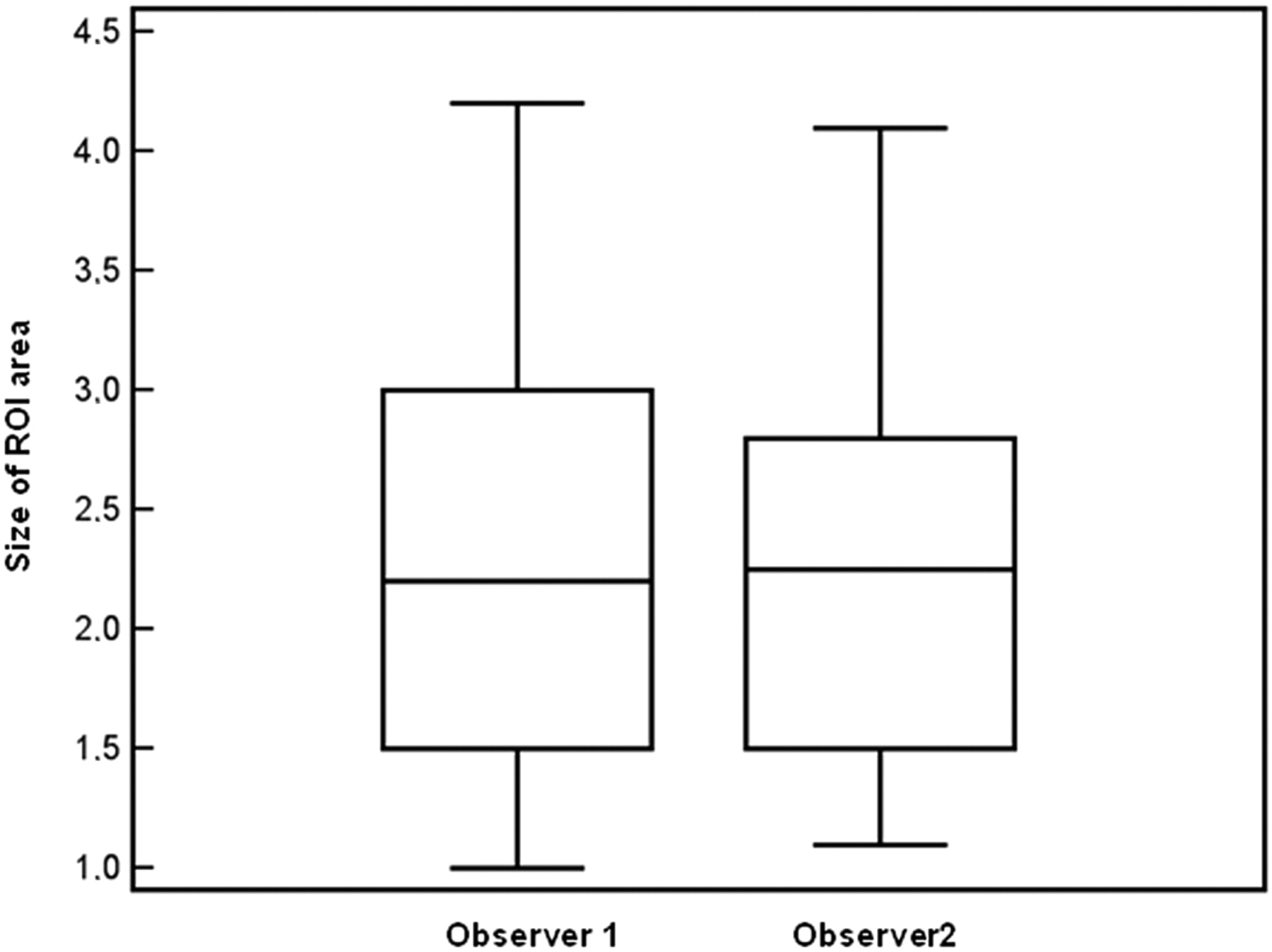
  • Fig 2.
    • Download figure
    • Open in new tab
    • Download powerpoint
    Fig 2.

    Box-and-whisker plot between the observers for the region-of-interest area quantification.

  • Fig 3.
    • Download figure
    • Open in new tab
    • Download powerpoint
    Fig 3.

    Interobserver variability in the HU plaque enhancement.

  • Fig 4.
    • Download figure
    • Open in new tab
    • Download powerpoint
    Fig 4.

    ROC curve analysis between HU plaque enhancement and cerebrovascular symptoms.

Tables

  • Figures
    • View popup
    Table 1:

    ROC curve analysis

    Criterion (HU value)Sensitivity95% CISpecificity95% CI+LR−LR
    ≥0100.0089.6–100.00.000.0–14.41.00NC
    >088.2472.5–96.650.0029.1–70.91.760.24
    >988.2472.5–96.658.3336.7–77.92.120.20
    >1085.2968.9–95.062.5040.6–81.22.270.24
    >1282.3565.5–93.266.6744.7–84.32.470.26
    >1376.4758.8–89.275.0053.3–90.23.060.31
    >15a76.47a58.8–89.2a83.33a62.6–95.2a4.59a0.28a
    >1670.5952.5–84.987.5067.6–97.25.650.34
    >1767.6549.5–82.687.5067.6–97.25.410.37
    >1964.7146.5–80.291.6773.0–98.77.760.39
    >2420.598.7–37.991.6773.0–98.72.470.87
    >2720.598.7–37.995.8378.8–99.34.940.83
    >3014.715.0–31.195.8378.8–99.33.530.89
    >315.880.9–19.7100.0085.6–100.0NC0.94
    >430.000.0–10.4100.0085.6–100.0NC1.00
    • ↵a CPE threshold value.

    • View popup
    Table 2:

    Multiple logistic regression analysis

    Independent VariableCoefficientStandard ErrortPR
    CPE0.011740.006131,914.0315a0.532
    Stenosisb0.27770.129162,15.0366a0.409
    Fatty plaque0.284710.149191,908.0423a0.449
    Age (yr)0.001150.004590.25.80330.036
    CAD0.104720.113980.919.36280.012
    Hypertension−0.249820.13419−1.862.06880.02
    Dyslipidemia0.104850.123310.85.39930.053
    Sex (male)0.125850.136050.925.35960.262
    Current smoker0.188220.120591.561.12510.208
    • ↵a Statistically significant value.

    • ↵b >70% NASCET stenosis.

PreviousNext
Back to top

In this issue

American Journal of Neuroradiology: 32 (10)
American Journal of Neuroradiology
Vol. 32, Issue 10
1 Nov 2011
  • Table of Contents
  • Index by author
Advertisement
Print
Download PDF
Email Article

Thank you for your interest in spreading the word on American Journal of Neuroradiology.

NOTE: We only request your email address so that the person you are recommending the page to knows that you wanted them to see it, and that it is not junk mail. We do not capture any email address.

Enter multiple addresses on separate lines or separate them with commas.
Carotid Plaque Enhancement and Symptom Correlations: An Evaluation by Using Multidetector Row CT Angiography
(Your Name) has sent you a message from American Journal of Neuroradiology
(Your Name) thought you would like to see the American Journal of Neuroradiology web site.
CAPTCHA
This question is for testing whether or not you are a human visitor and to prevent automated spam submissions.
Cite this article
L. Saba, G. Mallarini
Carotid Plaque Enhancement and Symptom Correlations: An Evaluation by Using Multidetector Row CT Angiography
American Journal of Neuroradiology Nov 2011, 32 (10) 1919-1925; DOI: 10.3174/ajnr.A2605

Citation Manager Formats

  • BibTeX
  • Bookends
  • EasyBib
  • EndNote (tagged)
  • EndNote 8 (xml)
  • Medlars
  • Mendeley
  • Papers
  • RefWorks Tagged
  • Ref Manager
  • RIS
  • Zotero
0 Responses
Respond to this article
Share
Bookmark this article
Carotid Plaque Enhancement and Symptom Correlations: An Evaluation by Using Multidetector Row CT Angiography
L. Saba, G. Mallarini
American Journal of Neuroradiology Nov 2011, 32 (10) 1919-1925; DOI: 10.3174/ajnr.A2605
del.icio.us logo Twitter logo Facebook logo Mendeley logo
  • Tweet Widget
  • Facebook Like
  • Google Plus One
Purchase

Jump to section

  • Article
    • Abstract
    • Abbreviations
    • Materials and Methods
    • Results
    • Discussion
    • Conclusions
    • Acknowledgments
    • References
  • Figures & Data
  • Info & Metrics
  • Responses
  • References
  • PDF

Related Articles

  • No related articles found.
  • PubMed
  • Google Scholar

Cited By...

  • Assessment of Attenuation in Pericarotid Fat among Patients with Carotid Plaque and Spontaneous Carotid Dissection
  • Perivascular Fat Density and Contrast Plaque Enhancement: Does a Correlation Exist?
  • Carotid Artery Wall Imaging: Perspective and Guidelines from the ASNR Vessel Wall Imaging Study Group and Expert Consensus Recommendations of the American Society of Neuroradiology
  • CT Attenuation Analysis of Carotid Intraplaque Hemorrhage
  • Correlation between Fissured Fibrous Cap and Contrast Enhancement: Preliminary Results with the Use of CTA and Histologic Validation
  • Carotid Artery Plaque Characterization Using CT Multienergy Imaging
  • Association between Carotid Artery Plaque Type and Cerebral Microbleeds
  • Carotid Artery Plaque Classification: Does Contrast Enhancement Play a Significant Role?
  • Crossref (32)
  • Google Scholar

This article has been cited by the following articles in journals that are participating in Crossref Cited-by Linking.

  • Contemporary carotid imaging: from degree of stenosis to plaque vulnerability
    Waleed Brinjikji, John Huston, Alejandro A. Rabinstein, Gyeong-Moon Kim, Amir Lerman, Giuseppe Lanzino
    Journal of Neurosurgery 2016 124 1
  • Carotid Artery Wall Imaging: Perspective and Guidelines from the ASNR Vessel Wall Imaging Study Group and Expert Consensus Recommendations of the American Society of Neuroradiology
    L. Saba, C. Yuan, T.S. Hatsukami, N. Balu, Y. Qiao, J.K. DeMarco, T. Saam, A.R. Moody, D. Li, C.C. Matouk, M.H. Johnson, H.R. Jäger, M. Mossa-Basha, M.E. Kooi, Z. Fan, D. Saloner, M. Wintermark, D.J. Mikulis, B.A. Wasserman
    American Journal of Neuroradiology 2018 39 2
  • Imaging of the Carotid Artery Vulnerable Plaque
    Luca Saba, Michele Anzidei, Beatrice Cavallo Marincola, Mario Piga, Eytan Raz, Pier Paolo Bassareo, Alessandro Napoli, Lorenzo Mannelli, Carlo Catalano, Max Wintermark
    CardioVascular and Interventional Radiology 2014 37 3
  • CT Attenuation Analysis of Carotid Intraplaque Hemorrhage
    L. Saba, M. Francone, P.P. Bassareo, L. Lai, R. Sanfilippo, R. Montisci, J.S. Suri, C.N. De Cecco, G. Faa
    American Journal of Neuroradiology 2018 39 1
  • Association between carotid plaque enhancement shown by multidetector CT angiography and histologically validated microvessel density
    Luca Saba, Maria Letizia Lai, Roberto Montisci, Elisabetta Tamponi, Roberto Sanfilippo, Gavino Faa, Mario Piga
    European Radiology 2012 22 10
  • Review of imaging biomarkers for the vulnerable carotid plaque
    Luca Saba, Nivedita Agarwal, Riccardo Cau, Clara Gerosa, Roberto Sanfilippo, Michele Porcu, Roberto Montisci, Giulia Cerrone, Yang Qi, Antonella Balestrieri, Pierleone Lucatelli, Carola Politi, Gavino Faa, Jasjit S. Suri
    JVS-Vascular Science 2021 2
  • Vulnerable Atherosclerotic Carotid Plaque Evaluation by Ultrasound, Computed Tomography Angiography, and Magnetic Resonance Imaging: An Overview
    Cyrille Naim, Maxime Douziech, Éric Therasse, Pierre Robillard, Marie-France Giroux, Frederic Arsenault, Guy Cloutier, Gilles Soulez
    Canadian Association of Radiologists Journal 2014 65 3
  • The role of imaging in acute ischemic stroke
    Elizabeth Tong, Qinghua Hou, Jochen B. Fiebach, Max Wintermark
    Neurosurgical Focus 2014 36 1
  • Perivascular Fat Density and Contrast Plaque Enhancement: Does a Correlation Exist?
    L. Saba, S. Zucca, A. Gupta, G. Micheletti, J.S. Suri, A. Balestrieri, M. Porcu, P. Crivelli, G. Lanzino, Y. Qi, V. Nardi, G. Faa, R. Montisci
    American Journal of Neuroradiology 2020 41 8
  • Carotid Artery Plaque Characterization Using CT Multienergy Imaging
    L. Saba, G.M. Argiolas, P. Siotto, M. Piga
    American Journal of Neuroradiology 2013 34 4

More in this TOC Section

  • Correlation of Apparent Diffusion Coefficient at 3T with Prognostic Parameters of Retinoblastoma
  • Parathyroid Lesions: Characterization with Dual-Phase Arterial and Venous Enhanced CT of the Neck
  • Efficacy of Diffusion-Weighted Imaging for the Differentiation between Lymphomas and Carcinomas of the Nasopharynx and Oropharynx: Correlations of Apparent Diffusion Coefficients and Histologic Features
Show more Head and Neck

Similar Articles

Advertisement

Indexed Content

  • Current Issue
  • Accepted Manuscripts
  • Article Preview
  • Past Issues
  • Editorials
  • Editors Choice
  • Fellow Journal Club
  • Letters to the Editor

Cases

  • Case Collection
  • Archive - Case of the Week
  • Archive - Case of the Month
  • Archive - Classic Case

Special Collections

  • Special Collections

Resources

  • News and Updates
  • Turn around Times
  • Submit a Manuscript
  • Author Policies
  • Manuscript Submission Guidelines
  • Evidence-Based Medicine Level Guide
  • Publishing Checklists
  • Graphical Abstract Preparation
  • Imaging Protocol Submission
  • Submit a Case
  • Become a Reviewer/Academy of Reviewers
  • Get Peer Review Credit from Publons

Multimedia

  • AJNR Podcast
  • AJNR SCANtastic
  • Video Articles

About Us

  • About AJNR
  • Editorial Board
  • Not an AJNR Subscriber? Join Now
  • Alerts
  • Feedback
  • Advertise with us
  • Librarian Resources
  • Permissions
  • Terms and Conditions

American Society of Neuroradiology

  • Not an ASNR Member? Join Now

© 2025 by the American Society of Neuroradiology All rights, including for text and data mining, AI training, and similar technologies, are reserved.
Print ISSN: 0195-6108 Online ISSN: 1936-959X

Powered by HighWire