Advertisement

Main menu

  • Home
  • Content
    • Current Issue
    • Accepted Manuscripts
    • Article Preview
    • Past Issue Archive
    • Video Articles
    • AJNR Case Collection
    • Case of the Week Archive
    • Case of the Month Archive
    • Classic Case Archive
  • Special Collections
    • AJNR Awards
    • Low-Field MRI
    • Alzheimer Disease
    • ASNR Foundation Special Collection
    • Photon-Counting CT
    • View All
  • Multimedia
    • AJNR Podcasts
    • AJNR SCANtastic
    • Trainee Corner
    • MRI Safety Corner
    • Imaging Protocols
  • For Authors
    • Submit a Manuscript
    • Submit a Video Article
    • Submit an eLetter to the Editor/Response
    • Manuscript Submission Guidelines
    • Statistical Tips
    • Fast Publishing of Accepted Manuscripts
    • Graphical Abstract Preparation
    • Imaging Protocol Submission
    • Author Policies
  • About Us
    • About AJNR
    • Editorial Board
    • Editorial Board Alumni
  • More
    • Become a Reviewer/Academy of Reviewers
    • Subscribers
    • Permissions
    • Alerts
    • Feedback
    • Advertisers
    • ASNR Home

User menu

  • Alerts
  • Log in

Search

  • Advanced search
American Journal of Neuroradiology

American Journal of Neuroradiology

ASHNR American Society of Functional Neuroradiology ASHNR American Society of Pediatric Neuroradiology ASSR
  • Alerts
  • Log in

Advanced Search

  • Home
  • Content
    • Current Issue
    • Accepted Manuscripts
    • Article Preview
    • Past Issue Archive
    • Video Articles
    • AJNR Case Collection
    • Case of the Week Archive
    • Case of the Month Archive
    • Classic Case Archive
  • Special Collections
    • AJNR Awards
    • Low-Field MRI
    • Alzheimer Disease
    • ASNR Foundation Special Collection
    • Photon-Counting CT
    • View All
  • Multimedia
    • AJNR Podcasts
    • AJNR SCANtastic
    • Trainee Corner
    • MRI Safety Corner
    • Imaging Protocols
  • For Authors
    • Submit a Manuscript
    • Submit a Video Article
    • Submit an eLetter to the Editor/Response
    • Manuscript Submission Guidelines
    • Statistical Tips
    • Fast Publishing of Accepted Manuscripts
    • Graphical Abstract Preparation
    • Imaging Protocol Submission
    • Author Policies
  • About Us
    • About AJNR
    • Editorial Board
    • Editorial Board Alumni
  • More
    • Become a Reviewer/Academy of Reviewers
    • Subscribers
    • Permissions
    • Alerts
    • Feedback
    • Advertisers
    • ASNR Home
  • Follow AJNR on Twitter
  • Visit AJNR on Facebook
  • Follow AJNR on Instagram
  • Join AJNR on LinkedIn
  • RSS Feeds

AJNR Awards, New Junior Editors, and more. Read the latest AJNR updates

Research ArticlePediatric NeuroimagingE

Deep Medullary Vein Involvement in Neonates with Brain Damage: An MR Imaging Study

F. Arrigoni, C. Parazzini, A. Righini, C. Doneda, L.A. Ramenghi, G. Lista and F. Triulzi
American Journal of Neuroradiology December 2011, 32 (11) 2030-2036; DOI: https://doi.org/10.3174/ajnr.A2687
F. Arrigoni
aFrom the Departments of Radiology and Neuroradiology (F.A., C.P., A.R., C.D., F.T.), Children's Hospital V. Buzzi-Instituti Clinic di Perfezionamento (I.C.P.), Milan, Italy
  • Find this author on Google Scholar
  • Find this author on PubMed
  • Search for this author on this site
C. Parazzini
aFrom the Departments of Radiology and Neuroradiology (F.A., C.P., A.R., C.D., F.T.), Children's Hospital V. Buzzi-Instituti Clinic di Perfezionamento (I.C.P.), Milan, Italy
  • Find this author on Google Scholar
  • Find this author on PubMed
  • Search for this author on this site
A. Righini
aFrom the Departments of Radiology and Neuroradiology (F.A., C.P., A.R., C.D., F.T.), Children's Hospital V. Buzzi-Instituti Clinic di Perfezionamento (I.C.P.), Milan, Italy
  • Find this author on Google Scholar
  • Find this author on PubMed
  • Search for this author on this site
C. Doneda
aFrom the Departments of Radiology and Neuroradiology (F.A., C.P., A.R., C.D., F.T.), Children's Hospital V. Buzzi-Instituti Clinic di Perfezionamento (I.C.P.), Milan, Italy
  • Find this author on Google Scholar
  • Find this author on PubMed
  • Search for this author on this site
L.A. Ramenghi
cNeonatal Intensive Care Unit, (L.A.R.) Fondazion IRCCS Ca'Granda-Ospedale Maggiore Policlinico, Milan, Italy.
  • Find this author on Google Scholar
  • Find this author on PubMed
  • Search for this author on this site
G. Lista
bNeonatal Intensive Care Unit (G.L.), Children's Hospital V. Buzzi-ICP, Milan, Italy
  • Find this author on Google Scholar
  • Find this author on PubMed
  • Search for this author on this site
F. Triulzi
aFrom the Departments of Radiology and Neuroradiology (F.A., C.P., A.R., C.D., F.T.), Children's Hospital V. Buzzi-Instituti Clinic di Perfezionamento (I.C.P.), Milan, Italy
  • Find this author on Google Scholar
  • Find this author on PubMed
  • Search for this author on this site
  • Article
  • Figures & Data
  • Info & Metrics
  • Responses
  • References
  • PDF
Loading

Article Figures & Data

Figures

  • Tables
  • Fig 1.
    • Download figure
    • Open in new tab
    • Download powerpoint
    Fig 1.

    Axial T2-weighted (A) and axial T1-weighted (B) sections in a premature neonate (gestational age, 31 weeks; case 4, Table). Periventricular WM fan-shaped linear lesions are evident bilaterally on both T2 and T1 sequences. Parietal hematoma (black arrows), GMH (arrowhead), and a lesion in the corpus callosum (white arrows) are also evident.

  • Fig 2.
    • Download figure
    • Open in new tab
    • Download powerpoint
    Fig 2.

    Axial T2-weighted (A) and coronal T1-weighted (B) sections in a full-term neonate (case 18, Table). Right frontal WM linear lesions suggestive of thrombotic DMVs are shown (black arrows). Arrowhead denotes a small right GMH.

  • Fig 3.
    • Download figure
    • Open in new tab
    • Download powerpoint
    Fig 3.

    Coronal (A) and axial (B) T2-weighted sections in a full-term neonate (case 13, Table). Periventricular linear lesions (black arrows) associated with linear cysts (arrowheads) are shown.

  • Fig 4.
    • Download figure
    • Open in new tab
    • Download powerpoint
    Fig 4.

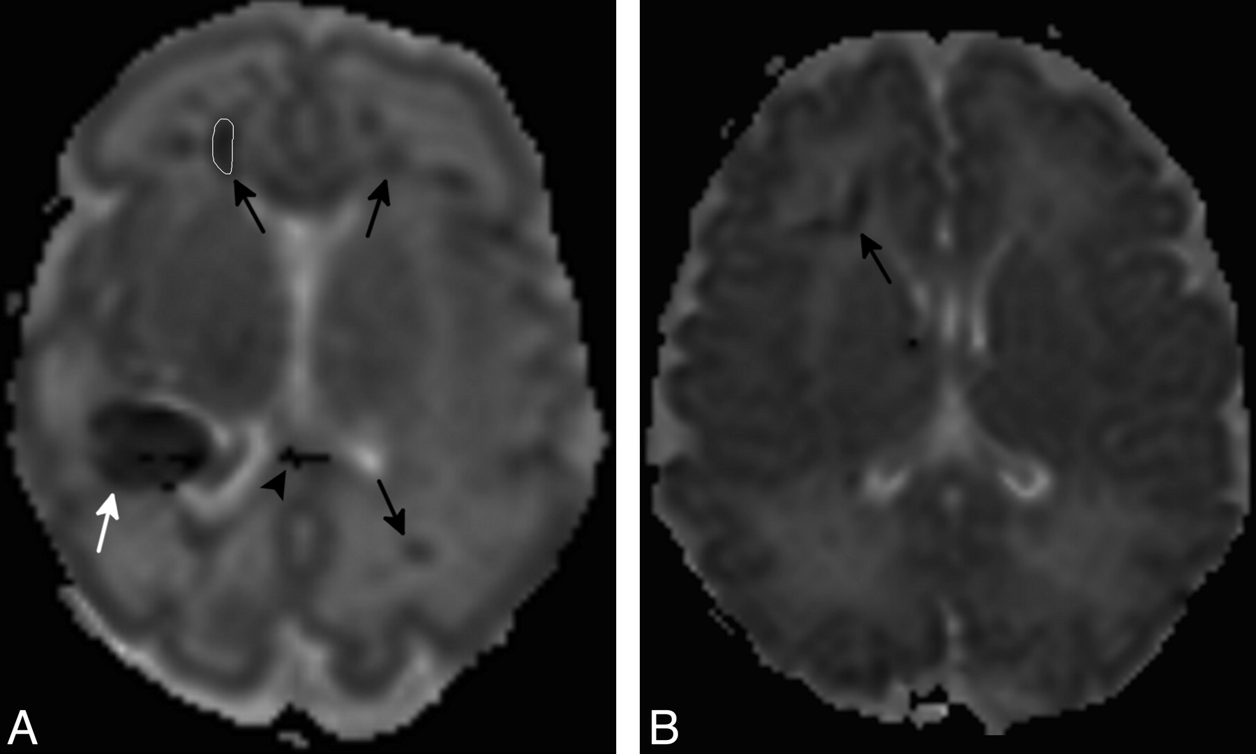
    Axial ADC maps in premature (A, same case as in Fig 1) and full-term (B, same case as in Fig 2) neonates. ADC reduction (arrows) is evident in the WM surrounding linear lesions described on T1- and T2-weighted images. A sample region of interest for ADC calculation (A) has been traced in the right frontal lobe. The white arrow and arrowhead show ADC alterations corresponding to parietal hematoma and a corpus callosum lesion.

  • Fig 5.
    • Download figure
    • Open in new tab
    • Download powerpoint
    Fig 5.

    Axial SWI in a full-term neonate (case 14, Table) shows small dilated vessels, compatible with DMV distribution, in the left periventricular WM (black arrows).

  • Fig 6.
    • Download figure
    • Open in new tab
    • Download powerpoint
    Fig 6.

    Coronal and axial T2-weighted sections in a full-term neonate (case 15, Table) examined at 9 days (A and B) and 18 months (C and D) of life. Bilateral anterior and posterior periventricular radial WM lesions are due to DMV pathology in the first examination (arrows in A and B). A PVL-like pattern with hyperintense signal intensity and reduced thickness of periventricular WM is evident on follow-up (C and D).

  • Fig 7.
    • Download figure
    • Open in new tab
    • Download powerpoint
    Fig 7.

    The graph shows the evolution of lesions with time (days in logarithmic scale).

Tables

  • Figures
  • Characteristics of the patients

    PatientGA (weeks)MRI IndicationMRI Timing (days from birth)MRI FindingsAssociated FindingsWM Injury LocationADC PatternSequences Used for Evaluation of Blood Products2D MR Venography
    130Difficult delivery10AGMHAF, FPFRT2 FFEYes
    231Seizures48BAFNSWINo
    331Difficult delivery9AGMH, PH, CCEAF, FP, PT, TFRSWINo
    58BAF, FP, PTN
    431Difficult delivery4AGMHAF, FP, PTFRT2 echo-planarYes
    80BAF, FP, PTN
    532Difficult delivery26AGMHAF, FP, PT, TNT2-FFEYes
    632Difficult delivery6BGMH, PH, TIAF, FPDRT2 echo-planarYes
    111BAF, FP
    734Difficult delivery44AAF, FP, PTNT2-FFEYes
    835Difficult delivery38BAF, FP, PTNSWINo
    935Difficult delivery45BGMHAF, FP, PT, TNT2 echo-planarNo
    1036Pathologic US3AGMH, CCEAF, FP, PTFRSWINo
    1136Difficult delivery0AAF, FP, PTDRSWIYes
    18AAF, FP, PTN
    628PVLAF, FP, PTN
    1237Seizures14BAF, FP, PT, TNT2 echo-planarYes
    287PVLAF, FP, PT, TN
    1337Seizures6AGMH, TI, CCEAF, FP, PT, TFRSWIYes
    48BAF, FP, PT, TN
    1438Difficult delivery9BAF, FP, PT, TFRSWI, T2-FFEYes
    567PVLAF, FP, PT, T
    1539Difficult delivery14BPH, TI, CCEAF, FP, PT, TDRT2 echo-planarNo
    730PVLAF, FP, PT, T
    1640Seizures4AAF, FP, PTFRSWIYes
    25BAF, FP, PTN
    1740Seizures3APH, SAHAFFRSWIYes
    36AAF
    1840Seizures10AAF, FP, PT, TDRT2 echo-planarNo
    26BAF, FP, PT, TN
    1940Seizures0ATI, SAHAF, FP, PTFRT2-FFEYes
    7AAF, FP, PTN
    2041Difficult delivery21BTI, CCEAF, FP, PT, TNSWINo
    452PVLAF, FP, PT, TN
    2141Seizures0AAF, FP, PTNSWIYes
    5AAFN
    416NormalN
    • Note:—GA indicates gestational age; MRI, MR imaging; TI, thalamic ischemia; CCE, corpus callosum edema; SAH, subarachnoid hemorrhage; A, pattern A; B, pattern B; AF, anterior frontal WM; FP, frontoparietal WM; PT, peritrigonal WM; T, temporal WM; DR, diffusely reduced ADC values; FR, focally reduced (surrounding lesions) ADC values; N, normal ADC values.

PreviousNext
Back to top

In this issue

American Journal of Neuroradiology: 32 (11)
American Journal of Neuroradiology
Vol. 32, Issue 11
1 Dec 2011
  • Table of Contents
  • Index by author
Advertisement
Print
Download PDF
Email Article

Thank you for your interest in spreading the word on American Journal of Neuroradiology.

NOTE: We only request your email address so that the person you are recommending the page to knows that you wanted them to see it, and that it is not junk mail. We do not capture any email address.

Enter multiple addresses on separate lines or separate them with commas.
Deep Medullary Vein Involvement in Neonates with Brain Damage: An MR Imaging Study
(Your Name) has sent you a message from American Journal of Neuroradiology
(Your Name) thought you would like to see the American Journal of Neuroradiology web site.
CAPTCHA
This question is for testing whether or not you are a human visitor and to prevent automated spam submissions.
Cite this article
F. Arrigoni, C. Parazzini, A. Righini, C. Doneda, L.A. Ramenghi, G. Lista, F. Triulzi
Deep Medullary Vein Involvement in Neonates with Brain Damage: An MR Imaging Study
American Journal of Neuroradiology Dec 2011, 32 (11) 2030-2036; DOI: 10.3174/ajnr.A2687

Citation Manager Formats

  • BibTeX
  • Bookends
  • EasyBib
  • EndNote (tagged)
  • EndNote 8 (xml)
  • Medlars
  • Mendeley
  • Papers
  • RefWorks Tagged
  • Ref Manager
  • RIS
  • Zotero
0 Responses
Respond to this article
Share
Bookmark this article
Deep Medullary Vein Involvement in Neonates with Brain Damage: An MR Imaging Study
F. Arrigoni, C. Parazzini, A. Righini, C. Doneda, L.A. Ramenghi, G. Lista, F. Triulzi
American Journal of Neuroradiology Dec 2011, 32 (11) 2030-2036; DOI: 10.3174/ajnr.A2687
del.icio.us logo Twitter logo Facebook logo Mendeley logo
  • Tweet Widget
  • Facebook Like
  • Google Plus One
Purchase

Jump to section

  • Article
    • Abstract
    • ABBREVIATIONS
    • Materials and Methods
    • Results
    • Discussion
    • Conclusions
    • References
  • Figures & Data
  • Info & Metrics
  • Responses
  • References
  • PDF

Related Articles

  • No related articles found.
  • PubMed
  • Google Scholar

Cited By...

  • The Needed Studies Trying to Untangle the Complex Nature of Neonatal Intracranial Bleeds Occurring around Birth
  • Deep Medullary Vein Thrombosis in a Neonate: A Peculiar MRI Pattern
  • Cerebral Venous Thrombosis Associated with COVID-19
  • MR Imaging Scoring System for White Matter Injury after Deep Medullary Vein Thrombosis and Infarction in Neonates
  • Teaching NeuroImages: Fetal deep medullary vein thrombosis presenting as progressive intracerebral hemorrhage
  • Acquisition Guidelines and Quality Assessment Tools for Analyzing Neonatal Diffusion Tensor MRI Data
  • Crossref
  • Google Scholar

This article has not yet been cited by articles in journals that are participating in Crossref Cited-by Linking.

More in this TOC Section

  • FRACTURE MR in Congenital Vertebral Anomalies
  • Comparing MRI Perfusion in Pediatric Brain Tumors
  • Sodium MRI in Pediatric Brain Tumors
Show more Pediatric Neuroimaging

Similar Articles

Advertisement

Indexed Content

  • Current Issue
  • Accepted Manuscripts
  • Article Preview
  • Past Issues
  • Editorials
  • Editor's Choice
  • Fellows' Journal Club
  • Letters to the Editor
  • Video Articles

Cases

  • Case Collection
  • Archive - Case of the Week
  • Archive - Case of the Month
  • Archive - Classic Case

More from AJNR

  • Trainee Corner
  • Imaging Protocols
  • MRI Safety Corner
  • Book Reviews

Multimedia

  • AJNR Podcasts
  • AJNR Scantastics

Resources

  • Turnaround Time
  • Submit a Manuscript
  • Submit a Video Article
  • Submit an eLetter to the Editor/Response
  • Manuscript Submission Guidelines
  • Statistical Tips
  • Fast Publishing of Accepted Manuscripts
  • Graphical Abstract Preparation
  • Imaging Protocol Submission
  • Evidence-Based Medicine Level Guide
  • Publishing Checklists
  • Author Policies
  • Become a Reviewer/Academy of Reviewers
  • News and Updates

About Us

  • About AJNR
  • Editorial Board
  • Editorial Board Alumni
  • Alerts
  • Permissions
  • Not an AJNR Subscriber? Join Now
  • Advertise with Us
  • Librarian Resources
  • Feedback
  • Terms and Conditions
  • AJNR Editorial Board Alumni

American Society of Neuroradiology

  • Not an ASNR Member? Join Now

© 2025 by the American Society of Neuroradiology All rights, including for text and data mining, AI training, and similar technologies, are reserved.
Print ISSN: 0195-6108 Online ISSN: 1936-959X

Powered by HighWire