Advertisement

Main menu

  • Home
  • Content
    • Current Issue
    • Accepted Manuscripts
    • Article Preview
    • Past Issue Archive
    • AJNR Case Collection
    • Case of the Week Archive
    • Classic Case Archive
    • Case of the Month Archive
  • Special Collections
    • Spinal CSF Leak Articles (Jan 2020-June 2024)
    • 2024 AJNR Journal Awards
    • Most Impactful AJNR Articles
  • Multimedia
    • AJNR Podcast
    • AJNR Scantastics
    • Video Articles
  • For Authors
    • Submit a Manuscript
    • Author Policies
    • Fast publishing of Accepted Manuscripts
    • Graphical Abstract Preparation
    • Manuscript Submission Guidelines
    • Imaging Protocol Submission
    • Submit a Case for the Case Collection
  • About Us
    • About AJNR
    • Editorial Board
  • More
    • Become a Reviewer/Academy of Reviewers
    • Subscribers
    • Permissions
    • Alerts
    • Feedback
    • Advertisers
    • ASNR Home
  • Other Publications
    • ajnr

User menu

  • Alerts
  • Log in

Search

  • Advanced search
American Journal of Neuroradiology

American Journal of Neuroradiology

ASHNR American Society of Functional Neuroradiology ASHNR American Society of Pediatric Neuroradiology ASSR
  • Alerts
  • Log in

Advanced Search

  • Home
  • Content
    • Current Issue
    • Accepted Manuscripts
    • Article Preview
    • Past Issue Archive
    • AJNR Case Collection
    • Case of the Week Archive
    • Classic Case Archive
    • Case of the Month Archive
  • Special Collections
    • Spinal CSF Leak Articles (Jan 2020-June 2024)
    • 2024 AJNR Journal Awards
    • Most Impactful AJNR Articles
  • Multimedia
    • AJNR Podcast
    • AJNR Scantastics
    • Video Articles
  • For Authors
    • Submit a Manuscript
    • Author Policies
    • Fast publishing of Accepted Manuscripts
    • Graphical Abstract Preparation
    • Manuscript Submission Guidelines
    • Imaging Protocol Submission
    • Submit a Case for the Case Collection
  • About Us
    • About AJNR
    • Editorial Board
  • More
    • Become a Reviewer/Academy of Reviewers
    • Subscribers
    • Permissions
    • Alerts
    • Feedback
    • Advertisers
    • ASNR Home
  • Follow AJNR on Twitter
  • Visit AJNR on Facebook
  • Follow AJNR on Instagram
  • Join AJNR on LinkedIn
  • RSS Feeds

Welcome to the new AJNR, Updated Hall of Fame, and more. Read the full announcements.


AJNR is seeking candidates for the position of Associate Section Editor, AJNR Case Collection. Read the full announcement.


Research ArticleBrain

Differentiation of Typical and Atypical Parkinson Syndromes by Quantitative MR Imaging

N.K. Focke, G. Helms, P.M. Pantel, S. Scheewe, M. Knauth, C.G. Bachmann, J. Ebentheuer, P. Dechent, W. Paulus and C. Trenkwalder
American Journal of Neuroradiology December 2011, 32 (11) 2087-2092; DOI: https://doi.org/10.3174/ajnr.A2865
N.K. Focke
aFrom the Department of Clinical Neurophysiology (N.K.F., P.M.P., S.S., C.G.B., W.P.)
dParacelsus Elena Klinik (N.K.F., J.E., C.T.), Specialized Movement Disorders Hospital, Kassel, Germany.
  • Find this author on Google Scholar
  • Find this author on PubMed
  • Search for this author on this site
G. Helms
bMR-Research in Neurology and Psychiatry (G.H., P.D.)
  • Find this author on Google Scholar
  • Find this author on PubMed
  • Search for this author on this site
P.M. Pantel
aFrom the Department of Clinical Neurophysiology (N.K.F., P.M.P., S.S., C.G.B., W.P.)
  • Find this author on Google Scholar
  • Find this author on PubMed
  • Search for this author on this site
S. Scheewe
aFrom the Department of Clinical Neurophysiology (N.K.F., P.M.P., S.S., C.G.B., W.P.)
  • Find this author on Google Scholar
  • Find this author on PubMed
  • Search for this author on this site
M. Knauth
cDepartment of Neuroradiology (M.K.), Georg-August University Göttingen, Göttingen, Germany
  • Find this author on Google Scholar
  • Find this author on PubMed
  • Search for this author on this site
C.G. Bachmann
aFrom the Department of Clinical Neurophysiology (N.K.F., P.M.P., S.S., C.G.B., W.P.)
  • Find this author on Google Scholar
  • Find this author on PubMed
  • Search for this author on this site
J. Ebentheuer
dParacelsus Elena Klinik (N.K.F., J.E., C.T.), Specialized Movement Disorders Hospital, Kassel, Germany.
  • Find this author on Google Scholar
  • Find this author on PubMed
  • Search for this author on this site
P. Dechent
bMR-Research in Neurology and Psychiatry (G.H., P.D.)
  • Find this author on Google Scholar
  • Find this author on PubMed
  • Search for this author on this site
W. Paulus
aFrom the Department of Clinical Neurophysiology (N.K.F., P.M.P., S.S., C.G.B., W.P.)
  • Find this author on Google Scholar
  • Find this author on PubMed
  • Search for this author on this site
C. Trenkwalder
dParacelsus Elena Klinik (N.K.F., J.E., C.T.), Specialized Movement Disorders Hospital, Kassel, Germany.
  • Find this author on Google Scholar
  • Find this author on PubMed
  • Search for this author on this site
  • Article
  • Figures & Data
  • Supplemental
  • Info & Metrics
  • Responses
  • References
  • PDF
Loading

Article Figures & Data

Figures

  • Tables
  • Fig 1.
    • Download figure
    • Open in new tab
    • Download powerpoint
    Fig 1.

    3D rendering of the regions of interest in a control subject: CN (red), PU (green), PA (blue), and SN (yellow). Regions of interest overlaid on the 3D T1-weighted image. A, Axial cut plane at the level of the striatum. B, Axial cut plane through the mesencephalon. C, Coronal oblique cut plane along the main axis of the SN region of interest.
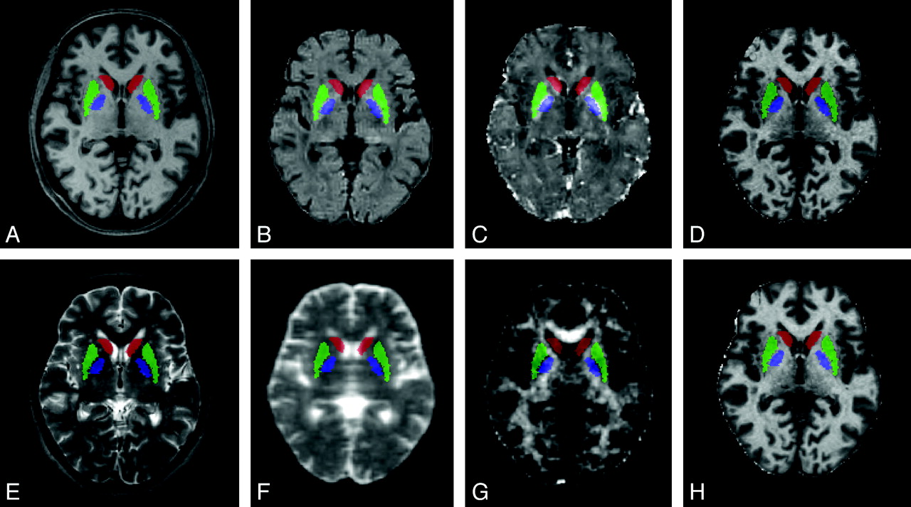
  • Fig 2.
    • Download figure
    • Open in new tab
    • Download powerpoint
    Fig 2.

    Regions of interest overlaid on the quantitative maps: CN (red), PU (green), and PA (blue). Regions of interest overlaid on axial sections of a 3D T1-weighted image (A), an R2 map (B), an R2* map (C), a magnetization transfer map (D), a 3D T2-weighted image (E), MD (F), FA (G), and R1 map (H).
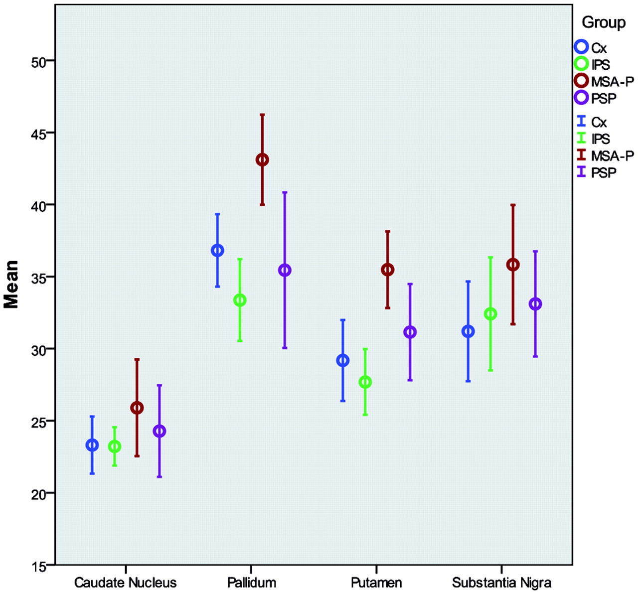
  • Fig 3.
    • Download figure
    • Open in new tab
    • Download powerpoint
    Fig 3.

    Group differences of R2* measurements. Error bars (95% confidence intervals) of R2* measurements (right side only) are shown clustered by diagnosis group.

Tables

  • Figures
  • Demographic details of controls and patient groups

    No.Age (mean)Sex (female/male)
    Controls1367.6 ± 10.58:5
    IPS1266.3 ± 7.82:10
    MSA-P1062.5 ± 8.55:5
    PSP967.0 ± 4.32:7
    Total4466.0 ± 8.317:27
    • Note:—MSA-P indicates multiple systems atrophy–type Parkinson.

PreviousNext
Back to top

In this issue

American Journal of Neuroradiology: 32 (11)
American Journal of Neuroradiology
Vol. 32, Issue 11
1 Dec 2011
  • Table of Contents
  • Index by author
Advertisement
Print
Download PDF
Email Article

Thank you for your interest in spreading the word on American Journal of Neuroradiology.

NOTE: We only request your email address so that the person you are recommending the page to knows that you wanted them to see it, and that it is not junk mail. We do not capture any email address.

Enter multiple addresses on separate lines or separate them with commas.
Differentiation of Typical and Atypical Parkinson Syndromes by Quantitative MR Imaging
(Your Name) has sent you a message from American Journal of Neuroradiology
(Your Name) thought you would like to see the American Journal of Neuroradiology web site.
CAPTCHA
This question is for testing whether or not you are a human visitor and to prevent automated spam submissions.
Cite this article
N.K. Focke, G. Helms, P.M. Pantel, S. Scheewe, M. Knauth, C.G. Bachmann, J. Ebentheuer, P. Dechent, W. Paulus, C. Trenkwalder
Differentiation of Typical and Atypical Parkinson Syndromes by Quantitative MR Imaging
American Journal of Neuroradiology Dec 2011, 32 (11) 2087-2092; DOI: 10.3174/ajnr.A2865

Citation Manager Formats

  • BibTeX
  • Bookends
  • EasyBib
  • EndNote (tagged)
  • EndNote 8 (xml)
  • Medlars
  • Mendeley
  • Papers
  • RefWorks Tagged
  • Ref Manager
  • RIS
  • Zotero
0 Responses
Respond to this article
Share
Bookmark this article
Differentiation of Typical and Atypical Parkinson Syndromes by Quantitative MR Imaging
N.K. Focke, G. Helms, P.M. Pantel, S. Scheewe, M. Knauth, C.G. Bachmann, J. Ebentheuer, P. Dechent, W. Paulus, C. Trenkwalder
American Journal of Neuroradiology Dec 2011, 32 (11) 2087-2092; DOI: 10.3174/ajnr.A2865
del.icio.us logo Twitter logo Facebook logo Mendeley logo
  • Tweet Widget
  • Facebook Like
  • Google Plus One
Purchase

Jump to section

  • Article
    • Abstract
    • ABBREVIATIONS
    • Materials and Methods
    • Results
    • Discussion
    • Conclusions
    • Footnotes
    • References
  • Figures & Data
  • Supplemental
  • Info & Metrics
  • Responses
  • References
  • PDF

Related Articles

  • PubMed
  • Google Scholar

Cited By...

  • Automated classification of neurodegenerative parkinsonian syndromes using multimodal magnetic resonance imaging in a clinical setting
  • Combined Diffusion Tensor Imaging and Apparent Transverse Relaxation Rate Differentiate Parkinson Disease and Atypical Parkinsonism
  • Diffusion tensor imaging in parkinsonian syndromes: A systematic review and meta-analysis
  • Individual Detection of Patients with Parkinson Disease using Support Vector Machine Analysis of Diffusion Tensor Imaging Data: Initial Results
  • Crossref (66)
  • Google Scholar

This article has been cited by the following articles in journals that are participating in Crossref Cited-by Linking.

  • Diffusion tensor imaging in parkinsonian syndromes
    Claire J. Cochrane, Klaus P. Ebmeier
    Neurology 2013 80 9
  • Magnetic resonance imaging for the diagnosis of Parkinson’s disease
    Beatrice Heim, Florian Krismer, Roberto De Marzi, Klaus Seppi
    Journal of Neural Transmission 2017 124 8
  • Imaging biomarkers in Parkinson’s disease and Parkinsonian syndromes: current and emerging concepts
    Usman Saeed, Jordana Compagnone, Richard I. Aviv, Antonio P. Strafella, Sandra E. Black, Anthony E. Lang, Mario Masellis
    Translational Neurodegeneration 2017 6 1
  • Free-water imaging in Parkinson’s disease and atypical parkinsonism
    Peggy J. Planetta, Edward Ofori, Ofer Pasternak, Roxana G. Burciu, Priyank Shukla, Jesse C. DeSimone, Michael S. Okun, Nikolaus R. McFarland, David E. Vaillancourt
    Brain 2016 139 2
  • Multiple System Atrophy: An Oligodendroglioneural Synucleinopathy
    Kurt A. Jellinger
    Journal of Alzheimer’s Disease 2018 62 3
  • Diffusion tensor imaging of nigral degeneration in Parkinson's disease: A region-of-interest and voxel-based study at 3T and systematic review with meta-analysis
    Stefan T. Schwarz, Maryam Abaei, Vamsi Gontu, Paul S. Morgan, Nin Bajaj, Dorothee P. Auer
    NeuroImage: Clinical 2013 3
  • Neurite orientation dispersion and density imaging in the substantia nigra in idiopathic Parkinson disease
    Koji Kamagata, Taku Hatano, Ayami Okuzumi, Yumiko Motoi, Osamu Abe, Keigo Shimoji, Kouhei Kamiya, Michimasa Suzuki, Masaaki Hori, Kanako K. Kumamaru, Nobutaka Hattori, Shigeki Aoki
    European Radiology 2016 26 8
  • Neuroimaging Advances in Parkinson's Disease and Atypical Parkinsonian Syndromes
    Usman Saeed, Anthony E. Lang, Mario Masellis
    Frontiers in Neurology 2020 11
  • Parkinsonism Beyond Parkinson's Disease
    Alessandra Fanciulli, Iva Stankovic, Florian Krismer, Klaus Seppi, Johannes Levin, Gregor K. Wenning
    2019 149
  • Individual Detection of Patients with Parkinson Disease using Support Vector Machine Analysis of Diffusion Tensor Imaging Data: Initial Results
    S. Haller, S. Badoud, D. Nguyen, V. Garibotto, K.O. Lovblad, P.R Burkhard
    American Journal of Neuroradiology 2012 33 11

More in this TOC Section

  • Statin Therapy Does Not Affect the Radiographic and Clinical Profile of Patients with TIA and Minor Stroke
  • Optimal MRI Sequence for Identifying Occlusion Location in Acute Stroke: Which Value of Time-Resolved Contrast-Enhanced MRA?
  • SWI or T2*: Which MRI Sequence to Use in the Detection of Cerebral Microbleeds? The Karolinska Imaging Dementia Study
Show more Brain

Similar Articles

Advertisement

Indexed Content

  • Current Issue
  • Accepted Manuscripts
  • Article Preview
  • Past Issues
  • Editorials
  • Editors Choice
  • Fellow Journal Club
  • Letters to the Editor

Cases

  • Case Collection
  • Archive - Case of the Week
  • Archive - Case of the Month
  • Archive - Classic Case

Special Collections

  • Special Collections

Resources

  • News and Updates
  • Turn around Times
  • Submit a Manuscript
  • Author Policies
  • Manuscript Submission Guidelines
  • Evidence-Based Medicine Level Guide
  • Publishing Checklists
  • Graphical Abstract Preparation
  • Imaging Protocol Submission
  • Submit a Case
  • Become a Reviewer/Academy of Reviewers
  • Get Peer Review Credit from Publons

Multimedia

  • AJNR Podcast
  • AJNR SCANtastic
  • Video Articles

About Us

  • About AJNR
  • Editorial Board
  • Not an AJNR Subscriber? Join Now
  • Alerts
  • Feedback
  • Advertise with us
  • Librarian Resources
  • Permissions
  • Terms and Conditions

American Society of Neuroradiology

  • Not an ASNR Member? Join Now

© 2025 by the American Society of Neuroradiology All rights, including for text and data mining, AI training, and similar technologies, are reserved.
Print ISSN: 0195-6108 Online ISSN: 1936-959X

Powered by HighWire