
Article Figures & Data
Figures
- Fig 1.
Manual segmentation of brain volumes.
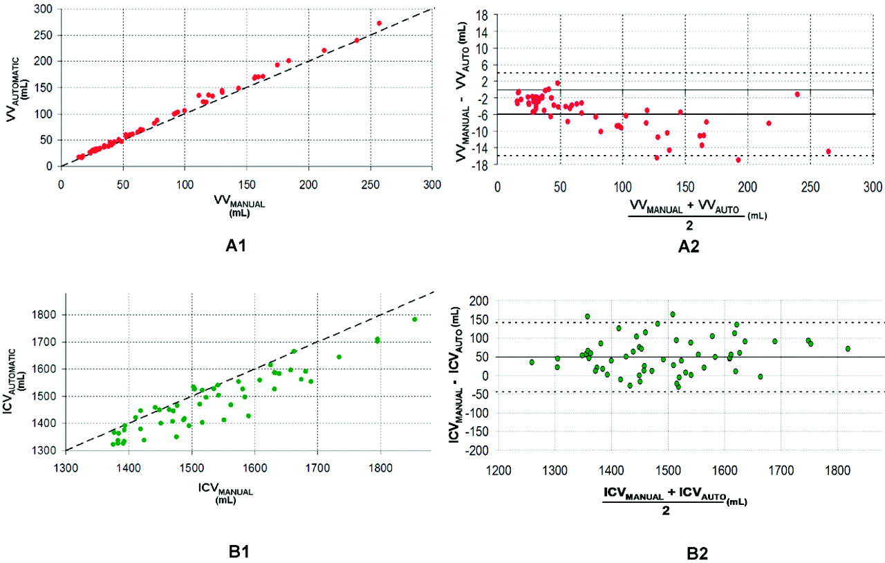
- Fig 2.
Comparison between manual and automatic volumes. A1 and B1, Plots of automatic versus manual for VV and ICV. The oblique dashed line represents the equality line. A2 and B2, The corresponding Bland-Altman plots for VV and ICV. The horizontal dashed lines are the confidence intervals of the difference between manual and automatic volumes.
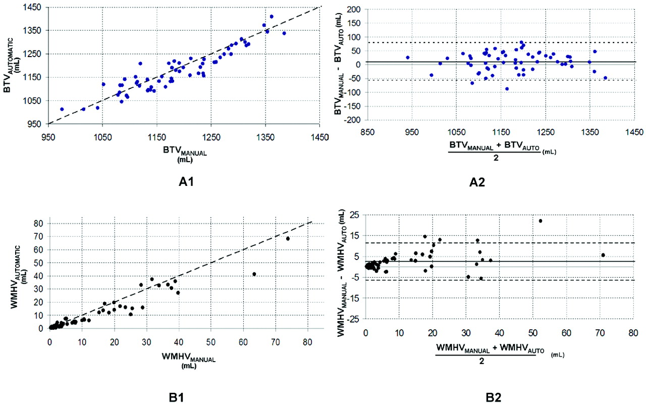
- Fig 3.
Comparison between manual and automatic volumes. A1 and B1, The plots of automatic versus manual for BTV and WMHV. The oblique dashed line represents the equality line. A2 and B2, The corresponding Bland-Altman plots for BTV and WMHV. The horizontal dashed lines are the confidence intervals of the difference between manual and automatic volumes.


Tables
Comparison MDVV (2SD, mL) MRDVV (2SD, %) MDBTV (2SD, mL) MRDBTV(2SD, %) MDICV(2SD, mL) MRDICV (2SD, %) MDWMHV (2SD, mL) MRDWMHV (2SD, %) Obs1 vs Obs2 −1 (8) −11 (34) −10 (45) −1 (6) (n = 10) −0.21 (14.45) −0.78 (3.59) −0.71 (3.12) −8.47 (47.54) Obs1 vs Obs1 1 (1) 4 (19) 6 (31) −1 (3) (n = 10) 0.75 (2.78) 1.00 (1.29) 0.37 (2.04) −6.10 (11.09) Obs1 vs Autoa −9 (8) 24 (65) 28 (90) 8 (10) (n= 10) −2.75 (9.66) 2.28 (5.62) 1.89 (6.08) 46.42 (11.88) Obs1 vs Autoa −6 (10) 11 (70) 49 (93) 2 (9) (n = 61) −9.14 (10.78) 1.00 (6.00) 3.20 (5.94) 15.99 (80.66) -
a Statistically different between the mean manual and the mean automatic (P < .05) for all brain volumes.
-
In this issue


Jump to section
Related Articles
Cited By...
- A Large-scale Comparison of Cortical and Subcortical Structural Segmentation Methods in Alzheimers Disease: a Statistical Approach
- Clinical Feasibility of Synthetic MRI in Multiple Sclerosis: A Diagnostic and Volumetric Validation Study
- Cerebral Microinfarcts in Primary Open-Angle Glaucoma Correlated With DTI-Derived Integrity of Optic Radiation
- Automated Determination of Brain Parenchymal Fraction in Multiple Sclerosis
- Evaluation of Automatic Measurement of the Intracranial Volume Based on Quantitative MR Imaging