Advertisement

Main menu

  • Home
  • Content
    • Current Issue
    • Accepted Manuscripts
    • Article Preview
    • Past Issue Archive
    • AJNR Case Collection
    • Case of the Week Archive
    • Classic Case Archive
    • Case of the Month Archive
  • Special Collections
    • Spinal CSF Leak Articles (Jan 2020-June 2024)
    • 2024 AJNR Journal Awards
    • Most Impactful AJNR Articles
  • Multimedia
    • AJNR Podcast
    • AJNR Scantastics
    • Video Articles
  • For Authors
    • Submit a Manuscript
    • Author Policies
    • Fast publishing of Accepted Manuscripts
    • Graphical Abstract Preparation
    • Manuscript Submission Guidelines
    • Imaging Protocol Submission
    • Submit a Case for the Case Collection
  • About Us
    • About AJNR
    • Editorial Board
  • More
    • Become a Reviewer/Academy of Reviewers
    • Subscribers
    • Permissions
    • Alerts
    • Feedback
    • Advertisers
    • ASNR Home
  • Other Publications
    • ajnr

User menu

  • Alerts
  • Log in

Search

  • Advanced search
American Journal of Neuroradiology

American Journal of Neuroradiology

ASHNR American Society of Functional Neuroradiology ASHNR American Society of Pediatric Neuroradiology ASSR
  • Alerts
  • Log in

Advanced Search

  • Home
  • Content
    • Current Issue
    • Accepted Manuscripts
    • Article Preview
    • Past Issue Archive
    • AJNR Case Collection
    • Case of the Week Archive
    • Classic Case Archive
    • Case of the Month Archive
  • Special Collections
    • Spinal CSF Leak Articles (Jan 2020-June 2024)
    • 2024 AJNR Journal Awards
    • Most Impactful AJNR Articles
  • Multimedia
    • AJNR Podcast
    • AJNR Scantastics
    • Video Articles
  • For Authors
    • Submit a Manuscript
    • Author Policies
    • Fast publishing of Accepted Manuscripts
    • Graphical Abstract Preparation
    • Manuscript Submission Guidelines
    • Imaging Protocol Submission
    • Submit a Case for the Case Collection
  • About Us
    • About AJNR
    • Editorial Board
  • More
    • Become a Reviewer/Academy of Reviewers
    • Subscribers
    • Permissions
    • Alerts
    • Feedback
    • Advertisers
    • ASNR Home
  • Follow AJNR on Twitter
  • Visit AJNR on Facebook
  • Follow AJNR on Instagram
  • Join AJNR on LinkedIn
  • RSS Feeds

Welcome to the new AJNR, Updated Hall of Fame, and more. Read the full announcements.


AJNR is seeking candidates for the position of Associate Section Editor, AJNR Case Collection. Read the full announcement.


Case ReportCase Report
Open Access

Diffusion-Weighted Imaging in Fetuses with Severe Congenital Heart Defects

J.I. Berman, S.E.G. Hamrick, P.S. McQuillen, C. Studholme, D. Xu, R.G. Henry, L.K. Hornberger and O.A. Glenn
American Journal of Neuroradiology February 2011, 32 (2) E21-E22; DOI: https://doi.org/10.3174/ajnr.A1975
J.I. Berman
  • Find this author on Google Scholar
  • Find this author on PubMed
  • Search for this author on this site
S.E.G. Hamrick
  • Find this author on Google Scholar
  • Find this author on PubMed
  • Search for this author on this site
P.S. McQuillen
  • Find this author on Google Scholar
  • Find this author on PubMed
  • Search for this author on this site
C. Studholme
  • Find this author on Google Scholar
  • Find this author on PubMed
  • Search for this author on this site
D. Xu
  • Find this author on Google Scholar
  • Find this author on PubMed
  • Search for this author on this site
R.G. Henry
  • Find this author on Google Scholar
  • Find this author on PubMed
  • Search for this author on this site
L.K. Hornberger
  • Find this author on Google Scholar
  • Find this author on PubMed
  • Search for this author on this site
O.A. Glenn
  • Find this author on Google Scholar
  • Find this author on PubMed
  • Search for this author on this site
  • Article
  • Figures & Data
  • Info & Metrics
  • Responses
  • References
  • PDF
Loading

Abstract

SUMMARY: Fetal diffusion MR imaging was performed in 3 fetuses with CHD. ADC values in the periatrial WM, thalamus, and basal ganglia were compared with those in a control population of fetuses. Diffusivity in the periatrial WM and thalamus was higher for the fetuses with CHD compared with controls. These observations support the finding of abnormal in utero brain development in fetuses with CHD.

Abbreviations

ADC
apparent diffusion coefficient
CHD
congenital heart disease
WM
white matter

Congenital heart anomalies have been linked to long-term motor, sensory, and cognitive difficulties.1,2 The origin and cause of these functional deficits are not entirely understood. Abnormal WM structure associated with CHD has been detected with MR imaging at an early stage of neurodevelopment in neonates before corrective surgery.3 This study sought to determine the feasibility of detecting structural abnormalities in utero with diffusion MR imaging. The diffusion of water is sensitive to subtle changes in WM architecture that may not be visible on traditional MR imaging. This study measured diffusivity in the basal ganglia, thalamus, and periatrial WM of 3 fetuses with CHD. Diffusion measurements were compared with those in a population of control fetuses.4

Case Reports

Three women were referred for MR imaging of the fetal brain after prenatal sonography showed congenital cardiac anomalies. All fetal brain MR imaging was performed as part of a study on CHD, which was approved by our institutional review board. Case 1 was a fetus at 32.14 weeks' gestational age at MR imaging with sonographic evidence of transposition of the great arteries, mild hydronephrosis, and a small cystic neck mass. T1- and T2-weighted imaging findings of the fetal brain were normal. Case 2 was a fetus at 34.57 weeks' gestational age with a hypoplastic left heart and mild pelviectasis on prenatal sonography. No abnormalities were evident on T1- or T2-weighted MR images. Case 3 was a fetus at 35.29 weeks' gestational age with a hypoplastic left heart identified on prenatal sonography. Fetal MR imaging demonstrated a hypoplastic corpus callosum. In Cases 1 and 2, a neonatal MR imaging was also performed, which demonstrated a normal appearance of the brain.

Diffusion-weighted imaging was performed on a 1.5T scanner with a b-value of 600 s/mm2 applied in 3 orthogonal directions in addition to a single b = 0 s/mm2 volume. Imaging parameters were TR = 4500 ms, TE = minimum, FOV = 32 × 32 cm, section thickness = 5 mm, and matrix = 128 × 128. To minimize motion artifacts, we acquired diffusion images during a maternal breath-hold. All acquired echo-planar images and the calculated ADC maps were visually examined, and those degraded by motion were excluded. Regions of interest were placed in the periatrial WM, thalamus, and basal ganglia, and ADC values were measured.

Fetuses with CHD were compared with a previously reported control fetal population comprising 28 fetuses with basal ganglia measurements, 33 fetuses with periatrial WM measurements, and 28 fetuses with thalamic measurements.4 The control population's gestational age at the time of the MR imaging ranged from 21 to 34 weeks. Statistical comparisons between the control population and the 3 fetuses with CHD were made with linear regression, including ADC and gestational age as variables. In addition, z scores of the individual patient measurements were calculated to determine their deviation from the control data.

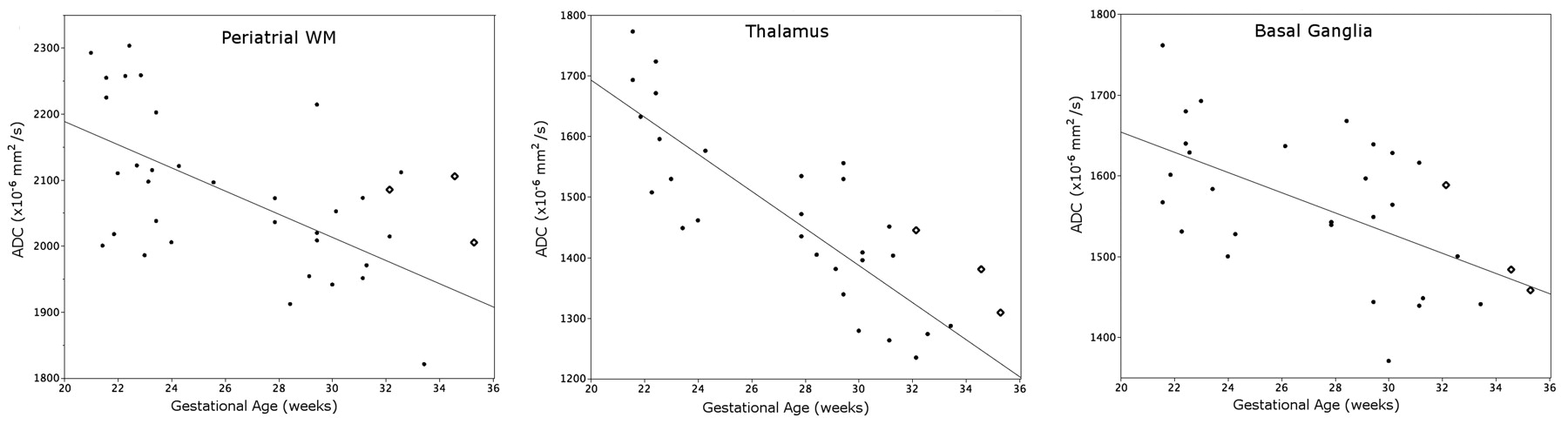
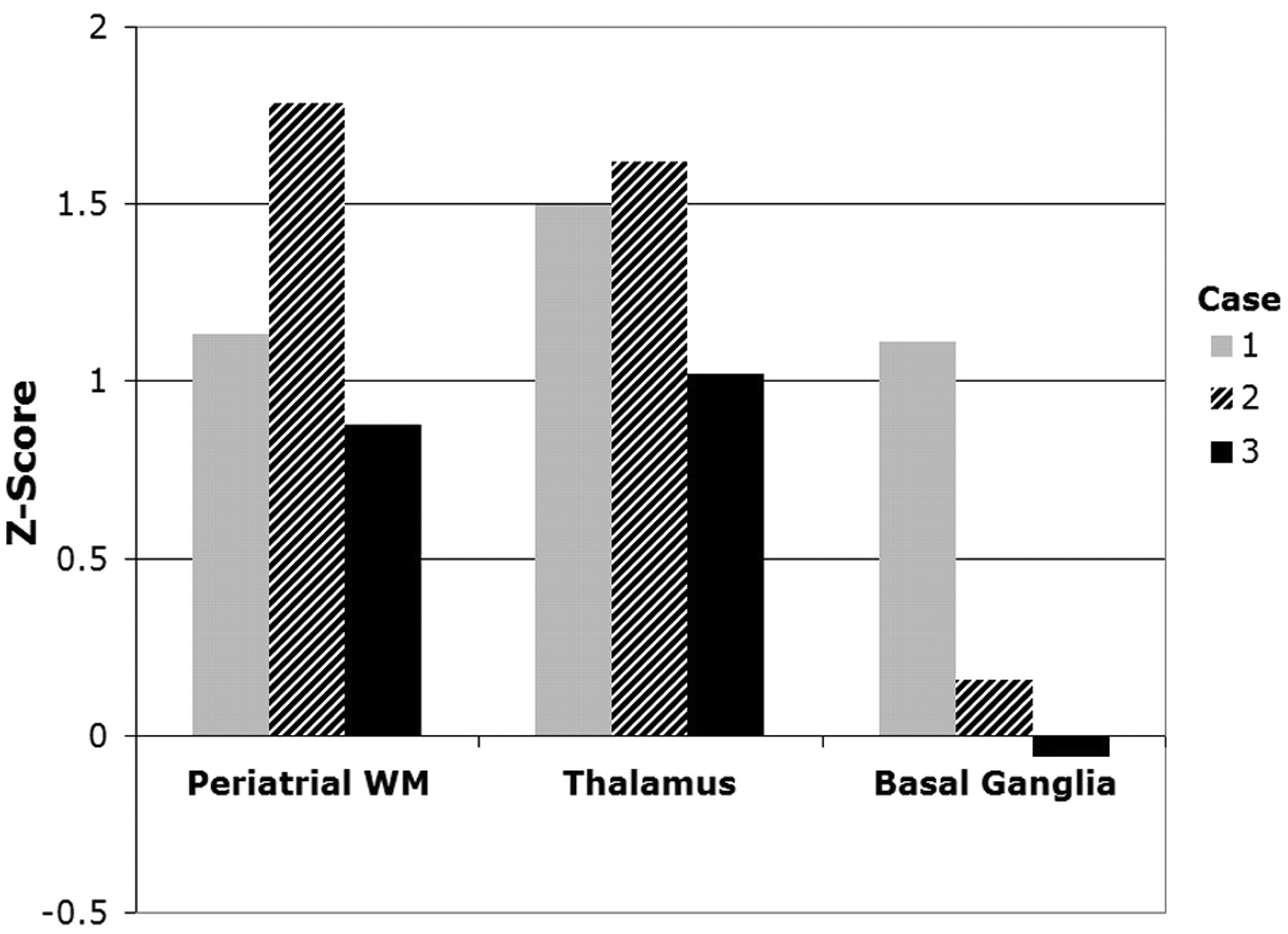
ADC values in the periatrial WM and thalamus were higher for all fetuses with CHD compared with the control population (Fig 1). The average z score was 1.26 for the periatrial WM, 1.38 for the thalamus, and 0.40 for the basal ganglia (Fig 2). Regression analysis indicated a significant difference between patient and control thalamic ADC values (P < .05) and a strong trend not reaching significance between patient and control periatrial WM ADC values (P = .071). Patient and control basal ganglia ADC values were not significantly different (P = .53).

Fig 1.
  • Download figure
  • Open in new tab
  • Download powerpoint
Fig 1.

Three fetuses with congenital heart disease (diamonds) are compared with a control cohort (solid dots). The regression line was fit to the control population.

Fig 2.
  • Download figure
  • Open in new tab
  • Download powerpoint
Fig 2.

Z scores of individual ADC measurements in fetuses with CHD show deviation from the control population. A negative z score indicates that the ADC measurement was below the expected value from the control population.

Discussion

We used fetal diffusion MR imaging to detect abnormally high water diffusion in the thalamus and periatrial WM of fetuses with CHD. High diffusivity indicates a difference in the structural organization of macromolecules during a critical period of neurodevelopment. Certain congenital heart lesions cause changes in cerebral perfusion and/or cerebral hypoxia, potentially leading to abnormal brain development and injury.5–7 This study reinforces the hypothesis that brain development is affected in utero by CHD.

In a recent study of neonates with CHD, Miller et al3 observed abnormally high diffusion in the neonatal brain before corrective heart surgery. In particular, the thalamus, perirolandic WM, posterior WM, and optic radiations appear to be most affected in neonates with CHD.3 We also observed abnormal diffusion in the thalamus and posterior WM of the fetuses in our study. We did not observe a significant abnormality in basal ganglia diffusivity in our patients; these results are similar to those of Miller et al. It is not precisely known why particular brain structures may be more susceptible to delayed development or damage in patients with CHD. However, different regions of the brain develop at different rates, and perfusion levels can vary throughout the brain. These developmental and anatomic differences may make certain parts of the brain more susceptible to damage and delayed development stemming from decreased perfusion in utero.

These case reports show that fetal diffusion MR imaging is a feasible method of detecting abnormal brain diffusivity associated with CHD. Further studies by using advanced in utero MR imaging techniques such as diffusion tensor imaging, spectroscopy, and possibly functional MR imaging may answer questions concerning the early structural and functional impact of CHD on the developing brain.

Footnotes

  • This work was supported by the Dana Foundation Brain and Immuno Imaging Track A, National Institutes of Health grant K23 NS52506, and NIH/NCRR UCSF-CTSI grant UL1 RR024131. Its contents are solely the responsibility of the authors and do not necessarily represent the official views of the NIH.

Indicates open access to non-subscribers at www.ajnr.org

References

  1. 1.↵
    1. Bellinger D,
    2. Jonas R,
    3. Rappaport L,
    4. et al
    . Developmental and neurologic status of children after heart surgery with hypothermic circulatory arrest or low-flow cardiopulmonary bypass. N Engl J Med 1995;332:549–55
    CrossRefPubMed
  2. 2.↵
    1. Partridge S,
    2. Vigneron D,
    3. Charlton N,
    4. et al
    . Pyramidal tract maturation after brain injury in newborns with heart disease. Ann Neurol 2006;59:640–51
    CrossRefPubMed
  3. 3.↵
    1. Miller S,
    2. McQuillen P,
    3. Hamrick S,
    4. et al
    . Abnormal brain development in newborns with congenital heart disease. N Engl J Med 2007;357:1928–38
    CrossRefPubMed
  4. 4.↵
    1. Schneider M,
    2. Berman J,
    3. Baumer F,
    4. et al
    . Normative apparent diffusion coefficient values in the developing fetal brain. AJNR Am J Neuroradiol 2009;30:1799–803. Epub 2009 Jun 25
    Abstract/FREE Full Text
  5. 5.↵
    1. Donofrio M,
    2. Bremer Y,
    3. Schieken R,
    4. et al
    . Autoregulation of cerebral blood flow in fetuses with congenital heart disease: the brain sparing effect. Pediatr Cardiol 2003;24:436–43
    CrossRefPubMed
  6. 6.↵
    1. Licht DJ,
    2. Wang J,
    3. Silvestre DW,
    4. et al
    . Preoperative cerebral blood flow is diminished in neonates with severe congenital heart defects. J Thorac Cardiovasc Surg 2004;128:841–49
    CrossRefPubMed
  7. 7.↵
    1. Shillingford AJ,
    2. Ittenbach RF,
    3. Marino BS,
    4. et al
    . Aortic morphometry and microcephaly in hypoplastic left heart syndrome. Cardiol Young 2007;17:189–95. Epub 2007 Mar 5
    CrossRefPubMed
  • Received October 16, 2009.
  • Accepted after revision October 20, 2009.
  • Copyright © American Society of Neuroradiology
View Abstract
PreviousNext
Back to top

In this issue

American Journal of Neuroradiology: 32 (2)
American Journal of Neuroradiology
Vol. 32, Issue 2
1 Feb 2011
  • Table of Contents
  • Index by author
Advertisement
Print
Download PDF
Email Article

Thank you for your interest in spreading the word on American Journal of Neuroradiology.

NOTE: We only request your email address so that the person you are recommending the page to knows that you wanted them to see it, and that it is not junk mail. We do not capture any email address.

Enter multiple addresses on separate lines or separate them with commas.
Diffusion-Weighted Imaging in Fetuses with Severe Congenital Heart Defects
(Your Name) has sent you a message from American Journal of Neuroradiology
(Your Name) thought you would like to see the American Journal of Neuroradiology web site.
CAPTCHA
This question is for testing whether or not you are a human visitor and to prevent automated spam submissions.
Cite this article
J.I. Berman, S.E.G. Hamrick, P.S. McQuillen, C. Studholme, D. Xu, R.G. Henry, L.K. Hornberger, O.A. Glenn
Diffusion-Weighted Imaging in Fetuses with Severe Congenital Heart Defects
American Journal of Neuroradiology Feb 2011, 32 (2) E21-E22; DOI: 10.3174/ajnr.A1975

Citation Manager Formats

  • BibTeX
  • Bookends
  • EasyBib
  • EndNote (tagged)
  • EndNote 8 (xml)
  • Medlars
  • Mendeley
  • Papers
  • RefWorks Tagged
  • Ref Manager
  • RIS
  • Zotero
0 Responses
Respond to this article
Share
Bookmark this article
Diffusion-Weighted Imaging in Fetuses with Severe Congenital Heart Defects
J.I. Berman, S.E.G. Hamrick, P.S. McQuillen, C. Studholme, D. Xu, R.G. Henry, L.K. Hornberger, O.A. Glenn
American Journal of Neuroradiology Feb 2011, 32 (2) E21-E22; DOI: 10.3174/ajnr.A1975
del.icio.us logo Twitter logo Facebook logo Mendeley logo
  • Tweet Widget
  • Facebook Like
  • Google Plus One
Purchase

Jump to section

  • Article
    • Abstract
    • Abbreviations
    • Case Reports
    • Discussion
    • Footnotes
    • References
  • Figures & Data
  • Info & Metrics
  • Responses
  • References
  • PDF

Related Articles

  • No related articles found.
  • PubMed
  • Google Scholar

Cited By...

  • DWI in Brains of Fetuses with Congenital Heart Disease: A Case-Control MR Imaging Study
  • Delayed maturation of the structural brain connectome in neonates with congenital heart disease
  • Association of Isolated Congenital Heart Disease with Fetal Brain Maturation
  • Prevalence and Spectrum of In Utero Structural Brain Abnormalities in Fetuses with Complex Congenital Heart Disease
  • Brain injury and development in newborns with critical congenital heart disease
  • Prenatal MR Imaging Detection of Deep Medullary Vein Involvement in Fetal Brain Damage
  • Crossref (34)
  • Google Scholar

This article has been cited by the following articles in journals that are participating in Crossref Cited-by Linking.

  • Delayed Cortical Development in Fetuses with Complex Congenital Heart Disease
    C. Clouchoux, A. J. du Plessis, M. Bouyssi-Kobar, W. Tworetzky, D. B. McElhinney, D. W. Brown, A. Gholipour, D. Kudelski, S. K. Warfield, R. J. McCarter, R. L. Robertson, A. C. Evans, J. W. Newburger, C. Limperopoulos
    Cerebral Cortex 2013 23 12
  • Brain injury and development in newborns with critical congenital heart disease
    Anastasia Dimitropoulos, Patrick S. McQuillen, Viyeka Sethi, Alisha Moosa, Vann Chau, Duan Xu, Rollin Brant, Anthony Azakie, Andrew Campbell, A. James Barkovich, Kenneth J. Poskitt, Steven P. Miller
    Neurology 2013 81 3
  • Severe Congenital Heart Defects Are Associated with Global Reduction of Neonatal Brain Volumes
    Michael von Rhein, Andreas Buchmann, Cornelia Hagmann, Hitendu Dave, Vera Bernet, Ianina Scheer, Walter Knirsch, Beatrice Latal, Vera Bernet, Christoph Bürki, Hitendu Dave, René Prêtre, Cornelia Hagmann, Walter Knirsch, Oliver Kretschmar, Christian Kellenberger, Ianina Scheer, Beatrice Latal, Rabia Liamlahi, Michael von Rhein, Barbara R. Plecko, Felix H. Sennhauser
    The Journal of Pediatrics 2015 167 6
  • Fetal MRI: A technical update with educational aspirations
    Ali Gholipour, Judith A. Estroff, Carol E. Barnewolt, Richard L. Robertson, P. Ellen Grant, Borjan Gagoski, Simon K. Warfield, Onur Afacan, Susan A. Connolly, Jeffrey J. Neil, Adam Wolfberg, Robert V. Mulkern
    Concepts in Magnetic Resonance Part A 2014 43 6
  • A Unified Approach to Diffusion Direction Sensitive Slice Registration and 3-D DTI Reconstruction From Moving Fetal Brain Anatomy
    Mads Fogtmann, Sharmishtaa Seshamani, Christopher Kroenke, Xi Cheng, Teresa Chapman, Jakob Wilm, Francois Rousseau, Colin Studholme
    IEEE Transactions on Medical Imaging 2014 33 2
  • Neonatal Neurobehavior and Diffusion MRI Changes in Brain Reorganization Due to Intrauterine Growth Restriction in a Rabbit Model
    Elisenda Eixarch, Dafnis Batalle, Miriam Illa, Emma Muñoz-Moreno, Ariadna Arbat-Plana, Ivan Amat-Roldan, Francesc Figueras, Eduard Gratacos, Olivier Baud
    PLoS ONE 2012 7 2
  • Prevalence and Spectrum of In Utero Structural Brain Abnormalities in Fetuses with Complex Congenital Heart Disease
    M. Brossard-Racine, A. J. du Plessis, G. Vezina, R. Robertson, D. Bulas, I. E. Evangelou, M. Donofrio, D. Freeman, C. Limperopoulos
    American Journal of Neuroradiology 2014 35 8
  • Delayed cortical gray matter development in neonates with severe congenital heart disease
    Nathalie H.P. Claessens, Pim Moeskops, Andreas Buchmann, Beatrice Latal, Walter Knirsch, Ianina Scheer, Ivana Išgum, Linda S. de Vries, Manon J.N.L. Benders, Michael von Rhein
    Pediatric Research 2016 80 5
  • Fetal Brain MRI Texture Analysis Identifies Different Microstructural Patterns in Adequate and Small for Gestational Age Fetuses at Term
    M. Sanz-Cortés, F. Figueras, E. Bonet-Carne, N. Padilla, V. Tenorio, N. Bargalló, I. Amat-Roldan, Eduard Gratacós
    Fetal Diagnosis and Therapy 2013 33 2
  • Long-Term Functional Outcomes and Correlation with Regional Brain Connectivity by MRI Diffusion Tractography Metrics in a Near-Term Rabbit Model of Intrauterine Growth Restriction
    Miriam Illa, Elisenda Eixarch, Dafnis Batalle, Ariadna Arbat-Plana, Emma Muñoz-Moreno, Francesc Figueras, Eduard Gratacos, Martin Gerbert Frasch
    PLoS ONE 2013 8 10

More in this TOC Section

  • Atypical Diffusion-Restricted Lesion in 5-Fluorouracil Encephalopathy
  • Dural Infantile Hemangioma Masquerading as a Skull Vault Lesion
  • Multimodal Imaging of Spike Propagation: A Technical Case Report
Show more Case Reports

Similar Articles

Advertisement

Indexed Content

  • Current Issue
  • Accepted Manuscripts
  • Article Preview
  • Past Issues
  • Editorials
  • Editors Choice
  • Fellow Journal Club
  • Letters to the Editor

Cases

  • Case Collection
  • Archive - Case of the Week
  • Archive - Case of the Month
  • Archive - Classic Case

Special Collections

  • Special Collections

Resources

  • News and Updates
  • Turn around Times
  • Submit a Manuscript
  • Author Policies
  • Manuscript Submission Guidelines
  • Evidence-Based Medicine Level Guide
  • Publishing Checklists
  • Graphical Abstract Preparation
  • Imaging Protocol Submission
  • Submit a Case
  • Become a Reviewer/Academy of Reviewers
  • Get Peer Review Credit from Publons

Multimedia

  • AJNR Podcast
  • AJNR SCANtastic
  • Video Articles

About Us

  • About AJNR
  • Editorial Board
  • Not an AJNR Subscriber? Join Now
  • Alerts
  • Feedback
  • Advertise with us
  • Librarian Resources
  • Permissions
  • Terms and Conditions

American Society of Neuroradiology

  • Not an ASNR Member? Join Now

© 2025 by the American Society of Neuroradiology All rights, including for text and data mining, AI training, and similar technologies, are reserved.
Print ISSN: 0195-6108 Online ISSN: 1936-959X

Powered by HighWire