Advertisement

Main menu

  • Home
  • Content
    • Current Issue
    • Accepted Manuscripts
    • Article Preview
    • Past Issue Archive
    • AJNR Case Collection
    • Case of the Week Archive
    • Classic Case Archive
    • Case of the Month Archive
  • Special Collections
    • Spinal CSF Leak Articles (Jan 2020-June 2024)
    • 2024 AJNR Journal Awards
    • Most Impactful AJNR Articles
  • Multimedia
    • AJNR Podcast
    • AJNR Scantastics
    • Video Articles
  • For Authors
    • Submit a Manuscript
    • Author Policies
    • Fast publishing of Accepted Manuscripts
    • Graphical Abstract Preparation
    • Manuscript Submission Guidelines
    • Imaging Protocol Submission
    • Submit a Case for the Case Collection
  • About Us
    • About AJNR
    • Editorial Board
  • More
    • Become a Reviewer/Academy of Reviewers
    • Subscribers
    • Permissions
    • Alerts
    • Feedback
    • Advertisers
    • ASNR Home
  • Other Publications
    • ajnr

User menu

  • Alerts
  • Log in

Search

  • Advanced search
American Journal of Neuroradiology

American Journal of Neuroradiology

ASHNR American Society of Functional Neuroradiology ASHNR American Society of Pediatric Neuroradiology ASSR
  • Alerts
  • Log in

Advanced Search

  • Home
  • Content
    • Current Issue
    • Accepted Manuscripts
    • Article Preview
    • Past Issue Archive
    • AJNR Case Collection
    • Case of the Week Archive
    • Classic Case Archive
    • Case of the Month Archive
  • Special Collections
    • Spinal CSF Leak Articles (Jan 2020-June 2024)
    • 2024 AJNR Journal Awards
    • Most Impactful AJNR Articles
  • Multimedia
    • AJNR Podcast
    • AJNR Scantastics
    • Video Articles
  • For Authors
    • Submit a Manuscript
    • Author Policies
    • Fast publishing of Accepted Manuscripts
    • Graphical Abstract Preparation
    • Manuscript Submission Guidelines
    • Imaging Protocol Submission
    • Submit a Case for the Case Collection
  • About Us
    • About AJNR
    • Editorial Board
  • More
    • Become a Reviewer/Academy of Reviewers
    • Subscribers
    • Permissions
    • Alerts
    • Feedback
    • Advertisers
    • ASNR Home
  • Follow AJNR on Twitter
  • Visit AJNR on Facebook
  • Follow AJNR on Instagram
  • Join AJNR on LinkedIn
  • RSS Feeds

Welcome to the new AJNR, Updated Hall of Fame, and more. Read the full announcements.


AJNR is seeking candidates for the position of Associate Section Editor, AJNR Case Collection. Read the full announcement.


LetterLetter

Nonalcoholic Wernicke Encephalopathy with Extensive Cortical Involvement: Cortical Laminar Necrosis and Hemorrhage Demonstrated with Susceptibility-Weighted MR Phase Images

D.B. Pereira, M.L. Pereira and E.L. Gasparetto
American Journal of Neuroradiology February 2011, 32 (2) E37-E38; DOI: https://doi.org/10.3174/ajnr.A2359
D.B. Pereira
  • Find this author on Google Scholar
  • Find this author on PubMed
  • Search for this author on this site
M.L. Pereira
  • Find this author on Google Scholar
  • Find this author on PubMed
  • Search for this author on this site
E.L. Gasparetto
  • Find this author on Google Scholar
  • Find this author on PubMed
  • Search for this author on this site
  • Article
  • Figures & Data
  • Info & Metrics
  • Responses
  • References
  • PDF
Loading

Wernicke encephalopathy (WE) is an acute neuropsychiatric condition due to an initially reversible brain lesion caused by depleted intracellular thiamine levels in neurons.1 Most of the affected patients are malnourished alcoholics; however, WE has also been associated with other conditions. The typical MR imaging features are symmetric hyperintensity on T2-weighted images in the medial thalami, mamillary bodies, tectal plate, periaqueductal area, and floor of the fourth ventricle.1 Atypical MR imaging findings, such as signal-intensity abnormalities in cranial nerve nuclei, cerebellum dentate nuclei, vermis, putamina, red nuclei, caudate nuclei, splenium, and cerebral cortex, have been more frequently described in nonalcoholic WE.1,2 The cortical involvement in WE is atypical and relatively rare, being associated with a worse prognosis due to potentially irreversible brain damage. Zuccoli et al1 reported only 1 case with cortical involvement in a series of 56 patients with WE.

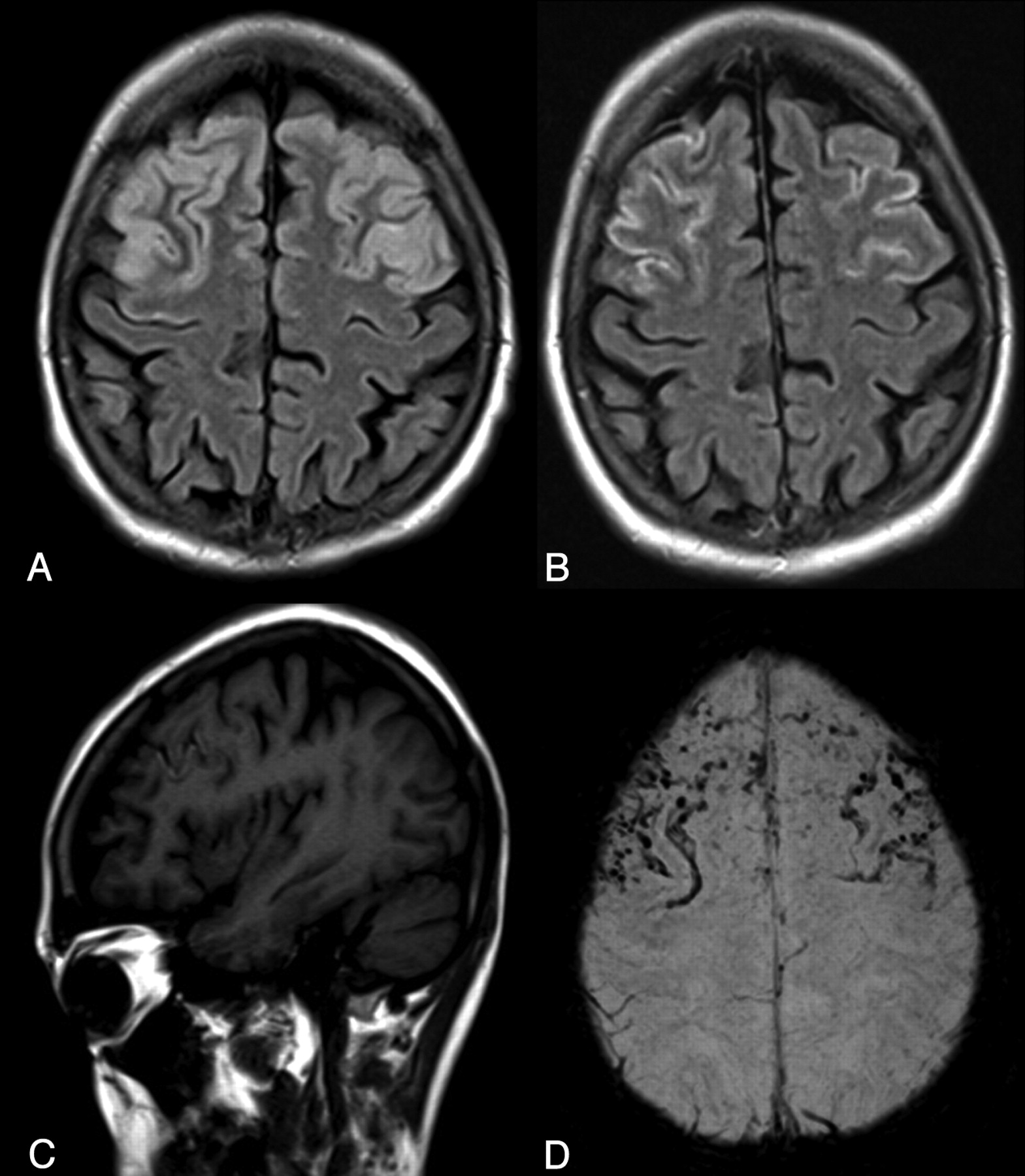
We report a 27-year-old woman with Crohn disease who underwent total colectomy due to partial intestinal occlusion and severe disease activity. Two weeks after total parenteral nutrition, she developed aphasia, diplopia, seizures, and acute confusional state. The neurologic examination revealed horizontal nystagmus, bilateral sixth and seventh nerve palsies, tetraparesis, and increased deep tendon reflexes with symmetric spasticity in both upper limbs. Bilateral Babinski reflexes were observed. MR imaging showed symmetric and bilateral hyperintensities on T2-weighted, fluid-attenuated inversion recovery (FLAIR), and diffusion-weighted images involving the hypoglossal, abducens and facial nuclei, medial right thalami, tectum of the midbrain, and extensive symmetric cortical abnormalities in the frontal lobes. The diagnosis of WE was suggested, and treatment with intravenous thiamine was started. Neurologic manifestations such as mental confusion, seizures, cranial nerve palsies, and nystagmus partially improved in the following 2 weeks, except for aphasia and tetraparesis, with spasticity mainly in the upper limbs, confirming the worse prognosis of the cortical lesions. Follow-up brain MR imaging 3 weeks later revealed improvement of most of the lesions. However, the areas of signal-intensity abnormality in the cortex of the frontal lobes persisted, revealing now peripheral high signal intensity on T1-weighted images (cortical laminar necrosis) and extensive areas of marked low-signal-intensity susceptibility-weighted MR phase images (SWI) (blood deposits) (Fig 1).

Fig 1.
  • Download figure
  • Open in new tab
  • Download powerpoint
Fig 1.

A, Axial FLAIR image shows extensive bilateral frontal cortical high signal intensity. B, Follow-up FLAIR image demonstrates significant improvement of the cortical lesions. C, Sagittal T1-weighted image shows cortical linear hyperintensities in the right frontal lobe (cortical laminar necrosis). D, SWI minimum-intensity-projection image shows extensive linear and focal areas of marked low signal intensity along the cortical margins in frontal lobes (hemosiderin/ferritin deposits).

This patient had nonalcoholic WE demonstrating atypical MR imaging findings and rare extensive cortical frontal lesions, progressing to cortical laminar necrosis and hemorrhage. To the best of our knowledge, cortical laminar necrosis and hemorrhage demonstrated with SWI in patients with WE has not been previously reported. SWI is highly sensitive and superior to T2*-weighted gradient-echo sequences to even traces of paramagnetic substances, such as blood products and calcifications.3 In a previous study of pediatric patients with cortical laminar necrosis evaluated with SWI, only 20% of the patients had signals of laminar or dotted hemorrhage.4 In conclusion, MR imaging follow-up is essential for monitoring patients with WE with cortical abnormalities due to the risk of irreversibility of lesions, which may progress to cortical laminar necrosis and hemorrhage.

References

  1. 1.↵
    1. Zuccoli G,
    2. Santa Cruz D,
    3. Bertolini M,
    4. et al
    . MR imaging findings in 56 patients with Wernicke encephalopathy: nonalcoholics may differ from alcoholics. AJNR Am J Neuroradiol 2009;30:171–76
    Abstract/FREE Full Text
  2. 2.↵
    1. Hygino da Cruz LC Jr.,
    2. Domingues RC,
    3. Vilanova I,
    4. et al
    . MR imaging findings in Wernicke encephalopathy: nonalcoholics may be similar to alcoholics. AJNR Am J Neuroradiol 2010;31:E54
    FREE Full Text
  3. 3.↵
    1. de Souza JM,
    2. Domingues RC,
    3. Cruz LC Jr.,
    4. et al
    . Susceptibility-weighted imaging for the evaluation of patients with familial cerebral cavernous malformations: a comparison with T2-weighted fast spin-echo and gradient-echo sequences. AJNR Am J Neuroradiol 2008;29:154–58
    Abstract/FREE Full Text
  4. 4.↵
    1. Niwa T,
    2. Aida N,
    3. Shishikura A,
    4. et al
    . Susceptibility-weighted imaging findings of cortical laminar necrosis in pediatric patients. AJNR Am J Neuroradiol 2008;29:1795–98
    Abstract/FREE Full Text
  • Copyright © American Society of Neuroradiology
PreviousNext
Back to top

In this issue

American Journal of Neuroradiology: 32 (2)
American Journal of Neuroradiology
Vol. 32, Issue 2
1 Feb 2011
  • Table of Contents
  • Index by author
Advertisement
Print
Download PDF
Email Article

Thank you for your interest in spreading the word on American Journal of Neuroradiology.

NOTE: We only request your email address so that the person you are recommending the page to knows that you wanted them to see it, and that it is not junk mail. We do not capture any email address.

Enter multiple addresses on separate lines or separate them with commas.
Nonalcoholic Wernicke Encephalopathy with Extensive Cortical Involvement: Cortical Laminar Necrosis and Hemorrhage Demonstrated with Susceptibility-Weighted MR Phase Images
(Your Name) has sent you a message from American Journal of Neuroradiology
(Your Name) thought you would like to see the American Journal of Neuroradiology web site.
CAPTCHA
This question is for testing whether or not you are a human visitor and to prevent automated spam submissions.
Cite this article
D.B. Pereira, M.L. Pereira, E.L. Gasparetto
Nonalcoholic Wernicke Encephalopathy with Extensive Cortical Involvement: Cortical Laminar Necrosis and Hemorrhage Demonstrated with Susceptibility-Weighted MR Phase Images
American Journal of Neuroradiology Feb 2011, 32 (2) E37-E38; DOI: 10.3174/ajnr.A2359

Citation Manager Formats

  • BibTeX
  • Bookends
  • EasyBib
  • EndNote (tagged)
  • EndNote 8 (xml)
  • Medlars
  • Mendeley
  • Papers
  • RefWorks Tagged
  • Ref Manager
  • RIS
  • Zotero
0 Responses
Respond to this article
Share
Bookmark this article
Nonalcoholic Wernicke Encephalopathy with Extensive Cortical Involvement: Cortical Laminar Necrosis and Hemorrhage Demonstrated with Susceptibility-Weighted MR Phase Images
D.B. Pereira, M.L. Pereira, E.L. Gasparetto
American Journal of Neuroradiology Feb 2011, 32 (2) E37-E38; DOI: 10.3174/ajnr.A2359
del.icio.us logo Twitter logo Facebook logo Mendeley logo
  • Tweet Widget
  • Facebook Like
  • Google Plus One
Purchase

Jump to section

  • Article
    • References
  • Figures & Data
  • Info & Metrics
  • Responses
  • References
  • PDF

Related Articles

  • No related articles found.
  • PubMed
  • Google Scholar

Cited By...

  • Cortical abnormalities on MRI: what a neurologist should know
  • Crossref (15)
  • Google Scholar

This article has been cited by the following articles in journals that are participating in Crossref Cited-by Linking.

  • MR Imaging Findings in Alcoholic and Nonalcoholic Acute Wernicke’s Encephalopathy: A Review
    Gaetana Manzo, Angela De Gennaro, Attilio Cozzolino, Antonietta Serino, Giacomo Fenza, Andrea Manto
    BioMed Research International 2014 2014
  • Wernicke's encephalopathy in Crohn's disease and ulcerative colitis
    Erik Oudman, Jan W. Wijnia, Misha J. Oey, Mirjam van Dam, Albert Postma
    Nutrition 2021 86
  • Cortical damage in Wernicke’s encephalopathy with good prognosis: a report of two cases and literature review
    Lei Wu, Di Jin, Xuan Sun, Liang Liang, Deihui Huang, Zhao Dong, Shengyuan Yu
    Metabolic Brain Disease 2017 32 2
  • Wernicke’s encephalopathy in a malnourished surgical patient: a difficult diagnosis
    Stefano Busani, Cinzia Bonvecchio, Arianna Gaspari, Marcella Malagoli, Alessandra Todeschini, Nicola Cautero, Massimo Girardis
    BMC Research Notes 2014 7 1
  • Wernicke’s encephalopathy in a patient with acute pancreatitis: unusual cortical involvement and marvelous prognosis
    Hong-Wei Cui, Bo-Ai Zhang, Tao Peng, Yu Liu, Yan-Ru Liu
    Neurological Sciences 2012 33 3
  • Cortical abnormalities on MRI: what a neurologist should know
    Dimitri Renard, Giovanni Castelnovo, Stephane Bouly, Anne Le Floch, Anne Waconge, Marie De Verdal, Eric Thouvenot
    Practical Neurology 2015 15 4
  • Epileptic seizures in nonalcoholic Wernicke’s encephalopathy: a case report and literature review
    Wenjin Shang, Xiuhui Chen, Xunhua Li, Hongbing Chen, Shujin Tang, Hua Hong
    Metabolic Brain Disease 2017 32 6
  • Non-alcoholic Wernicke’s encephalopathy with cortical involvement and polyneuropathy following gastrectomy
    Wei-Chia Tsao, Long-Sun Ro, Chiung-Mei Chen, Hong-Chiu Chang, Hung-Chou Kuo
    Metabolic Brain Disease 2017 32 5
  • A Case of Cortical Involvement in Marchiafava-Bignami Disease Accompanying Wernicke's Encephalopathy
    Michelle Youn, Jung-Ju Lee, Jong-Moo Park, Kyusik Kang, Ohyun Kwon, Woong-Woo Lee, Byung-Kun Kim
    Journal of Clinical Neurology 2021 17 3
  • Gayet Wernicke's encephalopathy with cortical damage following a subtotal gastrectomy: An uncommon association
    Meriam Benzalim, Soumaya Arharas, Soumaya Alj, Youssef Elouardi, Mohamed Khallouki
    Radiology Case Reports 2021 16 1

More in this TOC Section

  • Reply:
  • Brain AVM’s Nidus: What if We Hadn’t Understood Anything?
  • Letter to the Editor regarding “Automated Volumetric Software in Dementia: Help or Hindrance to the Neuroradiologist?”
Show more Letters

Similar Articles

Advertisement

Indexed Content

  • Current Issue
  • Accepted Manuscripts
  • Article Preview
  • Past Issues
  • Editorials
  • Editors Choice
  • Fellow Journal Club
  • Letters to the Editor

Cases

  • Case Collection
  • Archive - Case of the Week
  • Archive - Case of the Month
  • Archive - Classic Case

Special Collections

  • Special Collections

Resources

  • News and Updates
  • Turn around Times
  • Submit a Manuscript
  • Author Policies
  • Manuscript Submission Guidelines
  • Evidence-Based Medicine Level Guide
  • Publishing Checklists
  • Graphical Abstract Preparation
  • Imaging Protocol Submission
  • Submit a Case
  • Become a Reviewer/Academy of Reviewers
  • Get Peer Review Credit from Publons

Multimedia

  • AJNR Podcast
  • AJNR SCANtastic
  • Video Articles

About Us

  • About AJNR
  • Editorial Board
  • Not an AJNR Subscriber? Join Now
  • Alerts
  • Feedback
  • Advertise with us
  • Librarian Resources
  • Permissions
  • Terms and Conditions

American Society of Neuroradiology

  • Not an ASNR Member? Join Now

© 2025 by the American Society of Neuroradiology All rights, including for text and data mining, AI training, and similar technologies, are reserved.
Print ISSN: 0195-6108 Online ISSN: 1936-959X

Powered by HighWire