Advertisement

Main menu

  • Home
  • Content
    • Current Issue
    • Accepted Manuscripts
    • Article Preview
    • Past Issue Archive
    • Video Articles
    • AJNR Case Collection
    • Case of the Week Archive
    • Case of the Month Archive
    • Classic Case Archive
  • Special Collections
    • AJNR Awards
    • Low-Field MRI
    • Alzheimer Disease
    • ASNR Foundation Special Collection
    • Photon-Counting CT
    • View All
  • Multimedia
    • AJNR Podcasts
    • AJNR SCANtastic
    • Trainee Corner
    • MRI Safety Corner
    • Imaging Protocols
  • For Authors
    • Submit a Manuscript
    • Submit a Video Article
    • Submit an eLetter to the Editor/Response
    • Manuscript Submission Guidelines
    • Statistical Tips
    • Fast Publishing of Accepted Manuscripts
    • Graphical Abstract Preparation
    • Imaging Protocol Submission
    • Author Policies
  • About Us
    • About AJNR
    • Editorial Board
    • Editorial Board Alumni
  • More
    • Become a Reviewer/Academy of Reviewers
    • Subscribers
    • Permissions
    • Alerts
    • Feedback
    • Advertisers
    • ASNR Home

User menu

  • Alerts
  • Log in

Search

  • Advanced search
American Journal of Neuroradiology

American Journal of Neuroradiology

ASHNR American Society of Functional Neuroradiology ASHNR American Society of Pediatric Neuroradiology ASSR
  • Alerts
  • Log in

Advanced Search

  • Home
  • Content
    • Current Issue
    • Accepted Manuscripts
    • Article Preview
    • Past Issue Archive
    • Video Articles
    • AJNR Case Collection
    • Case of the Week Archive
    • Case of the Month Archive
    • Classic Case Archive
  • Special Collections
    • AJNR Awards
    • Low-Field MRI
    • Alzheimer Disease
    • ASNR Foundation Special Collection
    • Photon-Counting CT
    • View All
  • Multimedia
    • AJNR Podcasts
    • AJNR SCANtastic
    • Trainee Corner
    • MRI Safety Corner
    • Imaging Protocols
  • For Authors
    • Submit a Manuscript
    • Submit a Video Article
    • Submit an eLetter to the Editor/Response
    • Manuscript Submission Guidelines
    • Statistical Tips
    • Fast Publishing of Accepted Manuscripts
    • Graphical Abstract Preparation
    • Imaging Protocol Submission
    • Author Policies
  • About Us
    • About AJNR
    • Editorial Board
    • Editorial Board Alumni
  • More
    • Become a Reviewer/Academy of Reviewers
    • Subscribers
    • Permissions
    • Alerts
    • Feedback
    • Advertisers
    • ASNR Home
  • Follow AJNR on Twitter
  • Visit AJNR on Facebook
  • Follow AJNR on Instagram
  • Join AJNR on LinkedIn
  • RSS Feeds

AJNR Awards, New Junior Editors, and more. Read the latest AJNR updates

Research ArticleHead and Neck Imaging
Open Access

Identification of Intraplaque Hemorrhage on MR Angiography Images: A Comparison of Contrast-Enhanced Mask and Time-of-Flight Techniques

Y. Qiao, M. Etesami, S. Malhotra, B.C. Astor, R. Virmani, F.D. Kolodgie, H.H. Trout and B.A. Wasserman
American Journal of Neuroradiology March 2011, 32 (3) 454-459; DOI: https://doi.org/10.3174/ajnr.A2320
Y. Qiao
  • Find this author on Google Scholar
  • Find this author on PubMed
  • Search for this author on this site
M. Etesami
  • Find this author on Google Scholar
  • Find this author on PubMed
  • Search for this author on this site
S. Malhotra
  • Find this author on Google Scholar
  • Find this author on PubMed
  • Search for this author on this site
B.C. Astor
  • Find this author on Google Scholar
  • Find this author on PubMed
  • Search for this author on this site
R. Virmani
  • Find this author on Google Scholar
  • Find this author on PubMed
  • Search for this author on this site
F.D. Kolodgie
  • Find this author on Google Scholar
  • Find this author on PubMed
  • Search for this author on this site
H.H. Trout III
  • Find this author on Google Scholar
  • Find this author on PubMed
  • Search for this author on this site
B.A. Wasserman
  • Find this author on Google Scholar
  • Find this author on PubMed
  • Search for this author on this site
  • Article
  • Figures & Data
  • Info & Metrics
  • Responses
  • References
  • PDF
Loading

Article Figures & Data

Figures

  • Tables
  • Fig 1.
    • Download figure
    • Open in new tab
    • Download powerpoint
    Fig 1.

    Coregistration of MRA and histologic images. MIP image from the TOF-MRA that profiles the narrowed lumen contour (A) used to show the position of 8 reformatted TOF (B) and CE-MRA mask (C) sections (reconstructed thickness, 2 mm), to orient transverse 5-μm histologic sections through the specimen (D). IPH is identified as hyperintense signal intensity compared with adjacent muscle on MRA images (arrows, B and C) and is red on histologic sections (D) stained with Movat pentachrome.

  • Fig 2.
    • Download figure
    • Open in new tab
    • Download powerpoint
    Fig 2.

    Identifying IPH on CE-MRA mask images in a 72-year-old man with a right cerebral ischemic event. Coronal and reconstructed axial (inset) sections from the mask (A) series show hyperintense signal intensity (arrows) in the right carotid artery wall. The high signal intensity (arrows) is again seen on the corresponding postcontrast images (B) but is removed after the mask images are subtracted from this series (C). (Asterisk indicates the lumen.) MIP images are generated from the subtracted series (D), so IPH information is lost.
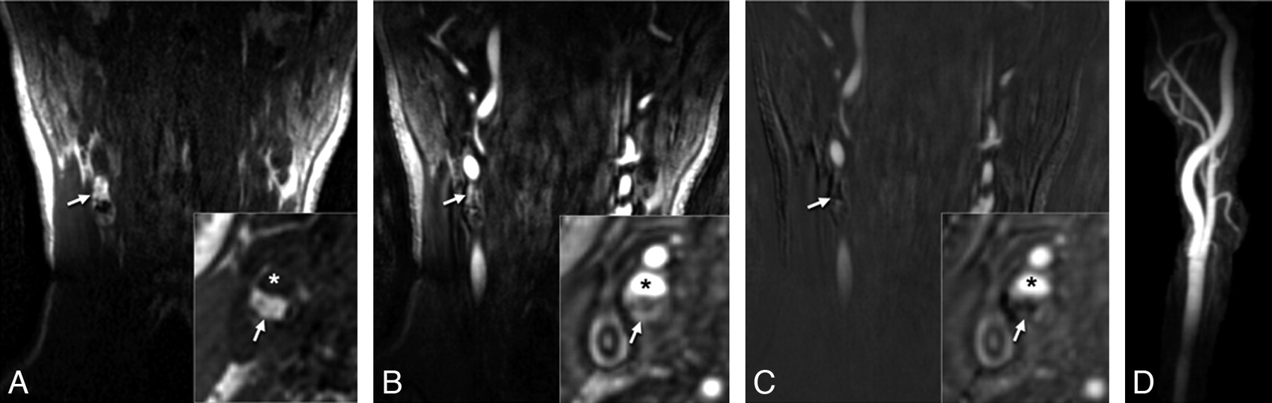
  • Fig 3.
    • Download figure
    • Open in new tab
    • Download powerpoint
    Fig 3.

    MRA images with matched histology for a 73-year-old man with a right cerebral ischemic event. Hyperintense signal intensity on the postcontrast (arrow, A) and mask (arrow, B) CE-MRA images (asterisk indicates the lumen) corresponds to red staining representing IPH on histology (C, Movat pentachrome stain). Immunostaining for glycophorin A (D) confirms the presence of erythrocytes (dark brown staining) mixed with cholesterol clefts (arrows, D) for a region within the core (box, C).
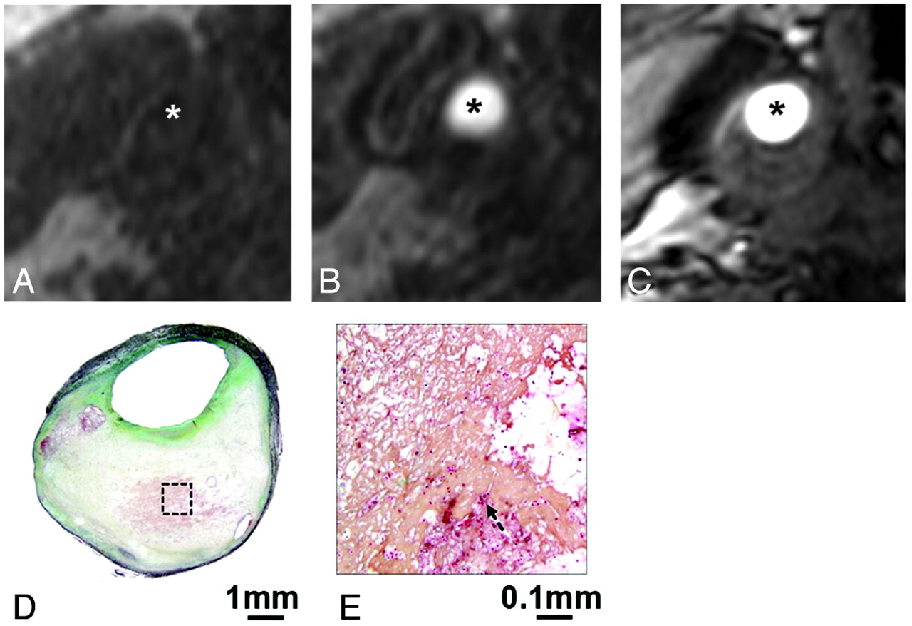
  • Fig 4.
    • Download figure
    • Open in new tab
    • Download powerpoint
    Fig 4.

    An example of IPH missed on both CE- and TOF-MRA. Reconstructed axial mask (A), postcontrast (B), and TOF-MRA (C) images in matched locations transversely through a carotid plaque show no hyperintense signal intensity to suggest IPH (asterisk indicates the lumen). The corresponding CEA specimen section stained with Movat pentachrome demonstrates an attenuated fibrin network. A high-power view (E) of a region of necrotic core (box, D) shows scattered erythrocytes (arrow, E).

Tables

  • Figures
  • Agreement between IPH detected by MR imaging and histology

    ReaderHistology +Histology −Total
    No. of Images6381144
    CE-MRA maska1+57259
    −67985
    2+53053
    −108191
    TOFb1+501161
    −137083
    2+501060
    −137184
    • a Reader 1: Sensitivity, specificity, PPV, and NPV (95% CI) are 90% (80%–96%), 98% (91%–100%), 97% (87%–99%), and 93% (85%–97%), respectively. Reader 2: Sensitivity, specificity, PPV, and NPV (95% CI) are 84% (72%–92%), 100% (94%–100%), 100% (92%–100%), and 89% (80%–94%), respectively.

    • b Reader 1: Sensitivity, specificity, PPV, and NPV (95% CI) are 79% (66%–88%), 86% (77%–93%), 82% (70%–90%), and 84% (74%–91%), respectively. Reader 2: Sensitivity, specificity, PPV, and NPV (95% CI) are 79% (67%–88%), 88% (78%–94%), 83% (71%–91%), and 85% (75%–91%), respectively.

PreviousNext
Back to top

In this issue

American Journal of Neuroradiology: 32 (3)
American Journal of Neuroradiology
Vol. 32, Issue 3
1 Mar 2011
  • Table of Contents
  • Index by author
Advertisement
Print
Download PDF
Email Article

Thank you for your interest in spreading the word on American Journal of Neuroradiology.

NOTE: We only request your email address so that the person you are recommending the page to knows that you wanted them to see it, and that it is not junk mail. We do not capture any email address.

Enter multiple addresses on separate lines or separate them with commas.
Identification of Intraplaque Hemorrhage on MR Angiography Images: A Comparison of Contrast-Enhanced Mask and Time-of-Flight Techniques
(Your Name) has sent you a message from American Journal of Neuroradiology
(Your Name) thought you would like to see the American Journal of Neuroradiology web site.
CAPTCHA
This question is for testing whether or not you are a human visitor and to prevent automated spam submissions.
Cite this article
Y. Qiao, M. Etesami, S. Malhotra, B.C. Astor, R. Virmani, F.D. Kolodgie, H.H. Trout, B.A. Wasserman
Identification of Intraplaque Hemorrhage on MR Angiography Images: A Comparison of Contrast-Enhanced Mask and Time-of-Flight Techniques
American Journal of Neuroradiology Mar 2011, 32 (3) 454-459; DOI: 10.3174/ajnr.A2320

Citation Manager Formats

  • BibTeX
  • Bookends
  • EasyBib
  • EndNote (tagged)
  • EndNote 8 (xml)
  • Medlars
  • Mendeley
  • Papers
  • RefWorks Tagged
  • Ref Manager
  • RIS
  • Zotero
0 Responses
Respond to this article
Share
Bookmark this article
Identification of Intraplaque Hemorrhage on MR Angiography Images: A Comparison of Contrast-Enhanced Mask and Time-of-Flight Techniques
Y. Qiao, M. Etesami, S. Malhotra, B.C. Astor, R. Virmani, F.D. Kolodgie, H.H. Trout, B.A. Wasserman
American Journal of Neuroradiology Mar 2011, 32 (3) 454-459; DOI: 10.3174/ajnr.A2320
del.icio.us logo Twitter logo Facebook logo Mendeley logo
  • Tweet Widget
  • Facebook Like
  • Google Plus One
Purchase

Jump to section

  • Article
    • Abstract
    • Abbreviations
    • Materials and Methods
    • Results
    • Discussion
    • Conclusions
    • Footnotes
    • References
  • Figures & Data
  • Info & Metrics
  • Responses
  • References
  • PDF

Related Articles

  • No related articles found.
  • PubMed
  • Google Scholar

Cited By...

  • Double lumens and flaps (and their mimics) on CT and MR angiography
  • Carotid Artery Wall Imaging: Perspective and Guidelines from the ASNR Vessel Wall Imaging Study Group and Expert Consensus Recommendations of the American Society of Neuroradiology
  • Asymptomatic moderate carotid artery stenosis with intraplaque hemorrhage: onset of new ischemic stroke
  • Magnetic Resonance Angiography Detection of Abnormal Carotid Artery Plaque in Patients With Cryptogenic Stroke
  • Carotid Plaque Characterization Using 3D T1-Weighted MR Imaging with Histopathologic Validation: A Comparison with 2D Technique
  • Is Carotid Intima-Media Thickness as Predictive as Other Noninvasive Techniques for the Detection of Coronary Artery Disease?
  • Predicting Carotid Plaque Characteristics Using Quantitative Color-Coded T1-Weighted MR Plaque Imaging: Correlation with Carotid Endarterectomy Specimens
  • Intraplaque High-Intensity Signal on 3D Time-of-Flight MR Angiography Is Strongly Associated with Symptomatic Carotid Artery Stenosis
  • Safety of Protected Carotid Artery Stenting in Patients with Severe Carotid Artery Stenosis and Carotid Intraplaque Hemorrhage
  • Carotid Plaque Neovascularization and Hemorrhage Detected by MR Imaging are Associated with Recent Cerebrovascular Ischemic Events
  • Crossref
  • Google Scholar

This article has not yet been cited by articles in journals that are participating in Crossref Cited-by Linking.

More in this TOC Section

  • Hydrops Herniation into the Semicircular Canals
  • ASL Sensitivity for Head and Neck Paraganglioma
  • Post SRS Peritumoral Hyperintense Signal of VSs
Show more HEAD AND NECK IMAGING

Similar Articles

Advertisement

Indexed Content

  • Current Issue
  • Accepted Manuscripts
  • Article Preview
  • Past Issues
  • Editorials
  • Editor's Choice
  • Fellows' Journal Club
  • Letters to the Editor
  • Video Articles

Cases

  • Case Collection
  • Archive - Case of the Week
  • Archive - Case of the Month
  • Archive - Classic Case

More from AJNR

  • Trainee Corner
  • Imaging Protocols
  • MRI Safety Corner
  • Book Reviews

Multimedia

  • AJNR Podcasts
  • AJNR Scantastics

Resources

  • Turnaround Time
  • Submit a Manuscript
  • Submit a Video Article
  • Submit an eLetter to the Editor/Response
  • Manuscript Submission Guidelines
  • Statistical Tips
  • Fast Publishing of Accepted Manuscripts
  • Graphical Abstract Preparation
  • Imaging Protocol Submission
  • Evidence-Based Medicine Level Guide
  • Publishing Checklists
  • Author Policies
  • Become a Reviewer/Academy of Reviewers
  • News and Updates

About Us

  • About AJNR
  • Editorial Board
  • Editorial Board Alumni
  • Alerts
  • Permissions
  • Not an AJNR Subscriber? Join Now
  • Advertise with Us
  • Librarian Resources
  • Feedback
  • Terms and Conditions
  • AJNR Editorial Board Alumni

American Society of Neuroradiology

  • Not an ASNR Member? Join Now

© 2025 by the American Society of Neuroradiology All rights, including for text and data mining, AI training, and similar technologies, are reserved.
Print ISSN: 0195-6108 Online ISSN: 1936-959X

Powered by HighWire