Advertisement

Main menu

  • Home
  • Content
    • Current Issue
    • Accepted Manuscripts
    • Article Preview
    • Past Issue Archive
    • AJNR Case Collection
    • Case of the Week Archive
    • Classic Case Archive
    • Case of the Month Archive
  • Special Collections
    • Spinal CSF Leak Articles (Jan 2020-June 2024)
    • 2024 AJNR Journal Awards
    • Most Impactful AJNR Articles
  • Multimedia
    • AJNR Podcast
    • AJNR Scantastics
    • Video Articles
  • For Authors
    • Submit a Manuscript
    • Author Policies
    • Fast publishing of Accepted Manuscripts
    • Graphical Abstract Preparation
    • Manuscript Submission Guidelines
    • Imaging Protocol Submission
    • Submit a Case for the Case Collection
  • About Us
    • About AJNR
    • Editorial Board
  • More
    • Become a Reviewer/Academy of Reviewers
    • Subscribers
    • Permissions
    • Alerts
    • Feedback
    • Advertisers
    • ASNR Home
  • Other Publications
    • ajnr

User menu

  • Alerts
  • Log in

Search

  • Advanced search
American Journal of Neuroradiology

American Journal of Neuroradiology

ASHNR American Society of Functional Neuroradiology ASHNR American Society of Pediatric Neuroradiology ASSR
  • Alerts
  • Log in

Advanced Search

  • Home
  • Content
    • Current Issue
    • Accepted Manuscripts
    • Article Preview
    • Past Issue Archive
    • AJNR Case Collection
    • Case of the Week Archive
    • Classic Case Archive
    • Case of the Month Archive
  • Special Collections
    • Spinal CSF Leak Articles (Jan 2020-June 2024)
    • 2024 AJNR Journal Awards
    • Most Impactful AJNR Articles
  • Multimedia
    • AJNR Podcast
    • AJNR Scantastics
    • Video Articles
  • For Authors
    • Submit a Manuscript
    • Author Policies
    • Fast publishing of Accepted Manuscripts
    • Graphical Abstract Preparation
    • Manuscript Submission Guidelines
    • Imaging Protocol Submission
    • Submit a Case for the Case Collection
  • About Us
    • About AJNR
    • Editorial Board
  • More
    • Become a Reviewer/Academy of Reviewers
    • Subscribers
    • Permissions
    • Alerts
    • Feedback
    • Advertisers
    • ASNR Home
  • Follow AJNR on Twitter
  • Visit AJNR on Facebook
  • Follow AJNR on Instagram
  • Join AJNR on LinkedIn
  • RSS Feeds

Welcome to the new AJNR, Updated Hall of Fame, and more. Read the full announcements.


AJNR is seeking candidates for the position of Associate Section Editor, AJNR Case Collection. Read the full announcement.


LetterLetter

Imaging of Bilateral Neck Paragangliomas with 68Ga-DOTATATE Positron-Emission Tomography/CT

R.C. Quah
American Journal of Neuroradiology April 2011, 32 (4) E71-E72; DOI: https://doi.org/10.3174/ajnr.A2472
R.C. Quah
  • Find this author on Google Scholar
  • Find this author on PubMed
  • Search for this author on this site
  • Article
  • Figures & Data
  • Info & Metrics
  • Responses
  • References
  • PDF
Loading

Paragangliomas of the neck are rare somatostatin receptor-expressing neuroendocrine tumors. Somatostatin-receptor scintigraphy is useful in diagnosing and staging these tumors, as well as assessing residual or recurrent disease posttreatment. We describe the gallium 68 DOTA, DPhe1, Tyr3-octreotate positron-emission tomography-CT (68Ga-DOTATATE PET-CT) findings of bilateral neck paragangliomas as seen in the following patient.

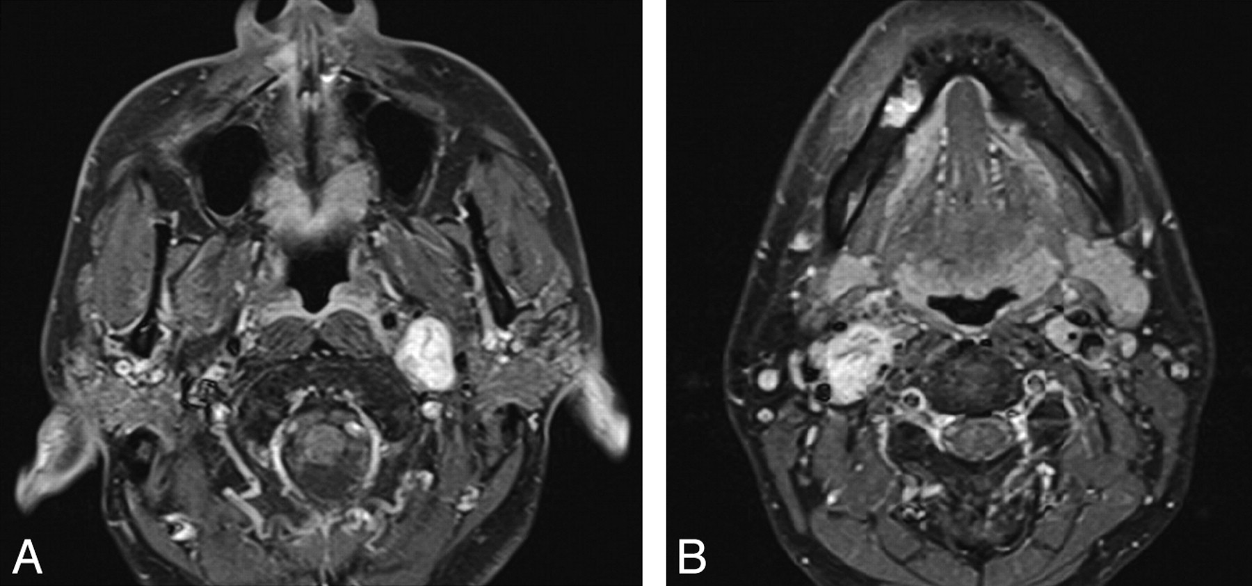
A previously healthy 40-year-old woman presented with asymmetric sensorineural hearing loss, right more than left, which was confirmed on an audiogram. There was no other neurologic deficit. MR imaging of the internal auditory meati did not reveal any abnormality at the cerebellopontine angles or internal auditory canals; however, a left carotid space mass was partially visualized. A dedicated MR image of the neck revealed avidly enhancing lobulated masses within bilateral carotid spaces (Figs 1 and 2 A). The right neck mass extended from the level of the carotid bifurcation to the level of the nasopharynx and splayed the right internal and external carotid arteries. The left neck mass extended from the level of the oropharynx to the left jugular foramen, splaying the left internal jugular vein and internal carotid artery. 68Ga-DOTATATE PET-CT demonstrated intense tracer uptake within the bilateral carotid space masses (Fig 2B). There was no other abnormal focus of increased tracer uptake in the rest of the body to suggest the presence of metastasis. These imaging findings confirmed a right carotid body tumor and a left glomus vagale. Biochemical screening revealed elevated urine norepinephrine (622 nmol/day; normal, 89–473 nmol/day) and normetanephrine (2895 nmol/day; normal, 600-1900 nmol/day) levels. The patient opted for and is scheduled to undergo radiation therapy.

Fig 1.
  • Download figure
  • Open in new tab
  • Download powerpoint
Fig 1.

Axial T1-weighted contrast-enhanced MR images reveals avidly enhancing lobulated masses in the bilateral carotid spaces of the neck, containing serpiginous signal-intensity voids.

Fig 2.
  • Download figure
  • Open in new tab
  • Download powerpoint
Fig 2.

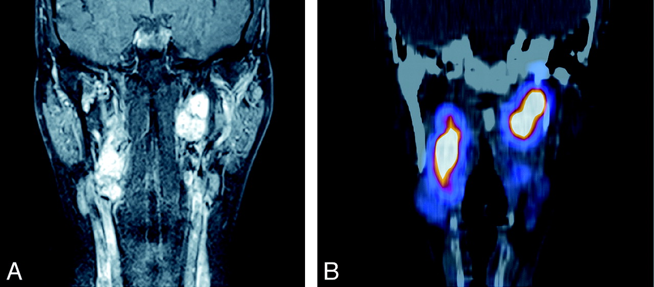
Coronal T1-weighted contrast-enhanced MR image (A) and coronal 68Ga-DOTATATE PET-CT image (B) reveal intense tracer uptake within the enhancing bilateral carotid space masses.

DOTATATE is a somatostatin analog, which can be radio-labeled with 68Ga. 68Ga is a positron emitter with a half-life of 68 minutes, produced by elution from 68Ge in a 68Ga generator. 68Ga-DOTATATE shows a high affinity for somatostatin type 2 receptors (SSR-2), is rapidly excreted from nontarget sites, and offers good target-to-nontarget imaging properties. Compared with indium 111-diethylene triamine pentaacetic acid-octreotide, 68Ga-DOTATATE demonstrates better sensitivity for neuroendocrine tumors1 and is useful for the diagnosis of small tumors and evaluation of disease response following therapy.2 The mainstay of treatment for paragangliomas in the head and neck region has been surgical resection, preoperative embolization, and/or external beam radiation therapy, though a role for stereotactic radiosurgery is emerging.3 There is also a promising role for peptide-receptor radionuclide therapy, where the somatostatin analog DOTATATE is radio-labeled with the β emitter lutetium 177 (177Lu) to deliver highly targeted doses to tumors expressing the SSR-2, with low doses to the normal organs.4

Our case highlights the intense and specific tracer uptake of 68Ga-DOTATATE in paragangliomas of the neck, which will be useful if findings on other imaging modalities are equivocal. There is also a promising role for using 177Lu-DOTATATE for highly targeted radionuclide therapy of these tumors.

References

  1. 1.↵
    1. Srirajaskanthan R,
    2. Kayani I,
    3. Quigley AM,
    4. et al
    . The role of 68Ga-DOTATATE PET in patients with neuroendocrine tumors and negative or equivocal findings on 111In-DTPA-octreotide scintigraphy. J Nucl Med 2010;51:875–82.Epub 2010 May 19
    Abstract/FREE Full Text
  2. 2.↵
    1. Haug AR,
    2. Auernhammer CJ,
    3. Wängler B,
    4. et al
    . 68Ga-DOTATATE PET/CT for the early prediction of response to somatostatin receptor-mediated radionuclide therapy in patients with well differentiated neuroendocrine tumors. J Nucl Med 2010;51:1349–56
    Abstract/FREE Full Text
  3. 3.↵
    1. Kupferman ME,
    2. Hanna EY
    . Paragangliomas of the head and neck. Curr Oncol Rep 2008;10:156–61
    CrossRefPubMed
  4. 4.↵
    1. Bodei L,
    2. Pepe G,
    3. Paganelli G
    . Peptide receptor radionuclide therapy (PRRT) of neuroendocrine tumors with somatostatin analogues. Eur Rev Med Pharmacol Sci 2010;14:247–51
  • Copyright © American Society of Neuroradiology
PreviousNext
Back to top

In this issue

American Journal of Neuroradiology: 32 (4)
American Journal of Neuroradiology
Vol. 32, Issue 4
1 Apr 2011
  • Table of Contents
  • Index by author
Advertisement
Print
Download PDF
Email Article

Thank you for your interest in spreading the word on American Journal of Neuroradiology.

NOTE: We only request your email address so that the person you are recommending the page to knows that you wanted them to see it, and that it is not junk mail. We do not capture any email address.

Enter multiple addresses on separate lines or separate them with commas.
Imaging of Bilateral Neck Paragangliomas with 68Ga-DOTATATE Positron-Emission Tomography/CT
(Your Name) has sent you a message from American Journal of Neuroradiology
(Your Name) thought you would like to see the American Journal of Neuroradiology web site.
CAPTCHA
This question is for testing whether or not you are a human visitor and to prevent automated spam submissions.
Cite this article
R.C. Quah
Imaging of Bilateral Neck Paragangliomas with 68Ga-DOTATATE Positron-Emission Tomography/CT
American Journal of Neuroradiology Apr 2011, 32 (4) E71-E72; DOI: 10.3174/ajnr.A2472

Citation Manager Formats

  • BibTeX
  • Bookends
  • EasyBib
  • EndNote (tagged)
  • EndNote 8 (xml)
  • Medlars
  • Mendeley
  • Papers
  • RefWorks Tagged
  • Ref Manager
  • RIS
  • Zotero
0 Responses
Respond to this article
Share
Bookmark this article
Imaging of Bilateral Neck Paragangliomas with 68Ga-DOTATATE Positron-Emission Tomography/CT
R.C. Quah
American Journal of Neuroradiology Apr 2011, 32 (4) E71-E72; DOI: 10.3174/ajnr.A2472
del.icio.us logo Twitter logo Facebook logo Mendeley logo
  • Tweet Widget
  • Facebook Like
  • Google Plus One
Purchase

Jump to section

  • Article
    • References
  • Figures & Data
  • Info & Metrics
  • Responses
  • References
  • PDF

Related Articles

  • No related articles found.
  • PubMed
  • Google Scholar

Cited By...

  • Reply: 68Ga-DOTATATE PET/CT Versus MRI: Why the Comparison of 68Ga-DOTATATE PET/CT to an Appropriate MRI Protocol Is Essential
  • Crossref (3)
  • Google Scholar

This article has been cited by the following articles in journals that are participating in Crossref Cited-by Linking.

  • Reply: 68Ga-DOTATATE PET/CT Versus MRI: Why the Comparison of 68Ga-DOTATATE PET/CT to an Appropriate MRI Protocol Is Essential
    David Taïeb, Nicholas J. Patronas, Karel Pacak
    Journal of Nuclear Medicine 2017 58 1
  • Molecular Imaging in the Head and Neck
    Brandon A. Howard
    Radiologic Clinics of North America 2020 58 6
  • Invited Commentary
    Jaydira Del Rivero, Steven K. Libutti
    RadioGraphics 2015 35 2

More in this TOC Section

  • Reply:
  • Brain AVM’s Nidus: What if We Hadn’t Understood Anything?
  • Letter to the Editor regarding “Automated Volumetric Software in Dementia: Help or Hindrance to the Neuroradiologist?”
Show more Letters

Similar Articles

Advertisement

Indexed Content

  • Current Issue
  • Accepted Manuscripts
  • Article Preview
  • Past Issues
  • Editorials
  • Editors Choice
  • Fellow Journal Club
  • Letters to the Editor

Cases

  • Case Collection
  • Archive - Case of the Week
  • Archive - Case of the Month
  • Archive - Classic Case

Special Collections

  • Special Collections

Resources

  • News and Updates
  • Turn around Times
  • Submit a Manuscript
  • Author Policies
  • Manuscript Submission Guidelines
  • Evidence-Based Medicine Level Guide
  • Publishing Checklists
  • Graphical Abstract Preparation
  • Imaging Protocol Submission
  • Submit a Case
  • Become a Reviewer/Academy of Reviewers
  • Get Peer Review Credit from Publons

Multimedia

  • AJNR Podcast
  • AJNR SCANtastic
  • Video Articles

About Us

  • About AJNR
  • Editorial Board
  • Not an AJNR Subscriber? Join Now
  • Alerts
  • Feedback
  • Advertise with us
  • Librarian Resources
  • Permissions
  • Terms and Conditions

American Society of Neuroradiology

  • Not an ASNR Member? Join Now

© 2025 by the American Society of Neuroradiology All rights, including for text and data mining, AI training, and similar technologies, are reserved.
Print ISSN: 0195-6108 Online ISSN: 1936-959X

Powered by HighWire